
Research article · 2024 · Full text
International Journal of Advanced Biochemistry ResearchBeyond nourishment: The impact of micronutrients on Magnaporthe grisea sporulation infecting finger millet
Volume 8(4) · Pages 715–719 · DOI: 10.33545/26174693.2024.v8.i4i.1033 ↗
Research areasAluminum toxicity and tolerance in plants and animalsRice Cultivation and Yield ImprovementGABA and Rice Research
Abstract
Author-writtenFinger millet blast, caused by Magnaporthe grisea is a devastating and recurrent issue across all finger millet cultivation areas globally. The main challenge linked to cultivating the M. grisea culture involve achieving sporulation on culture plates. Therefore, to investigate the impact of amino acids (L-tyrosine, arginine and phenylalanine) and vitamins (riboflavin and ascorbic acid) in promoting sporulation, experiment was conducted at three concentrations (0.01g /l, 0.02 g/l and 0.03 g/l) using three different cultural media, rice straw extract agar (RSEA), oat meat agar (OMA) and a combination of RSEA and OMA. Among all the amendments, L-tyrosine at the concentration of 0.03 g/l recorded highest mean radial growth of 9.00 cm, which was highest among all the treatments. Whereas, RSEA+OMA amended with phenyl alanine at 0.03 g/l produced highest number of conidia (33.33 conidia/40x microscopic view).
Summary
Source-checkedPlain-language reading of the full paper, prepared with AI and checked against the text. Each point links to the authors' own words.
Why it matters
- Finger millet blast, caused by the fungus Magnaporthe grisea, is a recurring and devastating problem for finger millet crops worldwide. View evidence ↗
- The blast disease can cause significant agricultural losses, including a 35.78% reduction in grain yield and a 43.72% reduction in fodder yield. View evidence ↗
- Developing disease-resistant crop varieties requires testing many plant accessions, which in turn requires a reliable way to produce large amounts of fungal spores (sporulation). View evidence ↗
The question
- The study aimed to investigate how specific amino acids (L-tyrosine, arginine, and phenylalanine) and vitamins (riboflavin and ascorbic acid) affect the sporulation of M. grisea. View evidence ↗
What they did
- The researchers used three different growth media: Rice Straw Extract Agar (RSEA), Oat Meal Agar (OMA), and a combination of the two (RSEA+OMA). View evidence ↗
- The three amino acids and two vitamins were tested at three different concentrations: 0.01 g/l, 0.02 g/l, and 0.03 g/l. View evidence ↗
- The growth of the fungal mycelium and the amount of spore production were measured seven days after the fungus was inoculated into the media. View evidence ↗
What they found
- Among the base media, RSEA+OMA supported the highest mean radial growth (7.03 cm), while RSEA supported the lowest (6.50 cm). View evidence ↗
- The combination of RSEA+OMA supplemented with 0.03 g/l of L-tyrosine resulted in the maximum radial growth of 9.00 cm across all tested treatments. View evidence ↗
- The lowest mean radial growth (3.92 cm) was observed in RSEA media amended with ascorbic acid. View evidence ↗
- RSEA+OMA was the most effective base medium for sporulation, yielding a mean of 21.70 conidia per 40x microscopic view. View evidence ↗
- The highest mean sporulation was achieved using RSEA+OMA amended with phenylalanine, resulting in 29.33 conidia per 40x microscopic view. View evidence ↗
- The lowest level of sporulation (11.33 conidia per 40x microscopic view) occurred in OMA media amended with 0.01 g/l of arginine. View evidence ↗
- Specifically, the highest single measurement of conidia production was 33.33 per 40x microscopic field, found in RSEA+OMA amended with 0.03 g/l of phenylalanine. View evidence ↗
What it means
- The authors concluded that RSEA+OMA is the most suitable medium for promoting both the growth and sporulation of M. grisea. View evidence ↗
- By providing a method for abundant sporulation, this research facilitates the screening of plant genotypes to develop finger millet varieties resistant to blast disease. View evidence ↗
Visual summary
2 visualsIllustrations explain the paper; charts are replotted from the paper's own table values.
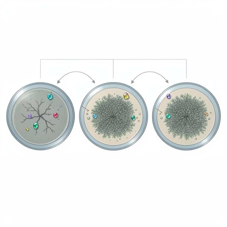
Experimental Design Overview
AI-generated conceptual illustration (Magnaporthe grisea fungus on finger millet growth media). It explains the idea and does not show study data.
View source evidence ↗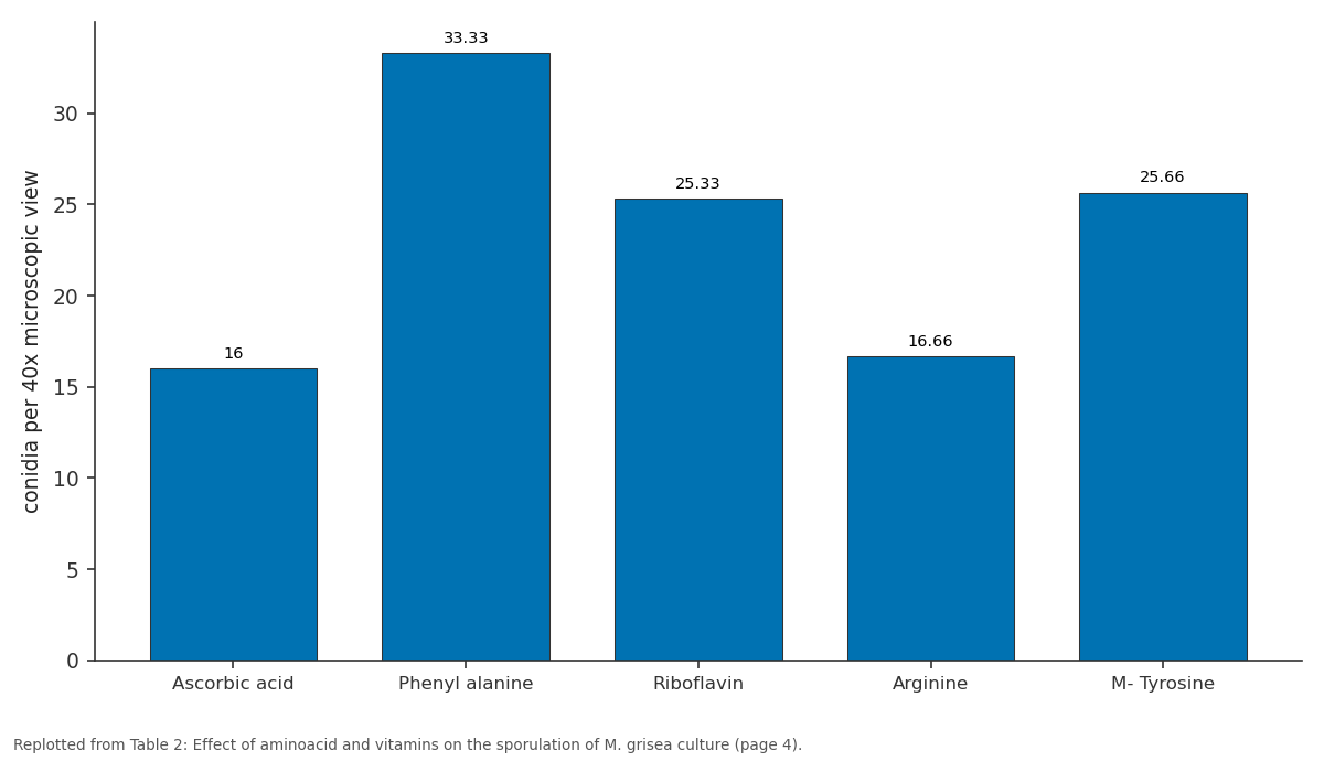
0.03 g/l: Ascorbic acid vs Phenyl alanine vs Riboflavin vs Arginine vs M- Tyrosine
Replotted from Table 2 (page 4) of the original paper. Every bar is the paper's own table value.
View source evidence ↗Figures & tables
From the original paper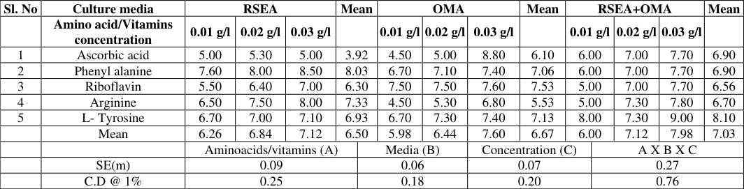 Table 1
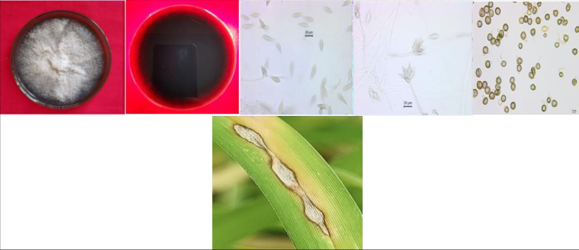 Figure 1
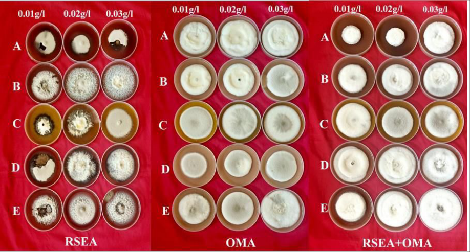 Figure 2
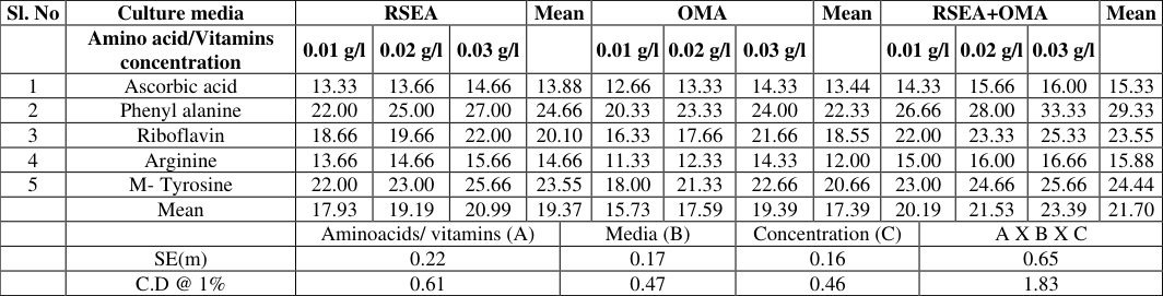 Table 2
Introduction
Finger millet is highly valued for its nutritional content, providing high levels of calcium, iron, protein and essential amino acids. Its gluten-free, making it a great choice for those with celiac disease. Its cultivation is also environmentally friendly due to its drought tolerance (Poonacha et al., 2023) [11] . Despite its resilience, the productivity of the plant is susceptible to various biotic and abiotic factors. Notably, the blast disease caused by Magnaporthe grisea incurs substantial yield losses. According to Prajapathi (2013) [12] the loss of grain yield occurred due to the blast disease was 35.78 per cent along with loss of fodder yield of 43.72 per cent. Nevertheless, the pathogen exhibits high variability, characterized by a pathogenic lifestyle distinguished by remarkable morphogenetic and biochemical specialization, resulting in the emergence of novel pathotypes. To develop resistant genotypes against these new pathotypes, it is imperative to evaluate a large number of accessions for blast disease resistance for which abundant sporulation of the pathogen being a crucial requirement. In experiments with three isolates of Pyricularia grisea originating from rice, finger millet and Panicum sp., prune agar and oatmeal agar were found to promote the highest levels of mycelial growth and sporulation for isolates from both rice and finger millet (Khadka et al., 2012) [6] . Rajashekara et al., 2016 [13] reported, the Rice straw extract oat meal agar media was suitable for inducing sporulation in M. oryzae cultures. Newly isolated cultures demonstrate a greater capacity to produce conidia in petri plates, but over time or during extended storage periods, their sporulation ability diminishes. So, use of media with common compositions will not be able to stimulate the sporulation in the cultures. The common challenge of achieving sporulation in Pyricularia sp. cultures is well recognized in studies (Narayanarao et al., 1972) [9] . Plant pathogens rely on amino acids as vital nutrients, crucial for regulating diverse biological functions within microorganisms. These functions encompass pivotal processes such as cell division, cell wall formation, cell growth, metabolism, inter-microbial communication (quorum sensing), and interactions with hosts (Idrees et al., 2020) [5] . Additionally, vitamins serve indispensable roles in the spore production of fungal plant pathogens by acting as co-factors in numerous enzymatic reactions essential for metabolism, growth, and development (Averianova et al., 2020) [1] . Thus, both amino acids and vitamins are intricately involved in the growth and development of plant pathogens. So, Keeping above problems in view, efforts have been undertaken to know the influence of micro-nutrients on the sporulation of M. grisea by supplementing the optimal media with micro-nutrients such as vitamins and amino acids.
Materials and Methods
Materials and Methods
Isolation of the pathogen and proving pathogenicity
Blast-infected finger millet tissues showing typical blast symptoms were cut into small bits. The standard spore drop technique was utilized for the isolation of mono-conidial isolate of M. grisae (Rajashekara et al., 2016) [13] . The pathogenicity of each isolate was tested using disease-free finger millet seeds. To develop healthy seedlings, seeds were sowed in pots filled with sterile soil and these seedling were sprayed with conidial suspension of M. grisae 20 days after sowing and observed for the development of the disease symptoms. The pathogen was re-isolated from the symptoms so developed on the inoculated plants and compared with the original culture to prove pathogenicity.
Identification of suitable media/substrate for spore production Three cultural media i.e., Rice straw extarct agar (RSEA), Oat meal agar (OMA) and Rice straw extract oat meal agar medium (RSEA+OMA) was prepared with the following compositions.
a) Rice Straw Extract Agar Medium (RSEA)
Hundred and fifty grams of rice straw was boiled in 800 mL of water for 30 minutes. After filtering the decoction through muslin cloth, 20 grams of sucrose was added, and the final volume was made up to 1000 mL by adding distilled water. The pH was adjusted to 6.5 and the broth was autoclaved at 1.1 kg cm -2 pressure for 15 minutes at 121 °C. Twenty grams of agar was added to the Rice Straw Extract (RSE) broth and the required quantity of the prepared medium was dispensed into required number of conical flasks, plugged with non-absorbent cotton and wrapped with paper. The flasks containing the medium were autoclaved at 1.1 kg cm -2 pressure for 15 minutes at 121 °C.
b) Oat meal agar (OMA)
Oat meal powder was dissolved in 500 mL of distilled water. Agar (20 g) was melted separately in 500 mL distilled water. Both the solutions were mixed thoroughly and the mixed solution was sterilized at 1.1 kg cm -2 pressure for 15 minutes and preserved for further use (Waller et al., 2001).
c) Rice straw extract oat meal agar medium (RSEA+OMA)
Rice straw extract oat meal agar medium was prepared by adding 20 g oat meal agar powder and 10 g agar powder in 1000 ml RSE Broth. The medium was thoroughly mixed by warming in microwave oven before autoclaving at 15 lbs pressure for 15 min at 121 °C. Three aminoacids (L-tyrosine, arginine and phenylalanine) and two vitamins (riboflavin and ascorbic acid) were added at three concentration i.e., 0.01 g/l, 0.02 g/l and 0.03 g/l were incorporated and mixed well in conical flask containing three different media at about 50 °C and poured 20 ml in each Petri dish of 9 cm diameter. The freshly growing mycelium from the selected culture plate was cut 5 mm with cork borer and inoculated at the centre of the Petri dish under aseptic condition in an isolation chamber. The mycelial growth and amount of sporulation was recorded seven days post inoculation.
Results and Discussion
Results and Discussion
Isolation of the pathogen
The pathogen was isolated by following spore drop technique. Microscopic analysis of the culture revealed the presence of pyriform conidia that were two-septate, threecelled, hyaline to brown and bore basal appendage at the point of attachment to the conidiophore. On RSEA, colony was whitish to greyish with black colour pigmentation on reverse side of the Petri plate (Fig. 1) Similar approach was adopted by Dhua (1986) [2] to isolate M. oryzae and Palanna et al. 2023 [10] used spore drop technique to isolate M. grisea infecting finger millet. The isolate was identified as M. grisea based on morphology of colony and conidial characters comparing with the original descriptions (Saccardo, 1880; Hebert, 1971) [14] . The morphological characters were found to be similar to the previous studies (Klaubauf et al. (2014) [7] , Shanmugapackiam et al. (2019) [16] and Palanna et al. (2023) [10] .
Pathogenicity test
The pathogenicity of the M. grisea isolate was confirmed by conducting experiments on finger millet susceptible variety (Uduru mallige) under greenhouse condition. Initial signs of infection appeared between 7 to 12 days post-inoculation, manifesting as small brown spots. These spots eventually developed into spindle-shaped lesions with pointed ends, measuring approximately 0.5 to 1.0 cm wide, characterized by a greyish center and surrounded by brownish margins. The pathogen was subsequently isolated from the infected leaves, and the morphology of the mycelium, conidia, and conidiophores closely matched the original descriptions (Fig. 1) Similar kind of results proving pathogenicity was done by Urashima and Silva (2011) [18] , Gowrisri et al. (2019) [3] and Shahriar et al. (2020) [15] . They observed symptoms ranging from tiny brown specks to spindle-shaped lesions measuring a few millimeters across. These lesions typically feature small grey or whitish centers bordered by brown margins upon inoculation with pathogen suspension.
Identification of suitable media/substrate for spore production
The impact of amino acids (L-tyrosine, arginine and phenylalanine) and vitamins (riboflavin and ascorbic acid) in promoting sporulation, experiment was conducted at three concentrations (0.01 g/l, 0.02 g/l and 0.03 g/l) using three different cultural media, Rice Straw Extract Agar (RSEA), Oat Meat Agar (OMA) and a combination of RSEA and OMA. The highest mean radial growth was recorded in RSEA+OMA (7.03cm) followed by OMA (6.67cm). The lowest mean radial growth was recorded in RSEA (6.50cm). Among all the media amended with different compounds, the RSEA+OMA amended with L-tyrosine recorded highest mean radial growth of 8.10cm followed by phenyl alanine amended RSEA media (8.03cm). Whereas, ascorbic acid amended RSEA media recorded least mean radial growth of 3.92cm (Table 1). The sporulation in the culture media supplimented with amendments was recorded after seven days post inoculation. Among all the culture media, the highest mean sporulation was recorded in RSEA+OMA with 21.70 conidia/40x microscopic view. Whereas, the least mean sporulation was recorded in the OMA (6.67 conidia/40x microscopic field). Among all the media amended with different compounds, The RSEA+OMA amended with phenyl alanine recorded highest mean sporulation of 29.33 conidia/40x microscopic view. The lowest mean sporulation was recorded in OMA amended with arginine (12.00 conidia/40x microscopic field) (Table 2). The RSEA+OMA amended with L-tyrosine at the concentration of 0.03 g/l recorded highest mean radial growth of 9.00cm, which was highest among all the treatments. Whereas, RSEA+OMA amended with phenyl alanine at 0.03 g/l produced highest number of conidia (33.33 conidia/40x microscopic field). Whereas, least sporulation was recorded in arginine (0.01 g/l) amended OMA media with 11.33 conidia/40x microscopic view. Among all cultural media, the RSEA+OMA was found to be most suitable for culture growth and sporulation (Fig 2). The Rajashekara et al. (2016) [13] documented that, after seven days of plating, a higher level of sporulation was observed in the rice straw extract oat meal agar medium compared to the other two media. Narayanarao et al., 1972 [9] found that the isolates from Eleusine coracana and Setaria italica sporulated better with shikimic acid and phenylalanine than with tyrosine. A crucial aspect of plantpathogen interactions involves the significant contribution of amino acids. Pathogens find it more energetically advantageous to obtain and metabolize amino acids directly from plants. Hence, various pathogens have the ability to specifically influence the activation of genes essential for amino acid transport (Sonawala et al., 2018 and Li et al., 2020). This strategic manipulation facilitates the direct acquisition of amino acids from the host plants. Which will enhance the ability of the plant pathogen proliferate. The phenyl alanine is an essential amino acid which has to be obtained from the food source. So amendment of Phenyl alanine helped in the increased sporulation of the pathogen.
| Sl. No | Culture media | RSEA | — | — | Mean | — | OMA | — | — | Mean | RSEA+OMA | — | — | — | Mean |
|---|---|---|---|---|---|---|---|---|---|---|---|---|---|---|---|
| Amino acid/Vitamins concentration | 0.01 g/l | 0.02 g/l | 0.03 g/l | 0.01 g/l | 0.02 g/l | 0.03 g/l | 0.01 g/l | 0.02 g/l | 0.03 g/l | ||||||
| 1 | Ascorbic acid | 13.33 | 13.66 | 14.66 | 13.88 | 12.66 | 13.33 | 14.33 | 13.44 | 14.33 | 15.66 | 16.00 | 15.33 | ||
| 2 | Phenyl alanine | 22.00 | 25.00 | 27.00 | 24.66 | 20.33 | 23.33 | 24.00 | 22.33 | 26.66 | 28.00 | 33.33 | 29.33 | ||
| 3 | Riboflavin | 18.66 | 19.66 | 22.00 | 20.10 | 16.33 | 17.66 | 21.66 | 18.55 | 22.00 | 23.33 | 25.33 | 23.55 | ||
| 4 | Arginine | 13.66 | 14.66 | 15.66 | 14.66 | 11.33 | 12.33 | 14.33 | 12.00 | 15.00 | 16.00 | 16.66 | 15.88 | ||
| 5 | M- Tyrosine | 22.00 | 23.00 | 25.66 | 23.55 | 18.00 | 21.33 | 22.66 | 20.66 | 23.00 | 24.66 | 25.66 | 24.44 | ||
| Mean | 17.93 | 19.19 | 20.99 | 19.37 | 15.73 | 17.59 | 19.39 | 17.39 | 20.19 | 21.53 | 23.39 | 21.70 | |||
| Aminoacids/ vitamins (A) | Media (B) | Concentration (C) | A X B X C | ||||||||||||
| SE(m) | 0.22 | 0.17 | 0.16 | 0.65 | |||||||||||
| C.D @ 1% | 0.61 | 0.47 | 0.46 | 1.83 |
Table text extracted from the original PDF; superscript letters are shown inline. The original table image is below.
View original table image
| Sl. No | Culture media | RSEA | — | — | Mean | — | OMA | — | — | Mean | RSEA+OMA | — | — | — | Mean |
|---|---|---|---|---|---|---|---|---|---|---|---|---|---|---|---|
| Amino acid/Vitamins concentration | 0.01 g/l | 0.02 g/l | 0.03 g/l | 0.01 g/l | 0.02 g/l | 0.03 g/l | 0.01 g/l | 0.02 g/l | 0.03 g/l | ||||||
| 1 | Ascorbic acid | 5.00 | 5.30 | 5.00 | 3.92 | 4.50 | 5.00 | 8.80 | 6.10 | 6.00 | 7.00 | 7.70 | 6.90 | ||
| 2 | Phenyl alanine | 7.60 | 8.00 | 8.50 | 8.03 | 6.70 | 7.10 | 7.40 | 7.06 | 6.00 | 7.00 | 7.70 | 6.90 | ||
| 3 | Riboflavin | 5.50 | 6.40 | 7.00 | 6.30 | 7.50 | 7.50 | 7.60 | 7.53 | 5.00 | 7.00 | 7.70 | 6.56 | ||
| 4 | Arginine | 6.50 | 7.50 | 8.00 | 7.33 | 4.50 | 5.30 | 6.80 | 5.53 | 5.00 | 7.30 | 7.80 | 6.70 | ||
| 5 | L- Tyrosine | 6.70 | 7.00 | 7.10 | 6.93 | 6.70 | 7.30 | 7.40 | 7.13 | 8.00 | 7.30 | 9.00 | 8.10 | ||
| Mean | 6.26 | 6.84 | 7.12 | 6.50 | 5.98 | 6.44 | 7.60 | 6.67 | 6.00 | 7.12 | 7.98 | 7.03 | |||
| Aminoacids/vitamins (A) | Media (B) | Concentration (C) | A X B X C | ||||||||||||
| SE(m) | 0.09 | 0.06 | 0.07 | 0.27 | |||||||||||
| C.D @ 1% | 0.25 | 0.18 | 0.20 | 0.76 |
Table text extracted from the original PDF; superscript letters are shown inline. The original table image is below.
View original table image
Conclusion
This study provides insights into how micronutrients enhance the sporulation and mycelial growth of the pathogen. Among all the amendments, L-tyrosine at the concentration of 0.03 g/l recorded highest mean radial growth of 9.00 cm, which was highest among all the treatments. Whereas, RSEA+OMA amended with phenyl alanine at 0.03 g/l produced highest number of conidia (33.33 conidia/40x microscopic view). This study helps in screening genotypes through artificial inoculation in both greenhouse and field conditions, as the sporulation of the pathogen is a prerequisite for this screening process. Consequently, it contributes to the development of diseaseresistant genotypes against blast disease in finger millet.
References
17 in the original paper- Averianova LA, Balabanova LA, Son OM, Podvolotskaya AB, Tekutyeva LA. Production of vitamin B2 (riboflavin) by microorganisms: an overview. Front Bioeng Biotechnol. 2020 Nov 12;8:570828.
- Gowrisri N, Kamalakannan A, Malathi VG, Rajendran L, Rajesh S. Morphological and molecular characterization of Magnaporthe oryzae B. Couch, inciting agent of rice blast disease. Madras Agric J. 2019 Jun 2;106.
- Hebert TT. The perfect stage of Pyricularia grisea. Phytopathology. 1971;61(1):83-87.
- Idrees M, Mohammad AR, Karodia N, Rahman A. Multimodal role of amino acids in microbial control and drug development. Antibiotics. 2020 Jun 17;9(6):330.
- Khadka RB, Shrestha SM, Manandhar HK, Gopal BK. Study on differential response of Pyricularia grisea isolates from rice, finger millet and Panicum sp. with local and alien media, and their host range. Nepal J Sci Technol. 2012;13(2):7-14.
- Klaubauf S, Tharreau D, Fournier E, Groenewald JZ, Crous PW, De Vries RP, Lebrun MH. Resolving the polyphyletic nature of Pyricularia (Pyriculariaceae). Stud Mycol. 2014 Sep 1;79(1):85-120.
- Li Y, Wang X, Zeng Y, Liu P. Metabolic profiling reveals local and systemic responses of kiwifruit to Pseudomonas syringae pv. actinidiae. Plant Direct. 2020 Dec;4(12):e00297.
- Narayanarao A, Manibhushanrao K, Suryanarayanan S. Sporulation of Pyricularia spp. in culture. Effect of some aromatic compounds. Proc Indian Acad Sci. 1972 Mar;75(3):105-110.
- Palanna KB, Vinaykumar HD, Koti PS, Jeevan B, Rajashekara H, Raveendra HR, et al. Geographic distribution, host preference and phylogenetic relationships among Pyricularia species inciting millet and rice blast disease in India. Plant Pathol. 2024 Apr;73(3):692-705.
- Poonacha TT, Bhavana CS, Farooqkhan, Ramesh GV, Gavayi N, Koti PS, et al. Blast disease of millets: present status and future perspectives. In: Millets - Rediscover Ancient Grains. 2023 May 25.
- Prajapati VP, Sabalpara AN, Pawar DM. Assessment of yield loss due to finger millet blast caused by Pyricularia grisea (Cooke) Sacc. Trends Biosci. 2013;6(3):876-788.
- Rajashekara H, Prakash G, Pandian RT, Sarkel S, Dubey A, Sharma P, et al. An efficient technique for isolation and mass multiplication of Magnaporthe oryzae from blast infected samples. Indian Phytopathol. 2017;69:260-5.
- Saccardo PA. Fungorum extra-europaeorum Pugillus. Michelia. 1880;2(6):136-49.
- Shahriar SA, Imtiaz AA, Hossain MB. Review: rice blast disease. Annu Res Rev Biol. 2020.
- Shanmugapackiam S, Irulandi S, Sankar PM, Nagaraja A, Raguchander T. Cultural, morphological, physiological and biochemical characteristics of Magnaporthe grisea isolates of finger millet. Int J Curr Microbiol App Sci. 2019;8(8):1830-45.
- Sonawala U, Dinkeloo K, Danna CH, McDowell JM, Pilot G. Functional linkages between amino acid transporters and plant responses to pathogens. Plant Sci. 2018 Dec 1;277:79-88.
- Urashima AS, Silva CP. Characterization of Magnaporthe grisea (Pyricularia grisea) from black oat in Brazil. J Phytopathol. 2011 Dec;159(11-12):789-95.
Cited by 2
From OpenAlex, OpenCitations and reference lists read by Scinovex
- Multi-environmental and multi-trait evaluation of elite finger millet genotypes for blast resistance using GGE biplot and stability analysis ↗
Farooqkhan, Kaki Boraiah Palanna, Gutha Venkata Ramesh, Prasanna S. Koti, H. R. Raveendra, Tssk Patro et al. · Physiological and Molecular Plant Pathology · 2025
❝ 3 - Identification of novel sources for grain yield and blast resistance in finger millet [Eleusine coracana (L.) Gaertn.] through multi-location evaluation ↗
Sujata Bhat, Kaki Boraiah Palanna, N. Anuradha, Ragimasalawada Madhusudhana, J. Meenakshi, Narayanasamy Marappa et al. · Indian Phytopathology · 2026
❝ 0