
Research article · 2024 · Full text
International Journal of Veterinary Sciences and Animal HusbandrySurgical management of femoral neck fracture in dog with trans-articular pinning: A case report
Volume 9(2) · Pages 1034–1036 · DOI: 10.22271/veterinary.2024.v9.i2n.1345 ↗
Research areasBone fractures and treatmentsVeterinary Orthopedics and NeurologyOrthopedic Surgery and Rehabilitation
Abstract
Author-writtenA 22 kg female Labrador presented with non-weight bearing lameness on her left hind limb two days after an automobile accident. Physical examination revealed swelling and pain at the thigh muscles and coxo-femoral joint, with crepitation. Radiographs confirmed a fracture of the femoral neck on the left side. Surgical correction was performed under general anesthesia, utilizing trans-articular pinning with 3 Kirschner wires. The surgery was successful, and the dog experienced an uneventful recovery.
Summary
Source-checkedPlain-language reading of the full paper, prepared with AI and checked against the text. Each point links to the authors' own words.
Why it matters
- Femoral neck fractures are frequent in growing cats and dogs, representing 20% to 25% of all fractures. View evidence ↗
- These injuries typically occur in dogs under one year of age and are usually caused by trauma. View evidence ↗
- Surgical correction is necessary to restore normal function to the limb. View evidence ↗
The question
- The study aimed to manage a femoral neck fracture in a dog using a trans-articular pinning technique with three Kirschner wires. View evidence ↗
What they did
- The subject was a 22 kg, 2-year-old female Labrador that presented with non-weight bearing lameness on the left hind limb two days after an automobile accident. View evidence ↗
- Radiographs identified a comminuted basilar femoral neck fracture on the left side, accompanied by cranio-dorsal luxation of the femoral neck. View evidence ↗
- The surgical team used a cranio-lateral approach to expose the coxo-femoral joint. View evidence ↗
- Three 2 mm Kirschner wires were employed to reduce and stabilize the fracture fragments. View evidence ↗
- The K-wires were inserted from the base of the greater trochanter, passing through the fracture site and into the femoral head. View evidence ↗
What they found
- The surgical procedure was successful, and the dog recovered without complications. View evidence ↗
- The case demonstrated that trans-articular pinning is an effective technique for treating femoral neck fractures in dogs. View evidence ↗
What it means
- The authors conclude that trans-articular pinning is a successful method for the surgical management of femoral neck fractures in dogs. View evidence ↗
- The authors state that early intervention, stable repair, and precise alignment generally lead to excellent or very good limb function. View evidence ↗
Visual summary
1 visualsIllustrations explain the paper; they do not show study data.
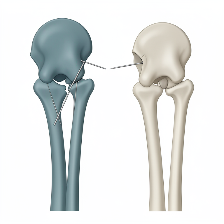
Trans-articular Pinning Path
AI-generated conceptual illustration (canine hip joint and femur). It explains the idea and does not show study data.
View source evidence ↗Figures & tables
From the original paper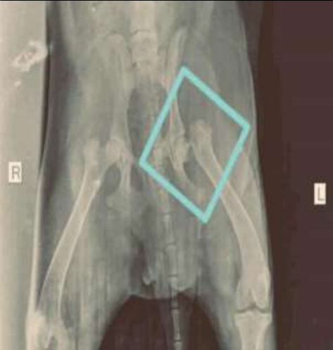 Figure 1
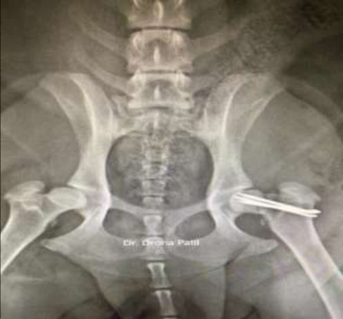 Figure 2
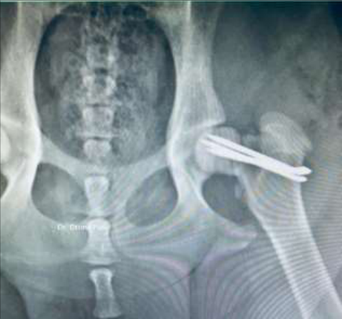 Figure 3
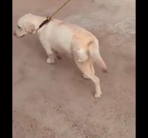 Figure 3
Introduction
Femoral neck fractures, including those of the femoral head, neck, and greater trochanter with physeal separations, are common in growing dogs and cats, accounting for 20%-25% of all fractures. Femoral neck fractures in dogs may occur within the joint capsule (intracapsular), such as sub capital or trans cervical fractures, or outside the capsule (extracapsular), known as basilar fractures. The most common type is the simple basilar fracture, although comminuted fractures can also occur (Hulse, 1974) [1] . These fractures typically affect young dogs under one year old and are often associated with trauma (Simpson, 2003) [2] . Restoring normal limb function requires surgical correction of the fracture. Internal fixation options for femoral neck fractures include placing a cortical bone screw in a lag fashion, along with an anti-rotational Kirschner wire, and inserting three divergent Kirschner wires (K-wires) (Piermattei, 2006) [5] . Early reduction, gentle handling of tissue to preserve the blood supply and stable internal fixation are the key points for favourable prognosis. In human patients the most common complication is persistent avascular necrosis of the femoral head unlikely complication in small animals (William, 1978) [4] .
Case history and observations
A 2 year old female Labrador weighing 22 kg was brought to the Jijai animal clinic and surgical centre, Mumbai with history of automobile accident 2 days back followed by swelling and non-weight bearing lameness on left hind limb. Clinical examination revealed that the basic physiological parameters were in normal range the dog was well hydrated and showing signs of pain on palpation at the level of coxo-femoral joint. The hemato-bio chemical analysis carried out was within the normal range. Radiographic evaluation revealed a comminuted basilar femoral neck fracture on left side with cranio-dorsal luxation of the femoral neck (Fig. 1).
Surgical treatment
The surgical correction was carried out by keeping the animal off feed and off water for 8 hours prior to the procedure. The animal was pre medicated with injection butorphanol @ 0.2 mg/kg IV, followed by injection dexmedetomidine @ 0.03 mg/kg IV. The anaesthetic induction was carried out using injection ketamine and midazolam (1:4) IV, combination till the effect.
Following smooth induction of the anaesthesia the endotracheal intubation what carried out with the appropriate size cuffed endotracheal tube and the maintenance of anaesthesia was done with (1-3%) Isoflurane and intraoperative pain management was carried out using fentanyl CRI @ 5-20 mcg/kg/hr (Eder J, 2023) [8] . The animal was positioned on its side with the affected leg elevated. The surgical area was prepared by clipping, scrubbing, and draping it in a sterile manner. The coxofemoral joint was exposed through cranio-lateral approach. By using the cranio-lateral approach, the surgeon was able to expose each fracture fragment adequately without damaging the caudal gluteal vessels (William, 1978) [4] . A bone forceps is used to reduce and stabilize the fracture. Three K-wires were passed from the base of the greater trochanter, traversing the fracture site and entering the femoral head. The outer wires were positioned as proximally and distally in the neck as feasible. Care was taken to prevent penetration of the articular cartilage during pin insertion (Hulse, 1974) [1] . Three 2 mm Kirschner wires, were used for reduction and stabilization of fracture fragments (Lea Lovric, 2020) [6] (Fig. 2). A routine closure was performed on the surgical wound.
Discussion
Trans-articular pinning has demonstrated efficacy as a viable technique for treating femoral neck fractures in dogs. This technique is applicable in young dogs, particularly in cases where surgical correction via osteotomy may not be feasible, and conservative management is not recommended due to suboptimal outcomes. In 1983, Brinker et al. introduced a method for repairing femoral neck fractures utilizing multiple Kirschner wires (K-wires). On other hand, femoral head excision is frequently linked to an extended post-operative recovery period before the animal achieves soundness, and complications such as secondary patellar luxation can arise (Duff & Campbell, 1977) [3] . Despite requiring surgical exposure of the hip joint, this technique is considered a relatively simple and quick procedure, particularly when compared to alternative methods such as hip toggling or femoral head excision (Bennett, 1980) [7] .
Conclusion
The surgical management of femoral neck fracture in dog was carried out successfully by trans articular pinning technique. Gentle handling, careful dissection of soft tissue, and control of haemorrhage while reducing and stabilizing the fracture were very important to reduce the post operative complications like wound dehiscence, seroma formation, delayed bone union, incision or bone infection, and hip joint arthritis and arthrosis. Early surgical intervention, coupled with stable fracture repair and precise anatomic alignment, typically yields excellent to very good function outcomes in most cases.
Acknowledgement
AcknowledgementI am grateful to all the professors for their guidance, academic encouragement, and constructive feedback. Additionally, I extend my sincere thanks to the Jijai Animal Clinic and Surgical Centre in Mumbai.
Figures and tables from the paper
References
6 in the original paper- Hulse DA, Wilson JW, Butler HC. Use of the lag screw principle for stabilization of Femoral neck and femoral capital epiphyseal fractures. J Am Anim Hosp Assoc. 1974;10:29-36.
- Simpson DJ, Lewis DD. Fractures of the femur. In: Slatter D, editor. Textbook of Small Animal Surgery. 3rd ed. Philadelphia: Saunders; c2003. p. 2059-2089.
- Duff R, Campbell JR. Long term results of excision arthroplasty of the canine hip. Vet Rec. 1977;101:181.
- Lovrić L, Kreszinger M, Pećin M. Surgical Treatment of Canine Femoral Fractures -A review. World Vet J. 2020;10(2):137-145.
- Bennett D, Duff SR. Trans articular pinning as a treatment for hip luxation in the dog and cat. J Small Anim Pract. 1980;21:313-379.
- Marques EJ, Monteiro ER, Herrera-Becerra JR, Tomazeli D, Rovaris IB, De Oliveira TF, et al. Influence of Constant Rate Infusions of Fentanyl Alone or in Combination With Lidocaine and Ketamine on the Response to Surgery and Postoperative Pain in Isoflurane Anesthetized Dogs Undergoing Unilateral Mastectomy: A Randomized Clinical Trial. Top Companion Anim Med. 2023;52.