
Research article · 2024 · Full text
International Journal of Advanced Biochemistry ResearchEvaluation of plasma lipid peroxidation and antioxidants activity in Plasmodium falciparum infected subjects
Volume 8(4) · Pages 545–548 · DOI: 10.33545/26174693.2024.v8.i4g.1012 ↗
Research areasDiverse Scientific Research StudiesMalaria Research and Control
Abstract
Author-writtenPlasmodium falciparum remains the deadliest among the malaria causing species of protozoan found mostly in female anopheles mosquito. Malaria is a serious health challenge in sub Saharan Africa. This protozoan destroy red blood cells causing anemia which complicate major tissues in the body. This research was done to evaluate the level of lipid peroxidation and antioxidant profile in the plasma of plasmodium falciparum infected subjects. The study includes 50 plasmodium falciparum infected malaria patients as test and 50 apparently healthy subjects without plasmodium falciparum infection as control group from the outpatient Department of Wesley Guide Hospital, Ilesa, Osun State. An amount of blood (5mls) was taken from both test and control and processed to obtain the plasma which was stored at room temperature before analysis. The activity of SOD, GSH, GPx and CAT and lipid peroxidation level were determined in the plasma of both test and control subjects using appropriate method. The study revealed no significant change (p
Summary
Source-checkedPlain-language reading of the full paper, prepared with AI and checked against the text. Each point links to the authors' own words.
Why it matters
- Plasmodium falciparum is the most lethal species of malaria-causing protozoan and represents a major health crisis in sub-Saharan Africa. View evidence ↗
- The parasite destroys red blood cells, leading to anemia and complications in vital body tissues. View evidence ↗
- Infection can trigger the immune system to release reactive oxygen species (ROS), which may cause oxidative damage and cell destruction. View evidence ↗
The question
- The study aimed to evaluate the antioxidant profile and lipid peroxidation levels in the plasma of individuals infected with Plasmodium falciparum. View evidence ↗
What they did
- The study compared 50 malaria patients positive for P. falciparum with 50 healthy control subjects from Wesley Guide Hospital in Ilesa, Osun State. View evidence ↗
- Plasma was obtained by centrifuging venous blood samples at 4000rpm for 3 minutes. View evidence ↗
- The researchers measured the activity of superoxide dismutase (SOD), reduced glutathione (GSH), glutathione peroxidase (GPx), and catalase (CAT), along with lipid peroxidation levels. View evidence ↗
- Data were analyzed using one-way ANOVA, with a p-value of less than 0.05 defined as statistically significant. View evidence ↗
What they found
- There was no statistically significant change in lipid peroxidation (MDA) levels between the control (5.0392×10⁻⁶ ± 2.6×10⁻⁶) and malaria positive (7.2157×10⁻⁶ ± 3.08×10⁻⁶) groups (p=0.112). View evidence ↗
- The activities of the antioxidant enzymes SOD, catalase (CAT), and glutathione (GSH) were significantly reduced (p<0.05) in the plasma of infected patients. View evidence ↗
- No significant difference was found in the activity of glutathione peroxidase (GPx) between the infected and control groups. View evidence ↗
What it means
- The authors conclude that P. falciparum infection is associated with oxidative stress and a reduced antioxidant defense system, which could threaten patient health if not managed. View evidence ↗
- The findings suggest that antioxidants may have therapeutic value as an adjunctive strategy for managing malaria. View evidence ↗
- The authors state that further research is needed to understand the mechanisms of antioxidant dysregulation and to test the efficacy of antioxidant supplements. View evidence ↗
Visual summary
1 visualsIllustrations explain the paper; they do not show study data.
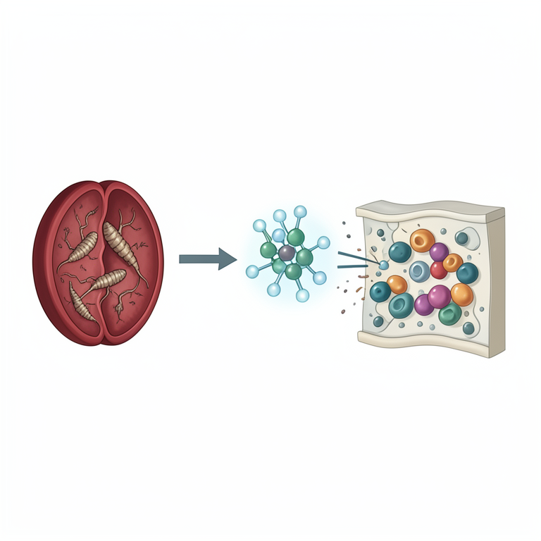
Oxidative Stress Cycle in Malaria
AI-generated conceptual illustration (human blood plasma containing Plasmodium falciparum parasites and red blood cells). It explains the idea and does not show study data.
View source evidence ↗Figures & tables
From the original paper Table 1
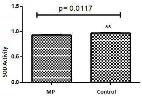 Figure 1
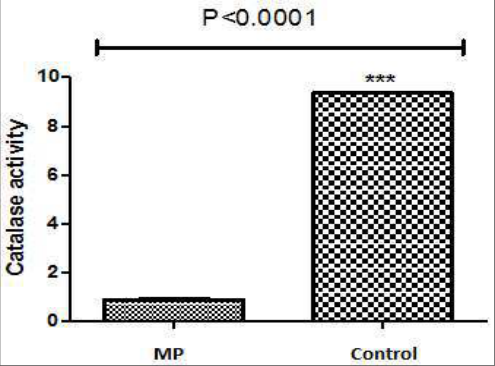 Figure 2
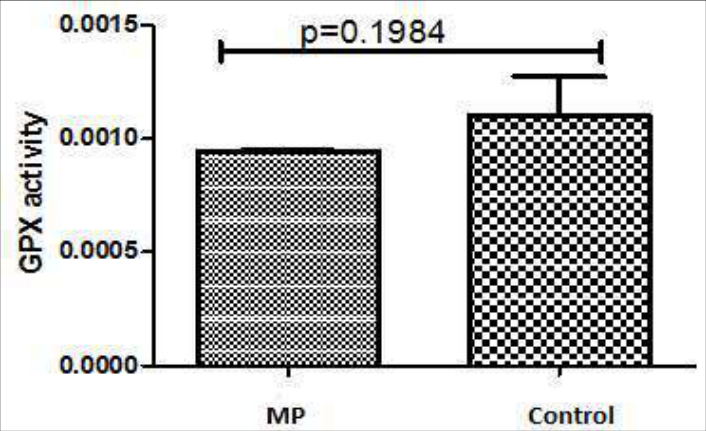 Figure 3
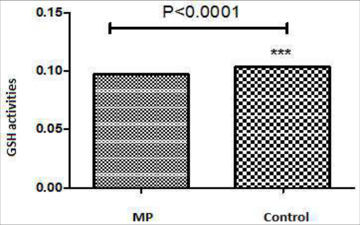 Figure 4
Introduction
Malaria fever constitutes a serious health challenge in many developing countries, resulting in almost 584,000 deaths, 90% of which are found in sub-Saharan Africa (WHO, 2019) [7] . It is endemic in most nations and territories around Africa, Asia, Latin Africa, the Middle East, and South Pacific. It results from a female anopheles mosquito infected with protozoan parasite of the genus Plasmodium biting an individual and is the deadliest among all the malaria causing species responsible for around 50% of all malaria cases (Klonis et al, 2013) [3] . P. falciparum breaks red blood cells, which may result in acute anemia and complications in certain body organs, such as the lungs, kidneys and brain The relationship between the host and the parasite in malaria infected individual can enhance the release of reactive oxygen species (ROS) through stimulation of the immune system; the raised level of ROS has the potential to induce oxidative damage and cell destruction, thus the erythrocytes of Plasmodium-infected patients are exposed to increased oxidative stress (Kremsner et al., 2000) [4] . The breakdown of these erythrocytes cells is the reason for most of the clinical symptoms of falciparum malaria infection and these include fever (>92% of cases), chills (79%), headaches (70%), sweating (64%). Others are dizziness, malaise, muscle pain, abdominal pain, nausea, vomiting, mild diarrhea, and dry cough. High heart rate, jaundice, pallor, orthostatic hypotension, enlarged liver, and enlarged spleen are also diagnosed (Dondorp et al., 2004) [5] . Antioxidants are molecules that mitigate against cellular damage as a result of the highly reactive free radicals activity. They are a complex system of molecules which includes some micronutrients such as vitamin C, E and β-carotene which the body cannot produce but must be supplemented in the diet as well as enzymes, such as catalase (CAT), glutathione peroxidase(GPx), superoxide dismutase (SOD), and various peroxidases (Hamid et al. 2010) [2] .
Materials and Methods
Materials and Methods
Subjects
A total of 50 malaria patients who tested positive for plasmodium falciparum and admitted at outpatients department of Wesley Hospital in Ilesha were used. The same numbers of apparently healthy subjects with no malaria infection were used as control. This study and the use of all bodily fluids were approved by the Committee on the Use of Human Subjects in Research at the Wesley Hospital, Ilesha, Osun state, Nigeria.
Sample collection and processing
Venous blood samples were collected from each of the test and control subjects into heparinized bottles. The sample was centrifuged using a bucket centrifuge at 4000rpm for 3 minutes to obtain the plasma which was used for analysis.
Biochemical analysis
Evaluation of superoxide dismutase (SOD) activity in plasma: SOD activity was determined using the method of Ookawara et al. (1998) [9] .
Assessment of catalase activity
Catalase activity in plasma was assessed using the method of Beers and Sizer, 1952 [11] .
Evaluation of the activity of Glutathione peroxidase.
Glutathione peroxidase activity was evaluated by the method described by Rotruck et al. (1973) [8] .
Estimation of the activity of reduced glutathione (GSH) in plasma
The activity of Reduced glutathione (GSH) was evaluated using the method described by Anderson (1985) [10] .
Data analysis
Values were presented as mean ± SD. Data were analyzed using one-way analysis of variance (ANOVA). P-value less than 0.05 (p<0.05) was considered to be statistically significant.
Results
Results
Table 1:
Present the result of the level of MDA in plasmodium falciparum infected malaria patients and control subjects.
Control
Control
Discussion
Malaria infection induced by plasmodium falciparum remains a significant health challenge in sub Saharan Africa characterize by physiologic and metabolic changes which could result in significant level of mortality and morbidity among the population. The result of the study in table 1 shows an increase but not statistically significant (p<0.05) in plasma level of MDA and hence lipid peroxidation levels in plasmodium falciparum infected malaria patients when compared with the control subjects. This raised level of lipid peroxidation might indicates high level of free radical productions and activities especially that of reactive oxygen species. There had been previous report of increased plasma MDA level in malaria patients. The increased level of free radicals arises from stimulating effect of the parasite on the host immune system (Kremsner et al., 2000) [4] . Furthermore, the outcome of the research shown in figures 1, 2 and 4 shows a significant decrease (p<0.05) in the activity of SOD, catalase and glutathione which are major antioxidant defense system of the body. The decrease activity of these antioxidants in these patients could be because of the increased level of reactive oxygen species which could mean they have been used up in neutralizing these oxidants. Similar outcome was previously reported about the activity of these antioxidants in plasmodium falciparum infected malaria patients, (Amit et al., 2017) [1] reported decreased activity of catalase and GSH in plasmodium falciparum infected malaria patients as reported in this study. However the activity of GPx reported in these patients was not significantly different (p<0.05) from the control subjects which contradict the outcome of a previous study by (Mohammed et al., 2014) [6] who reported a decrease activity of GPx in plasmodium falciparum infected malaria patients. The outcome of this research indicate possible presence of oxidative stress with reduced level of antioxidant defense system in plasmodium falciparum infected malaria patients which might present serious threat to the overall health of these patients if not well managed.
Conclusion
The findings of this study underscore the importance of understanding the role of antioxidants in mitigating the cellular damage caused by malaria infection, particularly Plasmodium falciparum. Our results indicate that malaria patients exhibit alterations in antioxidant enzyme activities, including superoxide dismutase, catalase, and glutathione peroxidase, as well as reduced glutathione levels, suggesting a heightened oxidative stress state. This oxidative stress, induced by the interaction between the host and the parasite, likely contributes to the pathogenesis of malaria and the manifestation of clinical symptoms. These insights highlight the potential therapeutic value of antioxidants in adjunctive malaria management strategies. However, further research is warranted to elucidate the specific mechanisms underlying antioxidant dysregulation in malaria and to evaluate the efficacy of antioxidant supplementation as a complementary approach to malaria treatment.
Acknowledgement
AcknowledgementsThe contributions of Dr. A. T. Ogundajo and other staffs of the Chemical pathology department of Wesley Guide Hospital Obafemi Awolowo University Teaching Hospital, Ilesa, Osun State, was crucial for the overall success of this research especially in the area of sampling and Biochemical analysis.
Figures and tables from the paper
References
11 in the original paper- Amit GT, Rupal AT, Prema RC, Jaidev SS. International Journal of Research in Medical Sciences. Int. J Res Med Sci. 2017;5(4):1649-165.
- Hamid AA, Aiyelaagbe OO, Usman LA, Ameen OM, Lawal A. Antioxidants: its medicinal and pharmacological applications. Afr. J Pure Appl. Chem. 2010;4(8):142-151.
- Klonis N, Creek DJ, Tilley L. Iron and heme metabolism in Plasmodium falciparum and the mechanism of action of artemisinins. Curr Opin Microbiol. 2013;16(6):722-727.
- Kremsner PG, Greve B, Lell B, Luckner D, Schmid D. Malarial anaemia in African children associated with high oxygen-radical production. Lancet. 2000;355(9197):40-41.
- Dondorp AM, Pongponratn E, White NJ. Reduced microcirculatory flow in severe falciparum malaria: pathophysiology and electron-microscopic pathology. Acta Trop. 2004;89(3):309-117.
- Mohammed HFS, Amar B3E, Omar EF, Ashraf N, Osman ME. Glutathione peroxidase (GPx) activity in platelets of patients with malaria parasitaemia caused by Plasmodium falciparum. Sky J Biochem Res. 2014;3(4):037-041.
- World Health Organization. World Malaria Report 2019. Switzerland: World Health Organization; c2019. pp. xii-xiii, 4-10.
- Rotruck JT, Pope AL, Ganther HE, Swanson AB, Hafeman DG, Hoekstra WG. Selenium: Biochemical Role as a Component of Glutathione Peroxidase. Science. 1973;179:588-590.
- Ookawara T, Imazeki N, Matsubara O, Kizaki T, OhIshi S, Nakao C, et al. Tissue distribution of immunoreactive mouse extracellular superoxide dismutase. Am J Physiol Cell Physiol. 1998;275(3):840-847.
- Anderson ME. Determination of glutathione and glutathione disulfide in biological samples. Methods Enzymol. 1985;113:548-555.
- Beers RF, Sizer IW. A spectrophotometric method for measuring the breakdown of hydrogen peroxide by catalase. J Biol. Chem. 1952;195(1):133-140.