
Research article · 2024 · Full text
International Journal of Advanced Biochemistry ResearchMilk DLC in early lactating Sahiwal cow for identification of udder health
Volume 8(4S) · Pages 374–377 · DOI: 10.33545/26174693.2024.v8.i4se.997 ↗
Research areasMilk Quality and Mastitis in Dairy CowsEffects of Environmental Stressors on Livestock
Abstract
Author-writtenMonitoring udder health in dairy herds is crucial for maintaining milk quality and animal welfare. Somatic cell count (SCC) serves as a primary indicator of udder inflammation, but its reliability has diminished with decreasing global SCC averages. This study investigates the utility of milk differential leukocyte count (DLC) in early lactating Karan Fries cows for identifying udder health status. A total of 36 cows were categorized into healthy, subclinical mastitis (SCM), and clinical mastitis (CM) groups based on SCC and California Mastitis Test scores. Milk samples were collected and analyzed for SCC and DLC using both automated and microscopic methods. Results revealed a significant increase in neutrophil percentage and a decrease in macrophage percentage as udder health deteriorated from healthy to SCM to CM. Lymphocyte percentage decreased but not significantly across the groups. The study highlights the potential of DLC as a complementary tool to SCC for precise udder health assessment in early lactating Karan Fries cows.
Summary
Source-checkedPlain-language reading of the full paper, prepared with AI and checked against the text. Each point links to the authors' own words.
Why it matters
- Monitoring the health of the udder in dairy herds is essential for ensuring animal welfare and the quality of the milk produced. View evidence ↗
- While somatic cell count (SCC) is the primary tool for detecting udder inflammation, it has become less reliable as global average SCC levels have decreased. View evidence ↗
- Because of the limitations of SCC, measuring the amount of polymorphonuclear neutrophils (PMN) is suggested as a more informative way to evaluate udder health. View evidence ↗
The question
- The study aims to determine if milk differential leukocyte count (DLC) can be used to identify the udder health status of early lactating Karan Fries cows. View evidence ↗
What they did
- The researchers studied 36 early lactating Karan Fries cows, divided equally into three groups of 12: healthy, subclinical mastitis (SCM), and clinical mastitis (CM), based on SCC and CMT scores. View evidence ↗
- Differential cell counting (DCC) was used to identify and quantify neutrophils, lymphocytes, and macrophages in the milk samples. View evidence ↗
- A one-way ANOVA test performed with IBM SPSS statistics 26 software was used to determine significant differences in DLC between the three cow groups. View evidence ↗
What they found
- SCC increased with disease severity: healthy cows (CMT 1-2) had up to 2x10^5 cells/ml, SCM cows (CMT 3) had 2x10^5 to 5x10^5 cells/ml, and CM cows (CMT >3) had more than 5x10^5 cells/ml. View evidence ↗
- Lymphocyte percentages decreased from 13.33±0.67% (healthy) to 12.16±0.98% (SCM) and 4.08±0.78% (CM), but a significant decrease (p<0.05) occurred only between the healthy and SCM groups. View evidence ↗
What it means
- The authors conclude that SCC alone does not provide a clear image of the cell types present; therefore, DLC is necessary to provide a detailed picture of the cell types in healthy, SCM, and CM milk. View evidence ↗
- The study recommends using DLC as a complementary tool to SCC for a more precise assessment of udder health in early lactating Karan Fries cows. View evidence ↗
Visual summary
1 visualsIllustrations explain the paper; charts are replotted from the paper's own table values.
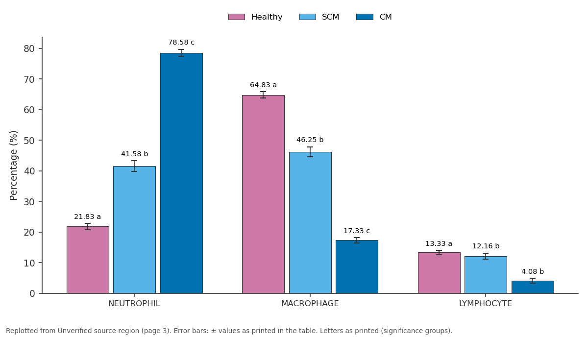
Healthy and SCM and more: NEUTROPHIL vs MACROPHAGE vs LYMPHOCYTE
Replotted from a table (page 3) of the original paper. Every bar is the paper's own table value.
View source evidence ↗Introduction
The most straightforward, practical, and sustainable approach currently utilized for monitoring udder health in dairy herds is individual somatic cell count (SCC). Although not as precise as microbiological analysis [1] , SCC serves to indicate inflammation rather than the presence of a specific pathogen. Generally a threshold value of 200,000 cells/ml is employed to detect subclinical mastitis under field conditions [2][3][4][5] . However, the global decrease in mean SCC across dairy herds has led to a reduced reliance on SCC for identifying diseased cows. There's a consensus regarding the correlation between increased SCC in milk and a shift in the proportion of inflammatory cells within the cell population. Consequently, the quantity of polymorphonuclear neutrophils (PMN) has been proposed as a potentially more informative indicator for evaluating udder health than SCC [6][7][8][9][10] . Indeed, alterations in milk composition have been observed even below the 100,000 cells/mL mark [11][12][13][14] . This body of evidence supports ongoing research with an aim of utilizing differential somatic cell count (DSCC) as a tool either in conjunction with SCC or independently to identify mastitis. In healthy dairy cows, the milk cell population typically consists of over 97% leukocytes and less than 3% of mammary epithelial cells (MECs) [11,15] . The presence of leukocytes in the milk of healthy cow udders is because they are able to traverse the blood-milk barrier (diapedesis) as a part of the natural immune survilleance to safeguard the mammary gland against infections. MECs are cast-off into the milk as a consequence of milk secretion process and also to carry out continuous renewal of the mammary gland epithelium. The milk secretion by the merocrine process in bovine aids in cell preservation, whereas the apocrine milk secretion process in human breasts is cell-destructive in nature and is responsible for comparative predominance of MECs in human milk [16] . Inflammation of the mammary gland i.e., mastitis can be characterized by the presence of leukocytes -primarily polymorphonuclear leukocytes (PMN) into the udder's lumen [11,17,18] . This migration of leukocytes causes the somatic cell count (SCC) to increase. Consequently, SCC is the internationally used predominant parameter for managing udder health in dairy herds. It's assessment can be done on-site and semi-quantitatively by visual inspection using California Mastitis Test (CMT), or by accurate counting them using a flow cytometer. [19] Materials and Methods This particular study was carried out at the Livestock Research Centre (LRC), of ICAR National Dairy Research Centre (NDRI), Karnal-132001, Haryana, India. The location of study comes under semi-arid region. In total 36 early lactating crossbred Karan Fries (Holstein -Friesian x Tharparkar) cows were selected and subdivided into three groups viz.; healthy group (n=12), subclinical mastitis group (n=12), and clinical mastitis group (n=12) in accordance with their Somatic Cell count (SCC) and California Mastitis Test (CMT). Milk collection was done under hygienic conditions involving udder cleaning of all cows and screening them for their mammary health using CMT. The colostrum of these animals was not taken into account and only normal milk SCC and DLC was done. All the selected animals were in parity number of 1 to 6. Loose housing system was adopted for maintenance of all the animals. The housing space for the cows was as specified under "BIS" standards. This type of housing system facilitates free movement and sufficient physical activity for the animals. Feeding standards were being followed at NDRI were based on Nutrient requirements of cattle and buffalo (ICAR, 2013) published by the Indian Council of Agricultural Research, New Delhi. During the postpartum period, cows were fed ad-lib available fodders and concentrates based on their milk production level. All the Karan Fries cows were machined milked thrice daily i.e., in the morning batch (5:00 to 6:00 am), in the noon batch (12:00 to 1:00 pm) and evening batch (5:00 to 6:00 pm) For machine milking the pulsators were adjusted to give a pulsation rate of around 50 pulsations per minute with a uniform vaccum level of 400 mm Hg. After washing the cows, each cow was stimulated for let-down for one minute before connecting to the milking machine. Chemicals, equipment used in this study were CMT reagent, for DLC-Xylene and methylene blue dye and for estimating milk composition-1 x PBS, Lactoscan SCC kit x 4 (manufactured by Milkotronic Ltd.) consisting of Lactochip, Sofia green lyophilized dye, Olympus Simple microscope CH20i and Lactoscan Milk analyser type MCC WS. SCC of milk samples were measured by two methods. Firstly, milk somatic cells were estimated by the Lctoscan milk SCC counter (Milkotronic Ltd. StaraZagora, Bulgaria) . The Lactoscan milk SCC analyzer is based on the fluorescent microscope technique of counting cells. Around 100 µl of fresh milk was diluted with distilled water at the ratio of 1:2 and then mixed with Sofia Green lyophilized dye in a microtube and the 8 µl was pipetted onto the single lactochip. After that, the chip was loaded into the machine. The analysis was done between 10 seconds and 2 minutes and this duration varied based on the number of filmed fields loaded. This Lactoscan SCC system focuses automatically on the chip and the dyed cells are filmed by the sensitive CCD camera. The algorithm analysis of digital images determines the number of fluorescent cells and counts their concenteration and size. The result was automatically displayed on the monitor. Secondly, milk smears were prepared on fresh slides to crosscheck the results of SCC obtained from the Lactoscan milk SCC counter and also to estimate milk differential leukocyte counts by a microscopic method using an inverted microscope as described by Dang et al. (2007) [20] . Differential cell counting (DCC) was carried out to determine the presence of different cell types like neutrophils, lymphocytes, and macrophages in milk. For milk slide preparation around 10 µl of fresh milk was spread on a 1cm 2 (1 x1 cm) arear of a degreased microscopic slide and was dried in a horizontal position. The films were airdried and then duplicate smears were fixed with 96% ethyl alcohol (min) air-dried, defatted with xylene (12 min), and rinsed smoothly with 60% ethyl alcohol, air-dried. Subsequently dyed with Methylene blue solution for 2 min and then rinse with water and air-dried. Milk SCC was corrected for the dilution done for final readings. SCC of milk samples was also measured by microscope at 40X and DCC of milk were carried out and 100X to determine the presence of different cell types like lymphocytes, neutrophils, and macrophages in milk. Milk neutrophils had multilobed nuclei with bridges, milk lymphocytes were distinguished by having a depply staining nucleus, which may be eccentric in location, and having a relatively small amount of cytoplasm. Milk macrophages were identified as the largest cell type seen in milk. The differential leukocyte count is expressed in percentage of total cell counts.
no. of that particular cell DLC of a particular cell type (%) =
x 100 Total no. of cells The differential leukocyte counts of healthy, sub-clinical mastitis and clinical mastitis animals were analyzed and tabulated as mean with standard error. For the determination of significant differences in DLC of healthy, sub-clinical mastitis and clinical mastitis KF cows, one-way ANOVA test was done using IBM SPSS statistics 26 software.
Results and Discussion
This study finds that early lactating healthy KF cows having CMT scores 1-2 with no visible symptoms have a somatic cell count of up to 2x 10 5 cells/ml of milk. Cows with subclinical mastitis with a CMT score of 3 and with no visible symptoms had a somatic cell count in a range of 2x 10 5 to 5x 10 5 cells/ml of milk while Clinical mastitis cows with a CMT score of more than 3 with symptoms like swelling, painful udder etc. had a somatic cell count of more than 5x 10 5 cells/ml of milk. The mean milk neutrophil percentage in early lactating healthy Sahiwal cows was 21.83±1.00 percent. These values increased significantly (p<0.05) as the animal went from healthy to subclinical mastitis to clinical mastitis form. It was observed the mean neutrophil percentage in sub-clinical mastitis and clinical mastitis in early lactating Sahiwal cows were 41.58±1.76 and 78.58±1.13. In the case of milk macrophages, their number decreased significantly (p<0.05) as udder condition deteriorated from healthy to subclinical to clinical mastitis in early lactating Sahiwal cows. In healthy, subclinical, and mastitis, the mean macrophage percentage was 64.83±1.05, 46.25±1.63, and 17.33±0.90 respectively. Mean milk lymphocyte percentage in healthy, sub-clinical mastitis and clinical mastitis cases in early lactating Sahiwal cows were 13.33±0.67, 12.16±0.98, and 4.08±0.78 respectively. Although the values decreased from healthy to subclinical mastitis to clinical mastitis but significant (p<0.05) decrease was observed only from healthy to subclinical mastitis. Due to inflammatory response due to invading pathogens or other antigenic factors, the immunological responses of the mammary glands were observed which resulted in change in milk SCC and milk DLC. Due to these invading pathogens or antigens, a newly recruited leucocytes are necessary to eliminate the invading pathogens which in case of mastitis enter through the teat canal thus DLC in milk in mastitis and sub clinical mastitis animal varies significantly from healthy animal.
Healthy
Healthy
Conclusion
It is concluded from this study that milk SCC increased in early lactating KF cows as the animals went from healthy to sub clinical mastitis to clinical mastitis. It was also found that the mean milk neutrophil percentage also increased i.e., there was significant increase in neutrophil count in both subclinical and mastitis milk. Mean milk macrophage percentage decreased as milk SCC increased i.e., there is significant decrease in macrophage count in both subclinical and mastitis milk. The mean lymphocyte percentage decreased from the healthy to subclinical mastitis group of cows significantly and decreased further in clinical mastitis cases but not significantly. It was also concluded from this study that one cannot get a clear image of type of cells from milk SCC in case of early lactating KF cows. For that, milk DLC is done which gives a clear picture of type of cell in different milk samples from healthy, sub-clinical mastitis and clinical mastitis animal milk.
References
20 in the original paper- Ferronatto JA, Ferronatto TC, Schneider M, Pessoa LF, Blagitz MG, Heinemann MB, Della Libera AMMP, Souza FN. Diagnosing mastitis in early lactation: Use of Somaticell®, California mastitis test and somatic cell count. Ital J Anim Sci. 2018;17:723-729. DOI: 10.1080/1828051X.2018.1427452
- Schukken Y, Wilson D, Welcome F, Garrison-Tikofsky L, Gonzalez R. Monitoring udder health and milk quality using somatic cell counts. Vet Res. 2003;34:579-596. DOI: 10.1051/vetres:2003027
- Piccinini R, Binda E, Belotti M, Dapra V, Zecconi A. Evaluation of milk components during whole lactation in healthy quarters. J Dairy Res. 2007;74:226-232. doi:10.1017/S0022029906002384
- Andersen S, Dohoo IR, Riekerink RO, Stryhn H, Mastitis Research Workers C. Diagnosing intramammary infections: Evaluating expert opinions on the definition of intramammary infection using conjoint analysis. J Dairy Sci. 2010;93:2966-2975. doi:10.3168/jds.2009-2850
- Dufour S, Dohoo IR. Monitoring herd incidence of intramammary infection in lactating cows using repeated longitudinal somatic cell count measurements. J Dairy Sci. 2013;96:1568-1580. doi:10.3168/jds.20125943
- Vangroenweghe F, Dosogne H, Burvenich C. Composition and milk cell characteristics in quarter milk fractions of dairy cows with low cell count. Vet J. 2002;164:254-260. doi:10.1053/tvjl.2002.0714
- Lindmark-Mansson H, Branning C, Alden G, Paulsson M. Relationship between somatic cell count, individual leukocyte populations and milk components in bovine udder quarter milk. Int Dairy J. 2006;16:717-727. doi:10.1016/j.idairyj.2005.08.001
- Schwarz D, Diesterbeck US, Konig S, Brugemann K, Schlez K, Zschock M, et al. Flow cytometric differential cell counts in milk for the evaluation of inflammatory reactions in clinically healthy and subclinically infected bovine mammary glands. J Dairy Sci. 2011;94:5033-5044. doi:10.3168/jds.2011-4250
- Zecconi A, Vairani D, Cipolla M, Rizzi N, Zanini L. Assessment of Subclinical Mastitis Diagnostic Accuracy by Differential Cell Count in Individual Cow Milk. Ital J Anim Sci. 2018;18:435-440. DOI: 10.1080/1828051X.2018.1437030
- Cuccuru C, Moroni P, Zecconi A, Casu S, Caria A, Contini A. Milk differential cell counts in relation to total counts in Sardinian ewes. Small Ruminant Res. 1997;25:169-173. doi:10.1016/S0921-4488(96)00992-5
- Schwarz D, Diesterbeck US, Konig S, Brugemann K, Schlez K, Zschock M, et al. Microscopic differential cell counts in milk for the evaluation of inflammatory reactions in clinically healthy and subclinically infected bovine mammary glands. J Dairy Res. 2011;78:448455. doi:10.1017/S0022029911000653
- Hamann J. Relationship between somatic cell counts and milk composition. Bull FIL-IDF. 2002;372:56-59.
- Merle R, Schroder A, Hamann J. Cell function in the bovine mammary gland: A preliminary study on interdependence of healthy and infected udder quarters. J Dairy Res. 2007;74:174-179. doi:10.1017/S0022029906002232
- Trevisi E, Zecconi A, Bertoni G, Piccinini R. Blood and milk immune and inflammatory profiles in periparturient dairy cows showing different liver activity index. J Dairy Res. 2010;77:310-317. doi:10.1017/S0022029910000048
- Koess C, Hamann J. Detection of mastitis in the bovine mammary gland by flow cytometry at early stages. J Dairy Res. 2008;75:225-232. doi:10.1017/S0022029908003268
- Boutinaud M, Jammes H. Potential uses of milk epithelial cells: A review. Reprod Nutr Dev. 2002;42:133-147. doi:10.1051/rnd:2002011
- Alhussien M, Kaur M, Manjari P, Kimothi SP, Mohanty AK, Dang AK. A comparative study on the blood and milk cell counts of healthy, subclinical, and clinical mastitis Karan Fries cows. Vet World. 2015;8:685-689. doi:10.14202/vetworld.2015.685-689
- Rivas AL, Quimby FA, Blue J, Coksaygan O. Longitudinal evaluation of bovine mammary gland health status by somatic cell counting, flow cytometry, and cytology. J Vet Diagn Investig. 2001;13:399-407. doi:10.1177/104063870101300507
- Adkins PRF, Middleton JR. Methods for Diagnosing Mastitis. Vet Clin N Am Food Anim Pract. 2018;34:479-491. doi:10.1016/j.cvfa.2018.06.002
- Dang AK, Kapila S, Tomar P, Singh C. Immunity of the buffalo mammary gland during different physiological stages. Asian-Australas J Anim Sci. 2007;20:1174-1181. doi:10.5713/ajas.2007.1174