
Research article · 2024 · Full text
International Journal of Veterinary Sciences and Animal HusbandryAptamers in diagnostic applications
Volume 9(2S) · Pages 341–351 · DOI: 10.22271/veterinary.2024.v9.i2se.1326 ↗
Research areasAdvanced biosensing and bioanalysis techniques
Abstract
Author-writtenAptamers are single-stranded oligonucleotides capable of folding into specific structures and binding to targets such as proteins, exhibit greater affinity and selectivity due to their inherent shape-forming characteristics. In this review, we elucidate recent advancements in aptamer-based technologies, delve into the fundamental properties of aptamers, explore chemical modifications, examine various in-vitro selection processes, and assess different analytical techniques.
Summary
Source-checkedPlain-language reading of the full paper, prepared with AI and checked against the text. Each point links to the authors' own words.
Why it matters
- Aptamers are short, single-stranded DNA or RNA molecules, generally consisting of 10 to 80 nucleotides with a molecular weight of 6 to 30 kDa. View evidence ↗
- Because they bind to targets similarly to antibodies, aptamers are often referred to as 'chemical antibodies'. View evidence ↗
- Aptamers are highly versatile and can be used in therapeutics, biosensors, targeted drug delivery, and diagnostics. View evidence ↗
The question
- The authors aim to review recent advancements in aptamer technologies, including their properties, chemical modifications, selection processes, and analytical techniques. View evidence ↗
What they did
- Aptamers are created using SELEX, a process that iteratively selects and amplifies oligonucleotides from a random pool to find those that bind a specific target. View evidence ↗
- The selection process in SELEX generally involves 8 to 20 rounds of iteration. View evidence ↗
What they found
- Aptamer production via improved SELEX is significantly faster than antibody production, taking 2 to 8 weeks or less compared to over 6 months. View evidence ↗
- Aptamers are smaller than antibodies, with a molecular weight of 6-30 kDa and a diameter of 1-2 nm, whereas antibodies range from 25-150 kDa and 10-15 nm. View evidence ↗
- Substituting a fluoro group at the 2' position (F-ANA) increases the stability of the quadruplex structure four-fold and nuclease resistance seven-fold. View evidence ↗
- Binding between an aptamer and its target typically results in a thermal shift of 2 to 10°C. View evidence ↗
- UV melting profiles show an increase in melting temperature (Tm) of 2 to 12°C upon aptamer-target binding, depending on interaction strength. View evidence ↗
- In MRI, aptamer-based probes increase the target-to-background ratio four-fold because unbound aptamers are removed rapidly. View evidence ↗
What it means
- Despite thousands of studies, only three aptamer-based diagnostics have been successfully commercialized to date. View evidence ↗
- The authors conclude that further research and development are necessary to create more sensitive, specific, and cost-effective diagnostic assays. View evidence ↗
Visual summary
1 visualsIllustrations explain the paper; they do not show study data.
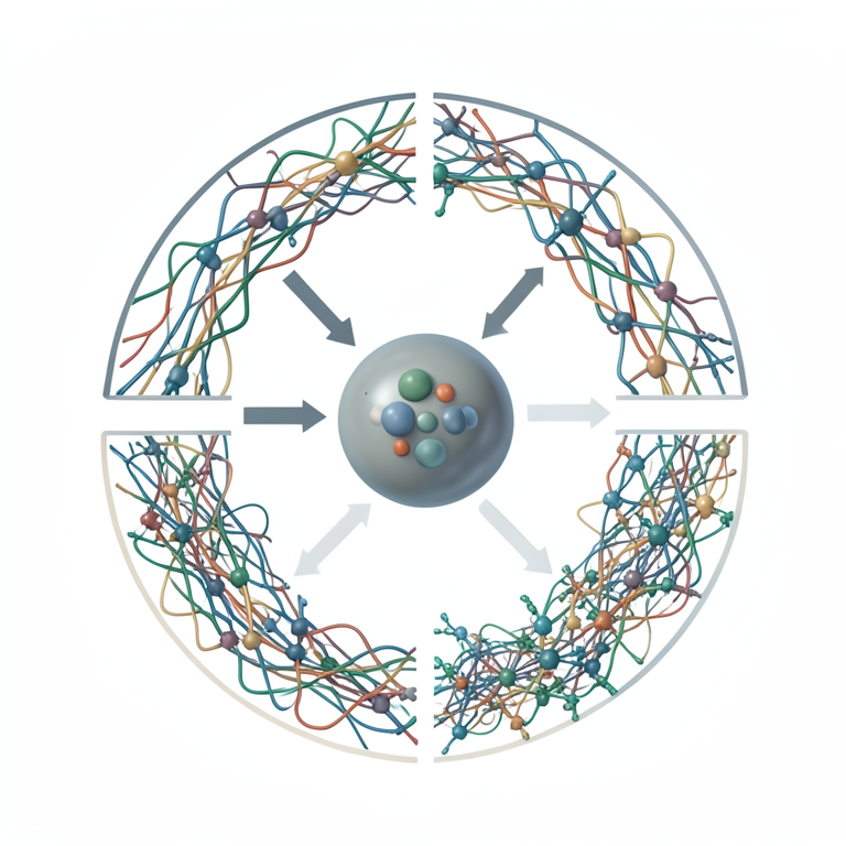
The SELEX Cycle
AI-generated conceptual illustration (single-stranded DNA oligonucleotides and a target protein molecule). It explains the idea and does not show study data.
View source evidence ↗Figures & tables
From the original paper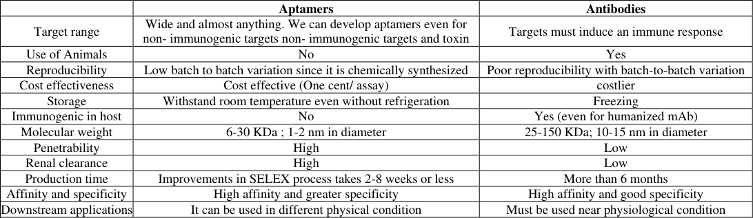 Table 1
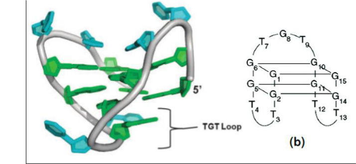 Figure 1
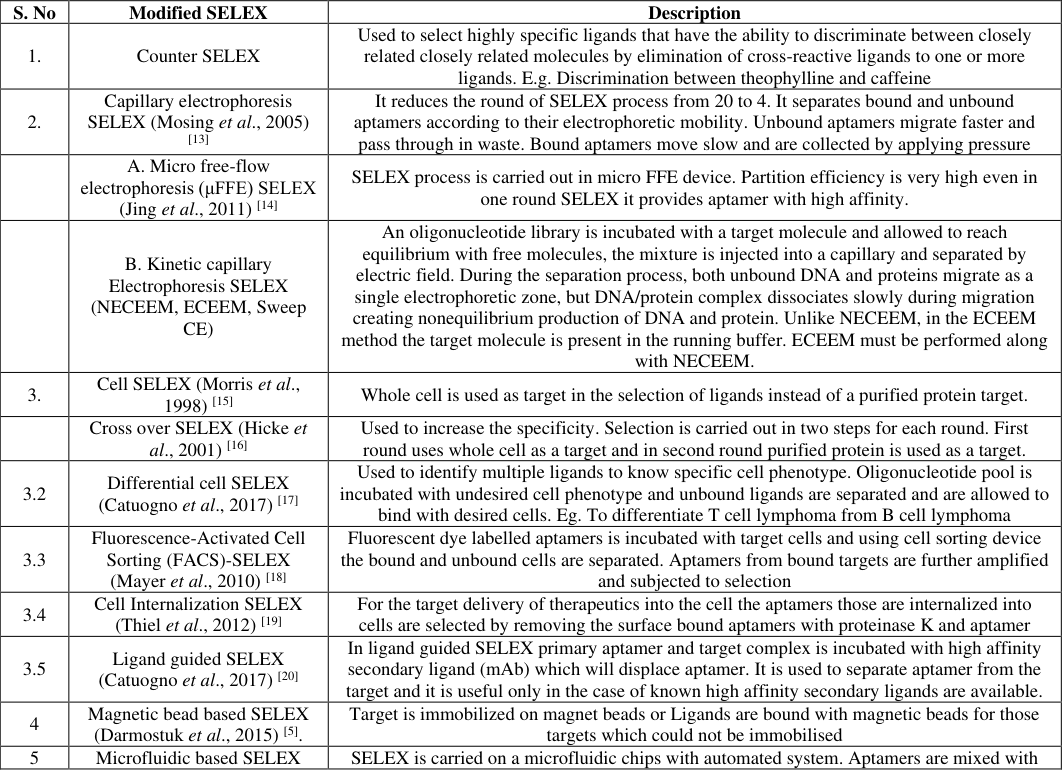 Table 2
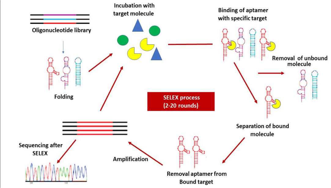 Figure 2
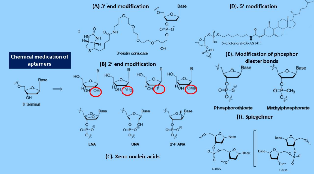 Figure 3
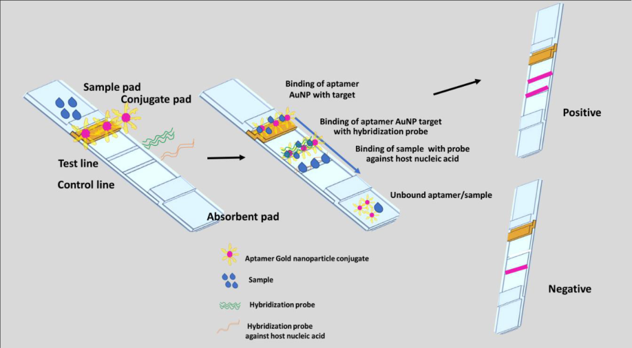 Figure 4
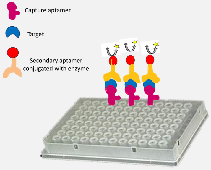 Figure 5
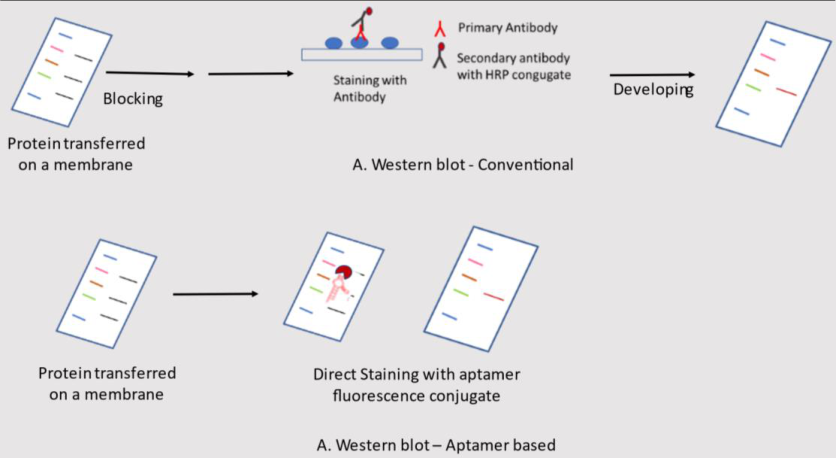 Figure 6
Introduction
Aptamers are single-stranded DNA or short RNA molecules typically comprising 10-80 nucleotides and weighing between 6-30 kDa (Sun et al., 2015) [1] (Hermann et al., 2000) [2] . The term "Aptamer" was coined by Andy Ellington and Jack Szostak in the year 1990 and it was derived from the Greek words "aptus," meaning 'to fit,' and "merus," meaning 'part' (Gold et al., 2021) [3] . Aptamers binds with their targets like antibodies and are also called as "chemical antibodies" (Baird et al., 2010) [4] . Aptamers are generated through a process called SELEX (Systematic Evolution of Ligands by Exponential Enrichment), where a large random pool of oligonucleotides is subjected to iterative rounds of selection and amplification to isolate sequences that bind to the target molecule of interest. Through this process, aptamers with high affinity and specificity for their targets can be obtained. Aptamers have a wide range of potential applications in diagnostics, therapeutics, biosensors, and targeted drug delivery '/ Aptamers are easy to produce and scale up. The production time is relatively less compared to antibodies. These aptamers are chemically synthesized and has greater variability and less variation between batches. Aptamers can be designed to target the bacteria and can be used in the treatment of multi-drug resistant bacteria / emerging viruses. Aptamers can maintain their structure and function over a wide range of temperatures, making them thermally stable and eliminating the need for refrigeration during storage and transportation. It can be reused in various or multiple applications since they can be denatured by applying heat and renatured by cooling (Bruno et al., 2015) [6] . Aptamers offer several advantages over antibodies. They are easy to produce and scale up, with shorter production times. Chemically synthesized, they exhibit greater consistency and less batch-to-batch variation. Unlike antibodies, aptamers do not provoke an immune response, have a broader target range, and do not require animals for production. They possess superior renal clearance and penetrability, and their production can be upscaled rapidly (Baird et al., 2010) [4] (Darmostuk et al., 2015) [5] (Bruno et al., 2015) [6] (Doggrell et al., 2018) [7] (Röthlisberger et al., 2017) [8] . The detailed comparison between antibodies and aptamers is provided in Table .1. [9] .
Structure of an Aptamer
Aptamers were discovered by Craig Tuerk and Larry Gold, as well as Andy Ellington and Jack Szostak, in 1990. The structure of the aptamer described in Figure 1 was the original work by Tuerk and Gold while investigating the translator switch of T4 bacteriophage 43 mRNA. This mRNA contains a hairpin and a Shine and Dalgarno domain at the 5' end of the start codon. Tuerk and Gold mutated the hairpin structure and identified eight nucleotides within it. Among the 65,000 combinations, they successfully isolated two aptamers, thereby established the SELEX process and obtained a patent for SELEX in 1995 (Baird et al., 2010) [4] . Despite over 5000 research articles following Tuerk and Gold's work, only one aptamer, Macugen, has been approved for therapeutic use. Macugen targets vascular endothelial growth factor (VEGF165) to treat age-related macular degeneration (Doggrell et al., 2018) [7] .
Aptamers adopt a stable G-quadruplex structure in guaninecytosine-rich environments (GC), contrasting with the stable double helical structure of DNA and RNA. Their threedimensional folding is crucial for target binding which is studied using techniques such as X-ray crystallography and NMR spectroscopy. Interactions between aptamers and their targets involve various forces, including Van der Waals forces, hydrogen bonding, electrostatic interactions, and shape complementarity (Zhou et al., 2006) [10] . The binding affinity of aptamers is quantified by the dissociation constant (Kd), which is derived from the ratio of dissociation rate constants to association rate constants (Koff/Kon). High-quality aptamers typically exhibit Kd values in the nanomolar (nM) to picomolar (pM) range. It is noteworthy that DNA aptamers are often more stable than RNA aptamers (Röthlisberger et al., 2017) [11] .
Selex: (Systematic Evolution of Ligand by Exponential Enrichment) Process SELEX is the process of selecting aptamers from an oligonucleotide pool of 10 [1] (Darmostuk et al., 2015) [5] (Blind et al., 2015) [12] (Mosing et al., 2005) [12] . 2. It reduces the round of SELEX process from 20 to 4. It separates bound and unbound aptamers according to their electrophoretic mobility. Unbound aptamers migrate faster and pass through in waste. Bound aptamers move slow and are collected by applying pressure A. Micro free-flow electrophoresis (μFFE) SELEX (Jing et al., 2011) [14] SELEX process is carried out in micro FFE device. Partition efficiency is very high even in one round SELEX it provides aptamer with high affinity.
B. Kinetic capillary Electrophoresis SELEX (NECEEM, ECEEM, Sweep CE)
An oligonucleotide library is incubated with a target molecule and allowed to reach equilibrium with free molecules, the mixture is injected into a capillary and separated by electric field. During the separation process, both unbound DNA and proteins migrate as a single electrophoretic zone, but DNA/protein complex dissociates slowly during migration creating nonequilibrium production of DNA and protein. Unlike NECEEM, in the ECEEM method the target molecule is present in the running buffer. ECEEM must be performed along with NECEEM.
3.
Cell SELEX (Morris et al., 1998) [15] Whole cell is used as target in the selection of ligands instead of a purified protein target.
Cross over SELEX (Hicke et al., 2001) [16] Used to increase the specificity. Selection is carried out in two steps for each round. First round uses whole cell as a target and in second round purified protein is used as a target.
3.2 Differential cell SELEX (Catuogno et al., 2017) [17] Used to identify multiple ligands to know specific cell phenotype. Oligonucleotide pool is incubated with undesired cell phenotype and unbound ligands are separated and are allowed to bind with desired cells. Eg. To differentiate T cell lymphoma from B cell lymphoma
3.3
Fluorescence-Activated Cell Sorting (FACS)-SELEX (Mayer et al., 2010) [18] Fluorescent dye labelled aptamers is incubated with target cells and using cell sorting device the bound and unbound cells are separated. Aptamers from bound targets are further amplified and subjected to selection 3.4 Cell Internalization SELEX (Thiel et al., 2012) [19] For the target delivery of therapeutics into the cell the aptamers those are internalized into cells are selected by removing the surface bound aptamers with proteinase K and aptamer 3.5 Ligand guided SELEX (Catuogno et al., 2017) [20] In ligand guided SELEX primary aptamer and target complex is incubated with high affinity secondary ligand (mAb) which will displace aptamer. It is used to separate aptamer from the target and it is useful only in the case of known high affinity secondary ligands are available.
4
Magnetic bead based SELEX (Darmostuk et al., 2015) [5] .
Target is immobilized on magnet beads or Ligands are bound with magnetic beads for those targets which could not be immobilised 5
Microfluidic based SELEX SELEX is carried on a microfluidic chips with automated system. Aptamers are mixed with (Qian et al., 2009) [21] magnetic beads-coated target cells and the magnetic-bead complexes are recovered by applying a magnetic field. Bound ssDNAs are then mixed with magnetic beads-coated control cells. Control cells-aptamer complexes are collected and the supernatant are transported to the PCR chamber for amplification.
3D cell SELEX or
In vivo SELEX (Souza et al., 2016) [22] Aptamers are first subjected to negative selection in-vitro. Then applied for in vivo either in 3D cell culture or in animal models 7
In silico selection (Darmostuk et al., 2015) [5] Selection and characterization of aptamers by docking using different bioinformatic tools like Rostta, FR3D, R3D align, Auto dock, DOVIS, Aptamotif, MPB bind and CLADE 8 SELEX with high throughput sequencing (Darmostuk et al., 2015) [5] The genomic DNA library is used in Genomic SELEX in contrast to chemically synthesized library in classic SELEX. The genomic library is first transcribed into an RNA and then incubated with the target protein. Then the aptamers bound to the target are chromatographically separated, recovered from the target by protein denaturation, reversely transcribed into cDNA and amplified by PCR. A final pool of oligonucleotides obtained from nine selection rounds is characterized by HTS 9
Minimal primer or primer free SELEX (Darmostuk et al., 2015) [5]. (Catuogno et al., 2017) [17] In genomic library primer binding sites with secondary structures self-anneal or anneal nonspecifically. To overcome this primer binding sites are either removed or reduced before endonuclease treatment and allowed to bind with the target. The bounded molecules are reannealed with primer and then amplification is carried out
Optimization of Aptamer
Aptamers have several advantages over antibodies and it is a promising alternative to antibody. But aptamers have limitations due to rapid renal clearance and inactivated by endonuclease. To overcome these limitations modifications of aptamers is practiced. The modifications that are commonly practiced are truncation, chemical modification, random mutation and bivalent and multivalent aptamers (Ni et al., 2017) [23] (Sharma et al., 2017) [24] .
Truncation
Truncation of aptamer is the removal of primer binding domains from either side of the aptamer which increases affinity, specificity and reduces cost.
Chemical modifications
The chemical modification of aptamers (Fig. 3 [25] (Nsairat et al., 2020) [26] . 3. Modification at 5' end -Addition of PEG (40 KDa) or cholesterol at 5' end enhances half-life of aptamers and reduces the clearance. When cholesterol was linked to the 5'-end of an aptamer via a phosphate spacer. This modified oligonucleotide, referred to as cholODN, which was then incubated with low-density lipoprotein (LDL), resulting in the formation of a complex termed cholODN-LDL aptamer. The cholODN-LDL aptamer has 10 times better half life than the original aptamer (De Smidt et al., 1991) [27] . 4. Phosphate backbone modification -Replacement of phosphodiester with methylphosphonate or phosphorothioate or Triazole increased the stability of aptamers (Sharma et al., 2017) [24] 5. Base modification (SOMAmers)-Base modification increase the affinity of aptamer binding. At 5' end of nucleotide hydrophilic and hydrophobic molecules having protein like chains or functional groups which are having inherent ability to bind targets are added.
Commonly used hydrophobic modifications include the incorporation of 5-(N-benzylcarboxyamide)-2deoxyuridine 5-BzdU), napthyl-dU (Nap-dU), Tryptamino -dU,(Trp-dU), Isobutyl-dU. These base modified aptamers are also called as SOMAmers (Slow Off-rate Modified Aptamers) (Ni et al., 2017) [23] (Sharma et al., 2017) [24] . 6. Spiegelmers -The normal form of DNA is in D-form.
The chiral transition from D-form to L-form of aptamers are not recognized by nucleases enzymes. L-form of aptamers are called as spigelmers. The disadvantage of Spiegelmer is the requirement of enantiomeric targets for SELEX process (Ni et al., 2017) [23] (Kong et al., 2013) [28] (Keefe et al., 2010) [29] .
Construction of bivalent or multivalent aptamer
Bivalent or Multivalent aptamer of homologous or heterologous aptamer for the same target with 15 mer dA linker increases affinity, avidity and therapeutic efficacy of aptamers (Guo et al., 2016) [30] Mutation Affinity of aptamers can be increased by site directed mutagenesis or by non-homologous recombination (Sharma et al., 2017) [24] (Nonaka n et al., 2013) [31] Characterization of Aptamer Homology assessment by sequencing is the first step in characterization of aptamer. It helps to determine whether the aptamers share common structural motifs or they have distinct sequences. Various assays are used to characterize the binding ability and specificity of the target. Aptamers are evaluated based on colorimetric assays, fluorescence-based assays, gelbased assays, PCR based assays and in silico assays (Sharma et al., 2017) [24] Colorimetric and fluorescence-based assays These assays are used to evaluate the binding of aptamer with target based on colour change. Colorimetric assays include Aptamer-Linked Immunosorbent Assay (ALISA) or Enzymelinked oligonucleotide assay (ELONA), also known as Enzyme linked Aptamer Assay (ELASA), dot blot, saltinduce Gold Nanoparticles (GNPs) aggregation assays and nanozyme (peroxidase like nanoparticles)-based assays, Dotblot (biotin-streptavidin and gold nanoparticles based).
Gel-based characterization
Polyacrylamide Gel Electrophoresis (PAGE) is used for the characterization of aptamer and it helps to study the structure of aptamer and target recognition site based on hydroxy radical footprinting and DNAse1 footprinting (Ditzler et al., 1998) [32] . Aptamers are polyanionic in nature and in the electric field unbound aptamer moves very fast and the aptamer-protein complex moves at a slower rate. Sometimes, the aptamer attains secondary and tertiary structure which reduces the migration of aptamer. This folded aptamer is misinterpreted as an aptamer-target complex. Hence, western blotting must be carried out to identify the protein aptamer complex (Joshi et al., 2009) [33] Biophysical characterization Aptamers are biophysically characterized based on Circular Dichroism(CD) test to predict secondary structures in aptamers. DNA has absorbance between 180-300 nm. B form of aptamers with stem-loop structure shows a positive peak between 260-280 nm. The AT rich DNA shows a negative peak from 245 to 250 nm. Aptamers in G4 form will show a positive peak at 260-265 nm and a negative peak at 240-245 nm. CD test is also used to differentiate wild aptamers from mutated aptamers and to assess the effect salts on aptamer's structure (Fujita et al., 2012) [34] (Kypr et al., 2009) [35] Thermal shift assay This test is used to measure the stability of aptamer. Aptamers are incubated with protein targets with a protein-dye SYPRO orange. When temperature is increased, the hydrophobic domains of proteins are exposed and release SYPRO orange thereby release increased fluorescence. Whereas, an aptamerbound protein complex doesn't release SYPRO orange and there is decreased fluorescence. Melting curve analysis is used to evaluate the interactions between the aptamer and the target. Tm value is increased (thermal shift) when the aptamer binds with its targets. Usually, a thermal shift of 2-10˚C is observed when the aptamer bounds to its target (Rupesh et al., 2014) [36] .
UV melting profile
Used to measure the stability of aptamer. It uses a UV spectrophotometer or CD instrument to measure absorbance at 260 nm. An increased Tm is observed when bound to its target and stable structure. An increase in Tm from 2-12 ˚C is observed when aptamer bounds to its target depending on the strength of interaction (Song et al., 2011) [37]
Computational tools
The following tools are used to predict aptamer structure (Sharma et al., 2017) [24] (Kinghorn et al., 2017) [38] (Gong et al., 2017) [39] (Caroli et al., 2016) [40] a. Mfold -Predict RNA folding and secondary structure of aptamer b. UNAfold -Predicts secondary structure and the propensity of G4 forming aptamers c. QGRS mapper -Predicts composition and distribution of G4 forming G-rich segments d. Autodock, ZDOCK, PatchDock, HADDOCK -Predicts interaction of aptamer with its target e. APTANIpredicts target specific aptamers from HTS data f. Rosetta, RNA composer -Gives 3D structure of aptamers
Aptamers in diagnostics
Aptamers have high affinity and specificity towards their target molecules. They are easy to synthesize and are not immunogenic. These unique properties made them more attaractive in the field of diagnostics. Aptamers are used in the pathogen detection, cancer detection, food safety assurance, environmental monitoring, water purification and in bio-imaging (Zhang et al., 2019) [41] . Several aptamers based diagnostics are in the pipeline and three companies, Neo Ventures Biotechnology Inc., SomaLogic, Inc (Boulder, CO, USA), Aptamer Sciences Inc. (AptSci, South Korea), and Base Pair (Houston, TX, USA) have commercialized their technology for the detection of Ochratoxin A (OTA sense ® ), Aflatoxin (AflaSense ® ) and for flowcytometry isolation of CD31, CD-31, EGFR, HGFR, ICAM-2, VEGFR-2 or HER-2 cells (AptoCyto ® ) (Kaur et al., 2018) [42] .
Pathogen detection
Aptamers have been used successfully in the detection of bacterial, viral, parasitic and protozoan pathogen. [41] (Vishwakarma et al., 2019) [43] .
Bacterial pathogen
Bacterial pathogen
Parasites
Aptamers have also been utilized to detect the parasitic pathogen Plasmodium spp., Leishmania infantum, Leishmania major, Trypanosoma brucei, Trypanosoma cruzi, Cryptosporidium parvum, Toxoplasma gondii, Schistosoma japonicum, Trichomonas vaginalis (Brosseau et al., 2023) [46] .
Aptamers in cancer detection
Various aptamers were developed in the diagnosis of cancer like breast cancer, colon cancer, T cell leukaemia, Burkitt lymphoma, hepatocellular carcinoma, malignant melanoma, prostate cancer. These aptamers were developed targeting cell surface markers, cancer specific proteins, Intracellular adhesion molecules and cell membrane associated antigens (Hu et al., 2022) [47] (Ruiz et al., 2018) [48] .
Aptamers in ensuring food safety
Several aptamers were developed to detect various food borne pathogens, adulterants, pesticides (Acetamiprid, malathion, atrazine, Edifenphos, Iprobenfos, Edifenphos, Phorate, Isocarbophos, phosalone, methamidophos, acephate, trichlorfon, dursban, Isocarbophos, profenofos, phorate, omethoate), drug residues (tetracycline, diclofenac, dopamine, ampicillin, neomycin B, enrofloxacin, penicillin G, florfenicol, sulfadimethoxine, lincomycin, diethylstilbesterol), food borne toxins (Aflatoxin, Zearalenone, Fumonisin, Anatoxin-a, Staphylococcus enterotoxins, okadoic acid), authentication and allergen (gluten) detection (Schmitz et al., 2020) [49] (Song et al., 2019) [50] .
Aptamers in environmental monitoring
Aptamers have been used to detect aquatic toxins (microcystin, nodularin, malachite green; saxitoxin), pesticides, insecticides, antibiotics, industrial by-products (2,3′,5,5′-tetrachlorobiphenyl -PCB72, 2-hydroxyfluorene, BPA), pharmaceuticals (ibuprofen, estradiol) in water sources. For the soil monitoring aptamers have been developed to detect soil contaminants like lead and mycotoxins such as aflatoxin ; and ochratoxin. Aptamers have also been developed to monitor air quality by detecting toxic gases (radon, nitrogen dioxide, sulphur dioxide, and hydrogen sulphide), 8-hydroxy-2′-deoxyguanosine (8-OHdG), a biomarker for oxidative stress in urine due to air pollution (Zhang et al., 2019) [41] (McConnell et al., 2019) [51] . Aptasensors (Reich et al., 2019) [52] (Kim et al., 2016) [53] (Song et al., 2012) [54] Biosensors are devices that transforms the recognition of targets into a physically detectable signal, such as optical, electronic mass or magnetic signal (Feng et al., 2014) [55] . Biosensors has two components: 1. Target recognition component 2. Signal transduction. Aptamer based biosensors are called as aptasensors. Aptasensors are constructed with different methodologies (Ruckman et al., 1998) [24] (Feng et al., 2014) [55] (Citartan et al., 2012) [56] viz. Colorimetric aptasensors, Fluorescence aptasensors, Chemiluminensce based aptasensors and Optical biosensors Aptamer Based Diagnostic Platforms Aptamer based biosensors using nanoparticles Gold and magnetic nanoparticles are extensively used to design aptasensors because of their unique properties and functionalization. Gold Nanoparticles (AuNPs) -AuNPs have excellent biocompatibility, high surface area-to-volume ratio, and unique optical properties, such as surface plasmon resonance (SPR). AuNPs can be easily functionalized with aptamers via thiol chemistry, where the aptamer is modified with a thiol group that binds strongly to the gold surface. The interaction between aptamers and their target molecules induces changes in the aggregation or dispersion of AuNPs, leading to detectable changes in their optical properties, which can be easily monitored. Magnetic Nanoparticles (MNPs) -MNPs, such as magnetite (Fe3O4) or maghemite (γ-Fe2O3), are commonly employed for their magnetic properties, allowing for easy manipulation and separation using an external magnetic field. In aptasensing applications, MNPs are often used for signal amplification or for separation purposes. For signal amplification, MNPs can be conjugated with detection molecules (e.g., enzymes, fluorescent tags) to increase the sensitivity of detection assays. In separation experiments, MNPs bound to aptamers can selectively capture target molecules from complex samples, enabling their isolation and subsequent analysis (Song et al., 2012) [54] . The AuNPs and MNPs in aptasensors when combined together offer synergistic advantages. MNPs can be used for target capture and concentration, while AuNPs can be utilized for signal transduction. This combination enhances the sensitivity and selectivity of the aptasensor (Zhao et al., 2020) [57] Carbon nanomaterial based Aptasensors -Single walled carbon nanotubes' (SWCNTs), graphene and graphene oxide (GO) are the most promising nanomaterials for the applications in biosensors due to their unique electronic, optical and mechanical properties. They have large surface area for binding. Functionalized graphene can be used for label-free detection of target analytes, enabling simple and cost-effective sensor designs. biocompatibility and enhanced signal amplification capabilities (Wang et al., 2016) [58] .
Aptamers in lateral flow assays
A reporter labelled antibody or aptamer is absorbed on the conjugate pad. A primary antibody or aptamer against the target analyte is immobilised on the test line and a secondary antibody or aptamer against the labelled bioconjugate is immobilised on the control line. Then the analyte is applied to the sample pad it migrates to the conjugate pad, where the target analyte binds to the labelled bioconjugate and this complex passes across the emmbrane via capillary movement to the test line where it is captured. The excess of labelled aptamer /antibody will then be captured at the control line. The appearance of both lines, test line and control line is indicative of a positive result, with a single line at the control line representing a negative result, whilst confirming correct assay function (figure 4). Different aptamer based lateral flow assays were develop for the diagnosis of different analyte viz. cocaine, aflatoxin, Arbovirus, ochratoxin, S. enteritidis, thrombin and β conglutinin (Jauset-Rubio et al., 2017) [59] . In the aptamer-based lateral flow assay, a target-specific aptamer conjugated with gold nanoparticles (AuNPs) is housed within the conjugate pad. The test line is impregnated with a hybridization probe specific to the target, while the control line contains a hybridization probe specific to host DNA. Upon loading the sample into the sample pad, it migrates into the conjugate pad. If the sample is specific to the aptamer, it binds with the AuNP-aptamer conjugate, causing this complex to move towards the test line. In the case of a positive sample, the AuNP conjugates with the hybridization probe, resulting in a pink coloration at the test line. Any remaining sample migrates towards the capture line, where it is captured by a host-specific probe, also producing a pink coloration. Consequently, the appearance of two pink lines indicates a positive result. If the sample is negative and fails to bind to the AuNP-aptamer, it will not be captured at the test line. However, the host DNA will be captured at the control line, leading to the appearance of only one pink line in negative samples.
Aptamer-Linked Immunosorbent Assay
Aptamer-Linked Immunosorbent Assay (ALISA), is a variation of the traditional ELISA (Enzyme-Linked Immunosorbent Assay) technique. In ALISA, instead of using antibodies as capture molecules, aptamers are employed. In ALISA format two different aptamers directed against the same target can be used. One aptamer with magnetic beads coated with magnetic beads as capture aptamers are immobilized onto the surface of a solid support, such as a microplate well. These aptamers selectively bind to the target molecules present in the sample. After washing away unbound substances, the secondary aptamer conjugated with an enzyme or other bio molecules. This secondary aptamer binds to the target molecules captured by the immobilized aptamers, enabling the detection and quantification of the target analyte (Figure 5). ALISA has several advantages over traditional ELISA, including increased stability of aptamers, lower production costs, and the potential for high specificity and affinity to target molecules.
Aptamers in western blotting
Conventional western blotting is a time-consuming process in which two types of antibodies are used. In aptamer based western blotting, RNA aptamer directed against His-tag and conjugated with quantum dots which are explained in figure 6 (Feng et al., 2014) [55] .
Aptamers in affinity purification
Aptamers are also used in affinity chromatography for purification of protein. Aptamers immobilized on polystyrene magnetic beads at 3'NH 2 terminal are used as functional affinity matrix. Once the target molecule is captured by the aptamers, it can be eluted from the solid support using specific conditions such as changes in pH, ionic strength, or competitive displacement by adding excess free aptamer or a molecule that competes with the target for binding (Schax et al., 2015) [60] .
Aptamer based point-of-care (POC) test Aptamer-based POC testing offers several advantages, including rapid results, high sensitivity and specificity, portability, ease of use, potential for multiplexing and on-site testing in dispensaries, hospitals and at home. Aptamers enabled personal glucometer has been explored in POC testing of various analytes like dopamine and lead. (Hun et al., 2015) [61] (Gu et al., 2015) [62] . Smartphone enabled aptasensors have been developed for the detection of cadmium, mercury, ochratoxin, carcinoembryonic antigen (CEA), Mycobacterium tuberculosis, Staphylococcus aureus, Salmonella Typhimurium and visualization will be carried out with smartphone torch and camera (Song et al., 2022) [63] (Dirkzwager et al., 2016) [64] .
3D printing of Aptamer assays
Functionalised aptamers are printed on 3mm Whatman filter paper using stereolithography -3D printer. In this format it contains a spin column containing cotton wool packed syringe filter device followed by aptamer-functionalized Whatmann 3MM punched paper. All reagents including samples & buffers are applied by syringe. It is a colorimetric based test. Positive samples are detected by based on colour development (Dirkzwager et al., 2016) [65] .
Aptamers in Bioimaging
Aptamers are used in bio-imaging study to understand the cell signalling pathways, molecular interactions, protein localization and to assess the dynamic changes in the cellular organelle (Le et al., 2017) [66] .
Aptamers in confocal imaging
Aptamers labelled with Fluorophores serves as a probe in live cell imaging especially with 3D Laser confocal imaging.
Aptamers in Magnetic Resonance Imaging (MRI)
The MRI is a non-invasive imaging technique that provides detailed three-dimensional (3D) images of living systems with high resolution. In conventional MRI, contrast agents containing gadolinium ions (Gd3+) are often used to enhance image contrast. These contrast agents influence the relaxation times of surrounding water molecules, leading to brighter or darker regions in the MRI image, depending on their distribution. In aptamer tagged contrast agents, Gd3+ molecules are tagged with aptamer specific to the target to be imaged and used in MRI. Upon binding of aptamer with the target, it causes dissociation of GD3+ (contrast agent), results in magnetic relaxation and brightened image. Aptamer tagged with Gd3+ can be used for targeted imaging of cancer cells (Zhou et al., 2006) [10] (Huizenga et al., 1995) [67] . Aptamer based probes increased the target to background ratio by 4fold due to rapid removal of unbound aptamers.
Aptamers in Positron emission tomography (PET) imaging
PET imaging is widely used in clinical oncology to diagnose cancer, neuro imaging, bio-distribution studies, and small animal imaging. In conventional PET imaging, radiopharmaceuticals (radioisotope usually [18] FDG or 15 O) is injected into live animals. When the radioisotope undergoes β decaying, releases positron which reacts with the inert electron in the tissue and releases two gamma rays that are detected by specialized gamma cameras to give 3D imaging. In aptamer assisted PET scanning instead of a drug aptamer targeting particular organelle is coupled with 18 F which will be injected into animal for capturing. The advantage of aptamer over antibodies in PET scanning is the unbound aptamers have good penetration, the unbound aptamers will be cleared rapidly from the system and it reduces background signal thereby improves image contrast (Wang et al., 2015) [68] .
Conclusion
Aptamers are ssDNA or RNA, offers distinct advantages over monoclonal antibodies and has been exploited in all forms of diagnosis due to their unique properties. Aptamers serve as a versatile tool in the detection of various diseases, environmental pollutants, food adulterants and in assessing the air quality. They can easily be applied to complex biological samples such as blood, urine, and tissue extracts. A commercial market report from the USA predicted the aptamer market will attain tremendous growth in near future. The improved practices of SELEX and optimization facilitated the growth of aptamer-based diagnostics. However, thousands of studies have been carried out on aptamer-based diagnostics development only three were successfully commercialized so far. Hence, continued research and development in aptamer technology are required to further expand and develop highly sensitive, specific, and costeffective diagnostic assays.
Competing Interests
Authors have declared that no competing interests exist.
Figures and tables from the paper
| — | Aptamers | Antibodies |
|---|---|---|
| Target range | Wide and almost anything. We can develop aptamers even for non- immunogenic targets non- immunogenic targets and toxin | Targets must induce an immune response |
| Use of Animals | No | Yes |
| Reproducibility | Low batch to batch variation since it is chemically synthesized | Poor reproducibility with batch-to-batch variation |
| Cost effectiveness | Cost effective (One cent/ assay) | costlier |
| Storage | Withstand room temperature even without refrigeration | Freezing |
| Immunogenic in host | No | Yes (even for humanized mAb) |
| Molecular weight | 6-30 KDa ; 1-2 nm in diameter | 25-150 KDa; 10-15 nm in diameter |
| Penetrability | High | Low |
| Renal clearance | High | Low |
| Production time | Improvements in SELEX process takes 2-8 weeks or less | More than 6 months |
| Affinity and specificity | High affinity and greater specificity | High affinity and good specificity |
| Downstream applications | It can be used in different physical condition | Must be used near physiological condition |
Table text extracted from the original PDF; superscript letters are shown inline. The original table image is below.
View original table image
References
68 in the original paper- Sun H, Zu Y. A highlight of recent advances in aptamer technology and its application. Molecules. 2015 Jun 30;20(7):11959-11980. https://doi.org/10.3390/molecules200711959
- Hermann T, Patel DJ. Adaptive recognition by nucleic acid aptamers. Science. 2000 Feb 4;287(5454):820-825.
- Gold L, Janjic N, Jarvis T, Schneider D, Walker JJ, Wilcox SK, Zichi D. Aptamers and the RNA world, past and present. Cold Spring Harbor perspectives in biology. 2012 Mar 1;4(3):a003582.
- Baird GS. Where are all the aptamers?. American journal of clinical pathology. 2010 Oct 1;134(4):529-531.
- Darmostuk M, Rimpelova S, Gbelcova H, Ruml T. Current approaches in SELEX: An update to aptamer selection technology. Biotechnology advances. 2015 Nov 1;33(6):1141-1161.
- Bruno JG. Predicting the uncertain future of aptamerbased diagnostics and therapeutics. Molecules. 2015 Apr 16;20(4):6866-6887.
- Doggrell SA. Pegaptanib: the first antiangiogenic agent approved for neovascular macular degeneration: GRAGOUNDAS ES, ADAMIS AP, Cunningham ET et al. Pegaptanib for neovascular age-related macular degeneration. N. Engl. J Med. 351: 2805-2816. Expert opinion on pharmacotherapy. 2005 Jul 1;6(8):1421-1423.
- Röthlisberger P, Hollenstein M. Aptamer chemistry. Advanced drug delivery reviews. 2018 Sep 1;134:3-21.
- Collie GW, Parkinson GN. The application of DNA and RNA G-quadruplexes to therapeutic medicines. Chemical Society Reviews. 2011;40(12):5867-5892.
- Zhou B, Wang B. Pegaptanib for the treatment of agerelated macular degeneration. Experimental eye research. 2006 Sep 1;83(3):615-619.
- Röthlisberger P, Gasse C, Hollenstein M. Nucleic acid aptamers: Emerging applications in medical imaging, nanotechnology, neurosciences, and drug delivery. International journal of molecular sciences. 2017 Nov 16;18(11):2430.
- Blind M, Blank M. Aptamer selection technology and recent advances. Molecular Therapy-Nucleic Acids. 2015 Jan 1;4.
- Mosing RK, Mendonsa SD, Bowser MT. Capillary electrophoresis-SELEX selection of aptamers with affinity for HIV-1 reverse transcriptase. Analytical chemistry. 2005 Oct 1;77(19):6107-6012.
- Jing M, Bowser MT. Isolation of DNA aptamers using micro free flow electrophoresis. Lab on a Chip. 2011;11(21):3703-3709.
- Morris KN, Jensen KB, Julin CM, Weil M, Gold L. High affinity ligands from in vitro selection: complex targets. Proceedings of the National Academy of Sciences. 1998 Mar 17;95(6):2902-2907.
- Hicke BJ, Marion C, Chang YF, Gould T, Lynott CK, Parma D, Schmidt PG, Warren S. Tenascin-C aptamers are generated using tumor cells and purified protein. Journal of Biological Chemistry. 2001 Dec 28;276(52):48644-48654.
- Catuogno S, Esposito CL, de Franciscis V. Developing aptamers by cell-based SELEX. Nucleic Acid Aptamers: Selection, Characterization, and Application. 2016:33-46.
- Mayer G, Ahmed MS, Dolf A, Endl E, Knolle PA, Famulok M. Fluorescence-activated cell sorting for aptamer SELEX with cell mixtures. Nature protocols. 2010 Dec;5(12):1993-2004.
- Thiel KW, Hernandez LI, Dassie JP, Thiel WH, Liu X, Stockdale KR, Rothman AM, Hernandez FJ, McNamara JO, Giangrande PH. Delivery of chemo-sensitizing siRNAs to HER2+-breast cancer cells using RNA aptamers. Nucleic acids research. 2012 Jul 1;40(13):6319-63137.
- Catuogno S, Esposito CL. Aptamer cell-based selection: overview and advances. Biomedicines. 2017 Aug 14;5(3):49.
- Qian J, Lou X, Zhang Y, Xiao Y, Soh HT. Generation of highly specific aptamers via micromagnetic selection. Analytical chemistry. 2009 Jul 1;81(13):5490-5495.
- Souza AG, Marangoni K, Fujimura PT, Alves PT, Silva MJ, Bastos VA, Goulart LR, Goulart VA. 3D CellSELEX: Development of RNA aptamers as molecular probes for PC-3 tumor cell line. Experimental cell research. 2016 Feb 15;341(2):147-156.
- Ni S, Yao H, Wang L, Lu J, Jiang F, Lu A, Zhang G. Chemical modifications of nucleic acid aptamers for therapeutic purposes. International journal of molecular sciences. 2017 Aug 2;18(8):1683.
- Sharma TK, Bruno JG, Dhiman A. ABCs of DNA aptamer and related assay development. Biotechnology advances. 2017 Mar 1;35(2):275-301.
- Ruckman J, Green LS, Beeson J, Waugh S, Gillette WL, Henninger DD, et al. 2′-Fluoropyrimidine RNA-based aptamers to the 165-amino acid form of vascular endothelial growth factor (VEGF165): Inhibition of receptor binding and VEGF-induced vascular permeability through interactions requiring the exon 7encoded domain. Journal of Biological Chemistry. 1998 Aug 7;273(32):20556-20567.
- Nsairat H, Mahmoud IS, Odeh F, Abuarqoub D, AlAzzawi H, Zaza R, et al. Grafting of anti-nucleolin aptamer into preformed and remotely loaded liposomes through aptamer-cholesterol post-insertion. RSC advances. 2020;10(59):36219-36229.
- De Smidt PC, Doan TL, Falco SD, Berkel TJ. Association of antisense oligonucleotides with lipoproteins prolongs the plasma half-life and modifies the tissue distribution. Nucleic Acids Research. 1991 Sep 11;19(17):4695-4700.
- Kong HY, Byun J. Nucleic acid aptamers: New methods for selection, stabilization, and application in biomedical science. Biomolecules & therapeutics. 2013 Nov;21(6):423.
- Keefe AD, Pai S, Ellington A. Aptamers as therapeutics. Nature reviews Drug discovery. 2010 Jul;9(7):537-550.
- Guo J, Li Y, Wang L, Xu J, Huang Y, Luo Y, et al. Aptamer-based fluorescent screening assay for acetamiprid via inner filter effect of gold nanoparticles on the fluorescence of CdTe quantum dots. Analytical and bioanalytical chemistry. 2016 Jan;408:557-566.
- Nonaka Y, Yoshida W, Abe K, Ferri S, Schulze H, Bachmann TT, et al. Affinity improvement of a VEGF aptamer by in silico maturation for a sensitive VEGFdetection system. Analytical chemistry. 2013 Jan 15;85(2):1132-1137.
- Ditzler MA, Bose D, Shkriabai N, Marchand B, Sarafianos SG, Kvaratskhelia M, et al. Broad-spectrum aptamer inhibitors of HIV reverse transcriptase closely mimic natural substrates. Nucleic acids research. 2011 Oct 1;39(18):8237-8247.
- Joshi R, Janagama H, Dwivedi HP, Kumar TS, Jaykus LA, Schefers J, et al. Selection, characterization, and application of DNA aptamers for the capture and detection of Salmonella enterica serovars. Molecular and cellular probes. 2009 Feb 1;23(1):20-28.
- Fujita S, Arinaga K, Fujihara T, Aki M, Kichise T. Novel protein detection system using DNA as a constituent material. Fujitsu Sci. Tech. J. 2012 Apr 1;48:237-243.
- Kypr J, Kejnovská I, Renčiuk D, Vorlíčková M. Circular dichroism and conformational polymorphism of DNA. Nucleic acids research. 2009 Feb 3;37(6):1713-1725.
- Rupesh KR, Smith A, Boehmer PE. Ligand induced stabilization of the melting temperature of the HSV-1 single-strand DNA binding protein using the thermal shift assay. Biochemical and biophysical research communications. 2014 Nov 28;454(4):604-608.
- Song KM, Cho M, Jo H, Min K, Jeon SH, Kim T, et al. Gold nanoparticle-based colorimetric detection of kanamycin using a DNA aptamer. Analytical biochemistry. 2011 Aug 15;415(2):175-181.
- Kinghorn AB, Fraser LA, Liang S, Shiu SC, Tanner JA. Aptamer bioinformatics. International journal of molecular sciences. 2017 Nov 24;18(12):2516.
- Gong S, Wang Y, Wang Z, Zhang W. Computational methods for modeling aptamers and designing riboswitches. International journal of molecular sciences. 2017 Nov 17;18(11):2442.
- Caroli J, Taccioli C, De La Fuente A, Serafini P, Bicciato S. APTANI: a computational tool to select aptamers through sequence-structure motif analysis of HT-SELEX data. Bioinformatics. 2016 Jan 15;32(2):161-164.
- Zhang Y, Lai BS, Juhas M. Recent advances in aptamer discovery and applications. Molecules. 2019 Mar 7;24(5):941.
- Kaur H, Bruno JG, Kumar A, Sharma TK. Aptamers in the therapeutics and diagnostics pipelines. Theranostics. 2018;8(15):4016.
- Vishwakarma A, Meganathan Y, Ramya M. Aptamerbased assay for rapid detection, surveillance, and screening of pathogenic Leptospira in water samples. Scientific Reports. 2023 Aug 17;13(1):13379.
- Choi SI, Cho Y, Ki M, Kim BH, Lee IJ, Kim TH, et al. Better survival of patients with hepatitis B virus-related hepatocellular carcinoma in South Korea: changes in 16years cohorts. PLoS One. 2022 Mar 24;17(3):e0265668.
- Chakraborty C, Bhattacharya M, Nandi SS, Mohapatra RK, Dhama K, Agoramoorthy G. Appearance and reappearance of zoonotic disease during the pandemic period: long-term monitoring and analysis of zoonosis is crucial to confirm the animal origin of SARS-CoV-2 and monkeypox virus. Veterinary Quarterly. 2022 Jun 7;42(1):119-124.
- Brosseau NE, Vallée I, Mayer-Scholl A, Ndao M, Karadjian G. Aptamer-Based Technologies for Parasite Detection. Sensors. 2023 Jan 4;23(2):562.
- Hu X, Zhang D, Zeng Z, Huang L, Lin X, Hong S. Aptamer-based probes for cancer diagnostics and treatment. Life. 2022 Nov 21;12(11):1937.
- Ruiz Ciancio D, Vargas MR, Thiel WH, Bruno MA, Giangrande PH, Mestre MB. Aptamers as diagnostic tools in cancer. Pharmaceuticals. 2018 Sep 11;11(3):86.
- Schmitz FR, Valério A, de Oliveira D, Hotza D. An overview and future prospects on aptamers for food safety. Applied microbiology and biotechnology. 2020 Aug;104:6929-6939.
- Song SH, Gao ZF, Guo X, Chen GH. Aptamer-based detection methodology studies in food safety. Food Analytical Methods. 2019 Apr 15;12:966-990.
- McConnell EM, Nguyen J, Li Y. Aptamer-based biosensors for environmental monitoring. Frontiers in chemistry. 2020 May 29;8:434.
- Reich P, Stoltenburg R, Strehlitz B, Frense D, Beckmann D. Development of an impedimetric aptasensor for the detection of Staphylococcus aureus. International journal of molecular sciences. 2017 Nov 21;18(11):2484.
- Kim YS, Raston NH, Gu MB. Aptamer-based nanobiosensors. Biosensors and Bioelectronics. 2016 Feb 15;76:2-19.
- Song KM, Lee S, Ban C. Aptamers and their biological applications. Sensors. 2012 Jan 9;12(1):612-631.
- Feng C, Dai S, Wang L. Optical aptasensors for quantitative detection of small biomolecules: A review. Biosensors and Bioelectronics. 2014 Sep 15;59:64-74.
- Citartan M, Gopinath SC, Tominaga J, Tan SC, Tang TH. Assays for aptamer-based platforms. Biosensors and Bioelectronics. 2012 Apr 15;34(1):1-1.
- Zhao Q, Tao J, Feng W, Uppal JS, Peng H, Le XC. Aptamer binding assays and molecular interaction studies using fluorescence anisotropy-a review. Analytica Chimica Acta. 2020 Aug 15;1125:267-278.
- Wang Z, Yu J, Gui R, Jin H, Xia Y. Carbon nanomaterials-based electrochemical aptasensors. Biosensors and Bioelectronics. 2016 May 15;79:136-149.
- Jauset-Rubio M, El-Shahawi MS, Bashammakh AS, Alyoubi AO, Ciara KO. Advances in aptamers-based lateral flow assays. TrAC Trends in Analytical Chemistry. 2017 Dec 1;97:385-398.
- Schax E, Lönne M, Scheper T, Belkin S, Walter JG. Aptamer-based depletion of small molecular contaminants: A case study using ochratoxin A. Biotechnology and bioprocess engineering. 2015 Nov;20:1016-1025.
- Hun X, Xu Y, Xie G, Luo X. Aptamer biosensor for highly sensitive and selective detection of dopamine using ubiquitous personal glucose meters. Sensors and Actuators B: Chemical. 2015 Mar 31;209:596-601.
- Gu C, Lan T, Shi H, Lu Y. Portable detection of melamine in milk using a personal glucose meter based on an in vitro selected structure-switching aptamer. Analytical chemistry. 2015 Aug 4;87(15):7676-7682.
- Liao SC, Peng J, Mauk MG, Awasthi S, Song J, Friedman H, et al. Smart cup: a minimally-instrumented, smartphone-based point-of-care molecular diagnostic device. Sensors and Actuators B: Chemical. 2016 Jun 28;229:232-238.
- Song W, Song Y, Li Q, Fan C, Lan X, Jiang D. Advances in aptamer-based nuclear imaging. European Journal of Nuclear Medicine and Molecular Imaging. 2022 Jul;49(8):2544-2559.
- Dirkzwager RM, Liang S, Tanner JA. Development of aptamer-based point-of-care diagnostic devices for malaria using three-dimensional printing rapid prototyping. Acs Sensors. 2016 Apr 22;1(4):420-426.
- Le TT, Chang P, Benton DJ, McCauley JW, Iqbal M, Cass AE. Dual recognition element lateral flow assay toward multiplex strain specific influenza virus detection. Analytical chemistry. 2017 Jun 20;89(12):6781-6786.
- Huizenga DE, Szostak JW. A DNA aptamer that binds adenosine and ATP. Biochemistry. 1995 Jan 1;34(2):656665.
- Wang K, Fan D, Liu Y, Wang E. Highly sensitive and specific colorimetric detection of cancer cells via dualaptamer target binding strategy. Biosensors and Bioelectronics. 2015 Nov 15;73:1-6.
Cited by 1
From OpenAlex, OpenCitations and reference lists read by Scinovex
- Recent Developments in Aptamer-Based Sensors for Diagnostics ↗
Muhammad Sheraz, Sun Xiao, Yongke Wang, Jiayi Chen, Le Sun · Sensors · 2024
Chronic and non-communicable diseases (NCDs) account for a large proportion of global disorders and mortality, posing significant burdens on healthcare systems. Early diagnosis and timely interference are critical for effective management and disease prevention. However, the traditional methods of diagnosis still suffer from high costs, time delays in processing, and infrastructure requirements that are usually unaffordable in resource-constrained settings. Aptamer-based…
❝ 30PDF