
Research article · 2024 · Full text
International Journal of Veterinary Sciences and Animal HusbandryA pathomorphological report on traumatic diaphragmatic hernia in a mongrel pup
Volume 9(2) · Pages 699–701 · DOI: 10.22271/veterinary.2024.v9.i2j.1313 ↗
Research areasCongenital Diaphragmatic Hernia Studies
Abstract
Author-writtenDiaphragmatic hernia is a condition frequently met with dogs, especially during road traffic accidents. It results in severe changes in abdominal and thoracic pressures. Rupture of the diaphragm and the resultant herniation of abdominal contents into the thoracic cavity ultimately leading to entrapment of abdominal organs and respiratory compromise. The focus of this report is to describe in detail the gross pathological findings of diaphragmatic hernia due to trauma in 7 months old mongrel pup with a history of sudden collapse after being met with a road traffic accident. This case report aims at providing knowledge in easy recognition of the gross pathoanatomical morphological lesions visually associated with a diaphragmatic hernia and in differentiating from eventration.
Summary
Source-checkedPlain-language reading of the full paper, prepared with AI and checked against the text. Each point links to the authors' own words.
Why it matters
- Diaphragmatic hernia is a common condition in dogs, often caused by road traffic accidents, which leads to severe pressure changes in the thoracic and abdominal cavities. View evidence ↗
- When the diaphragm ruptures, abdominal organs can move into the thoracic cavity, causing them to become trapped and compromising the animal's ability to breathe. View evidence ↗
- In small animals, traumatic causes account for 85% of diaphragmatic hernias, while 10% are congenital. View evidence ↗
The question
- The study aims to provide a detailed description of the gross pathological findings in a 7-month-old mongrel pup that suffered a traumatic diaphragmatic hernia following a road accident. View evidence ↗
- The report intends to help clinicians recognize the visual morphological lesions of diaphragmatic hernia and distinguish them from eventration. View evidence ↗
What they did
- The researchers used standard necropsy techniques to observe and record gross morphological features and pathoanatomical abnormalities. View evidence ↗
What they found
- External examination of the dog's body showed distension in the lateral thoraco-abdominal region. View evidence ↗
- A circumferential tear approximately 3 cm in diameter was found in the diaphragm, allowing the right kidney, pancreas, right lateral liver lobe, and intestinal loops to enter the thoracic cavity. View evidence ↗
- The herniated abdominal organs compressed the heart and lungs within the thoracic cavity. View evidence ↗
- All lobes of the right lung were found to be congested and collapsed. View evidence ↗
- The stomach was distended with gas, its serosa was highly congested, and it contained approximately 10 ml of liquid. View evidence ↗
- The heart showed signs of constriction and venous stasis. View evidence ↗
- The cause of death was asphyxia resulting from severe respiratory compromise, caused by the loss of diaphragmatic function and the compression of lung lobes by herniated organs. View evidence ↗
What it means
- The authors conclude that acute diaphragmatic herniation has a guarded prognosis, highlighting the necessity for rapid recognition and medical intervention. View evidence ↗
- Surgical intervention was not possible in this case because the dog collapsed immediately and arrived dead, illustrating the difficulty of managing such acute trauma. View evidence ↗
Visual summary
1 visualsIllustrations explain the paper; they do not show study data.
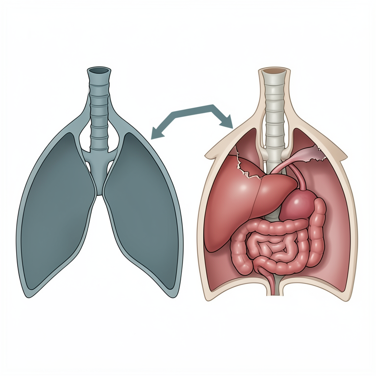
Mechanism of Traumatic Diaphragmatic Hernia
AI-generated conceptual illustration (mongrel pup). It explains the idea and does not show study data.
View source evidence ↗Figures & tables
From the original paper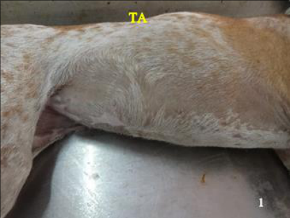 Figure 1
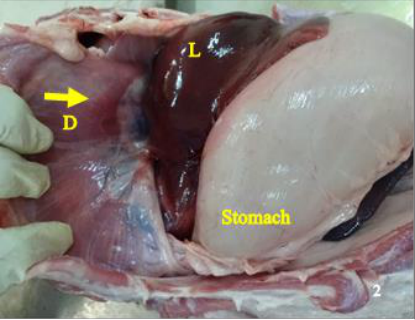 Figure 2
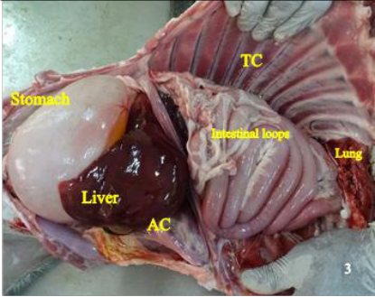 Figure 3
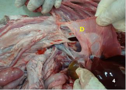 Figure 4
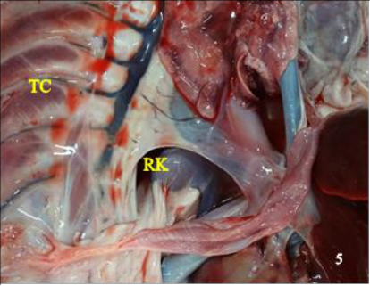 Figure 5
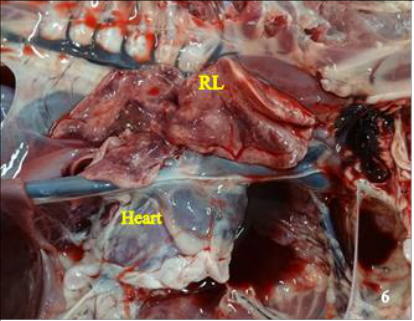 Figure 6
Introduction
The diaphragm is a sheet of muscle that separates the abdominal and thoracic cavities (Hermanson et al., 2018) [1] . A hernia is a pathologic disorder in which a particular part or part of the body project abnormally through a tear or opening into an adjacent part. Diaphragmatic hernia is of two types viz, True and false diaphragmatic hernias. True diaphragmatic or pleuroperitoneal hernias are defects in the diaphragmatic in which the serosa on the thoracic surface of the diaphragm remains intact, which further prevents direct communication between the thoracic and abdominal cavities. The false diaphragmatic hernia is a protrusion of abdominal viscera through an opening in the diaphragm and is caused mainly by trauma such as an automobile accident-traumatic diaphragmatic hernia, and rarely by congenital defects (Salci et al., 2009; Igna et al., 2014) [2,3] . In most cases, diaphragmatic hernia in dogs is caused due to trauma resultant from motor vehicle accidents. Traumatic diaphragmatic in small animals has been statistically reported to constitute 85%, while 10% are congenital and the rest are of unknown etiology (Wilson et al., 1971; Wilson and Hayes, 1986; Boudrieau, 1993) [4,5,6] . There is no breed disposition in dogs towards traumatic diaphragmatic hernias. Sex predilection towards male dogs was noted in few reports (Ranganath et al., 2000) [7] . However, further studies have indicated no sex predilection in dogs later (Fossum, 2006) [8] . The size of the diaphragmatic tear and its location depends on the position of the animal at the time of impact and the location of the viscera (Fossum, 2005) [9] . The type of tear depends on the location on the diaphragm in which the central tendinous area is stronger than the lateral paired costal area and occur along with the fiber orientation of the musculature (Levine, 1987) [10] . This case report aims at describing traumatic diaphragmatic hernia in a mongrel dog and its sudden collapse and the corresponding pathomorphological findings.
Materials and Methods
A seven months old female mongrel dog was presented for necropsy with a history of sudden collapse after being met with a road traffic accident. The necropsy examination was conducted, keeping in a record the history and a set of differentials.
Necropsy examination was carried out based on the standard necropsy techniques; the gross morphological features and the pathoanatomical abnormalities observed were described and noted down.
Results and Discussion
External examination of the carcass revealed distension of the lateral thoraco-abdominal region (Fig 1). The left lateral and medial lobe of the liver was highly congested and slightly adherent to the intestinal loops. Blood oozed out on a cut section of the liver. Spleen was highly congested. Kidneys were congested with congestion of cortico-medullary junction. The stomach was distended with gas. Serosa was highly congested, and the lumen contained about 10 ml of liquid contents. The intestinal serosa was congested. Pancreas revealed congestion and the urinary bladder was empty. The heart appeared constricted, and venous stasis was observed. All the lobes of the right lung appeared collapsed and congested (Fig 6). The above postmortem findings were in correlation to the previous reports (Sullivan and Reid, 1990; Oviawe et al., 2016) [11,12] . The cause of sudden death, in this case, was due to asphyxia as a result of severe respiratory compromise due to loss of mechanical function of the diaphragm, the pleural space-occupying effect of abdominal organ herniation further exacerbated by accumulation of air and compression of lung lobes by herniated organs with resultant atelectasis. The immediate increase in intra-abdominal pressure accompanying forceful blows to the abdominal wall causes the lungs to rapidly deflate when the glottis is open, as a result of which there is an increased pressure grade in the pleuroperitoneal space. On the other hand, the pressure gradient that occurs between the thorax and the abdomen may cause the diaphragm to tear (Fossum, 2006) [8] . The generalized effects include pain, haemorrhage and hypovolemic shock. All these factors combine together to produce hypoventilation, alveolar ventilation, perfusion mismatch and shunting, which results in hypoxia (Worth and Machon, 2005) [13] . In this case, the diaphragmatic tear was circumferential, which correlated with a few of the previous findings by (Igna et al., 2014; Sullivan and Reid, 1990) [3,11] , in which mostly circumferential tears were encountered in dogs. No surgical intervention was possible in this case as the dog immediately collapsed after being met with a road traffic accident and was submitted for necropsy (Dead on arrival). A diaphragmatic herniation is most frequently met with due to motor vehicle accidents, mostly having a guarded prognosis in acute cases (Fossum, 2006; Worth and Machon, 2005) [8,13] .
Conclusion
In conclusion, the external and internal examinations of the carcass revealed significant findings consistent with severe respiratory compromise resulting from a circumferential tear of the diaphragm, leading to herniation of abdominal organs into the thoracic cavity. This condition ultimately led to asphyxia and sudden death. The observed postmortem findings align with previous reports and underscore the devastating consequences of diaphragmatic herniation, particularly in cases of acute trauma such as road traffic accidents. Unfortunately, due to the immediate collapse of the dog, surgical intervention was not feasible, highlighting the challenges in managing such cases. The prognosis for diaphragmatic herniation in acute cases remains guarded, emphasizing the critical need for prompt recognition and intervention in traumatic injuries involving the diaphragm.
Acknowledgements
The author would like to thank the Tamil Nadu Veterinary and Animal Sciences University for providing support in conducting the necropsy of the case.
Figures and tables from the paper
References
9 in the original paper- Salci H, Cecen GÖ, OS G, Akin I. Multiple thoracic and thoracoabdominal trauma: Case report. Kafkas Üniversitesi Veteriner Fakültesi Dergisi. 2009;15(3):473476. DOI:10.9775/kvfd.2009.053-G.
- Igna C, Schuszler L, Sala A, Bumb D, Proteasa A, Dascalu R. Diaphragmatic hernia in dogs and cats: a report of 43 cases (2001-2013). Lucrari Stiintifice - Universitatea de Stiinte Agricole a Banatului Timisoara, Medicina Veterinara. 2014;47(4):48-53.
- Wilson GP, Newton CD, Burt JK. A review of 116 diaphragmatic hernias in dogs and cats. Journal of American Veterinary Medical Association. 1971;159(9):1142-1145.
- Wilson GP, Hayes HM Jr. Diaphragmatic hernia in the dog and cat: a 25-year overview. Seminars in Veterinary Medicine and Surgery (Small Animal). 1986;1(4):318326.
- Ranganath L, Vijayakumar AK, Rao NM, Jayadevappa SM. Intas Polivet. 2000;1(1):119-120.
- Levine SH. Diaphragmatic hernia. Veterinary Clinics of North America Small Animal Practice. 1987;17(2):411430. DOI: 10.1016/s0195-5616(87)50033-x.
- Sullivan M, Reid J. Management of 60 cases of diaphragmatic rupture. Journal of Small Animal Practice. 1990;31(9):425-430. DOI: 10.1111/j.1748-5827.1990.tb00503.x.
- Oviawe EI, Umar SA, Abubakar N, Yakubu AS, Buhari S. Traumatic Diaphragmatic Hernia in a 2-year-old Nigerian Female Mongrel Dog. Nigerian Veterinary Journal. 2016;37(2):117-122.
- Worth AJ, Machon RG. Traumatic diaphragmatic herniation: pathophysiology and management. Compendium: Continuing Education Practicing Veterinarians. 2005;27:178-190.