
Research article · 2024 · Full text
International Journal of Veterinary Sciences and Animal HusbandryMolecular identification of bacterial pathogen in suspected cases of respiratory tract infection from chicken
Volume 9(2) · Pages 877–880 · DOI: 10.22271/veterinary.2024.v9.i2l.1301 ↗
Research areasMicrobial infections and disease research
Abstract
Author-writtenThe respiratory illness is characterized by weakness, gasping, pump-handled respiration, dyspnea, mucous discharge, mortality, sinus swelling, facial edema, tracheitis, exudative pneumonia, pleuritis, pericarditis, sinusitis, decrease in egg production and poor egg quality (Zorman et al., 2000; Canal et al. 2005). Air sacculitis, higher mortality rates, higher medical expenses, and decreased egg production are the most common outcomes of respiratory illnesses in poultry. Total of 52 tissue samples of lungs and tracheae were collected from 52 birds suffering from respiratory tract infections during the period from December 2022 to March 2023. Out of 52 tissue samples, 98.08% tissue samples were found positive for bacterial pathogen isolation, while 1.92% wound swab samples were negative. Molecular detection of bacteria from tissue samples 98.07%, 59.61%, 9.61%, 7.69%, 3.84% and 1.92% isolated E. coli, Staphylococcus spp., M. gallisepticum, O. rhinotracheale, A. paragallinarum, and M. synoviae respectively. In this study, none of the tissue samples detected B. avium, P. aeruginosa, and P. multocida.
Summary
Source-checkedPlain-language reading of the full paper, prepared with AI and checked against the text. Each point links to the authors' own words.
Why it matters
- Respiratory tract illnesses are a primary cause of disease in poultry and result in substantial global economic losses for the industry. View evidence ↗
- Respiratory infections in chickens manifest through various symptoms, including respiratory distress, facial edema, and a decrease in both the quantity and quality of egg production. View evidence ↗
- Polymerase chain reaction (PCR) methods are considered more sensitive and specific than other diagnostic tools for identifying respiratory infections. View evidence ↗
The question
- The study aimed to identify the specific bacterial organisms present in the respiratory tracts of chickens using both molecular methods and direct culture. View evidence ↗
What they did
- The researchers collected lung and trachea tissue samples from 52 birds with respiratory infections between December 2022 and March 2023. View evidence ↗
- The samples were obtained from 40 different chicken flocks on commercial farms during post-mortem examinations. View evidence ↗
- Bacterial genomic DNA was isolated from the lung and trachea tissues using the DNeasy blood & tissue kit. View evidence ↗
- The study used PCR to screen for several pathogens, including E. coli, Staphylococcus spp., M. gallisepticum, O. rhinotracheale, A. paragallinarum, and M. synoviae. View evidence ↗
What they found
- PCR analysis showed that 98.07% (51 out of 52) of the tissue samples were positive for at least one bacterial pathogen. View evidence ↗
- Escherichia coli was the most prevalent pathogen, detected in 98.08% (51/52) of the samples. View evidence ↗
- Staphylococcus spp. was the second most common pathogen, found in 59.61% (31/52) of the samples. View evidence ↗
- Other detected pathogens included M. gallisepticum (9.61%), O. rhinotracheale (7.69%), A. paragallinarum (3.85%), and M. synoviae (1.92%). View evidence ↗
- Bordetella avium, Pasteurella multocida, and Pseudomonas aeruginosa were not detected in any of the samples. View evidence ↗
What it means
- The authors conclude that thorough surveillance and targeted control of prevalent bacteria are necessary to reduce the impact of respiratory infections in poultry. View evidence ↗
- The authors suggest that future research should investigate the virulence factors and molecular characteristics of the identified pathogens to improve control strategies. View evidence ↗
Visual summary
1 visualsIllustrations explain the paper; they do not show study data.
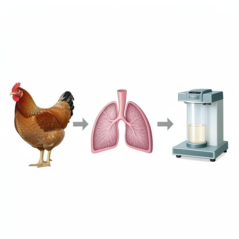
Diagnostic Workflow
AI-generated conceptual illustration (broiler chickens and respiratory tract tissues). It explains the idea and does not show study data.
View source evidence ↗Figures & tables
From the original paper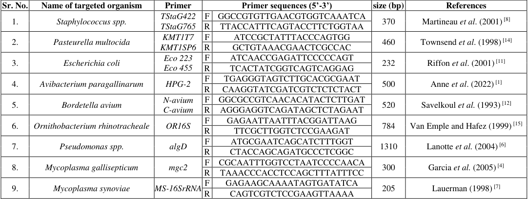 Table 1
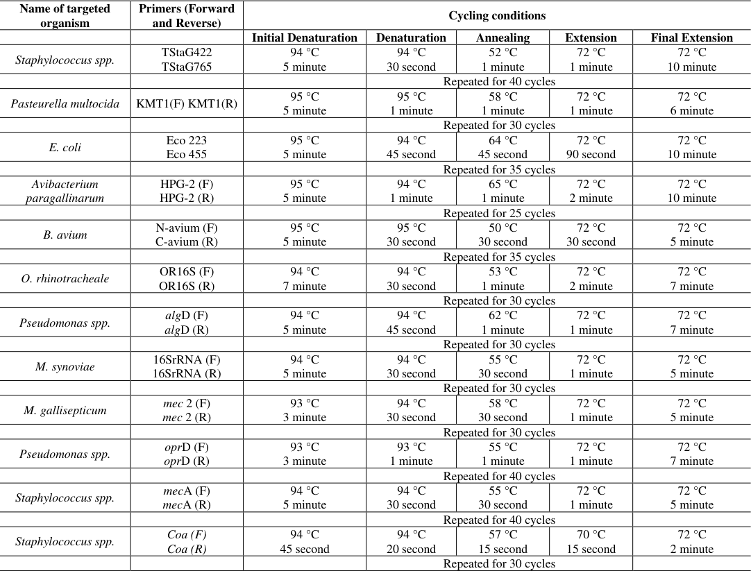 Table 2
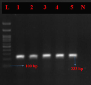 Figure 1
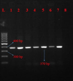 Figure 2
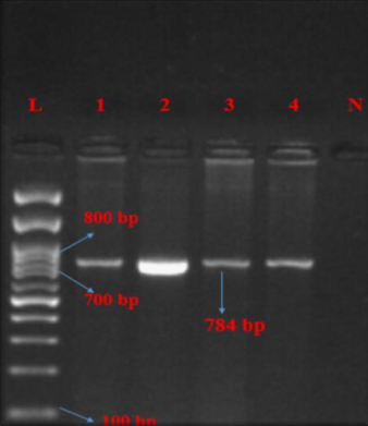 Figure 3
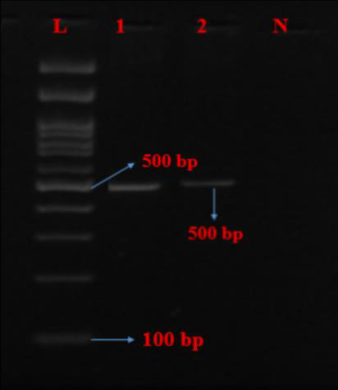 Figure 4
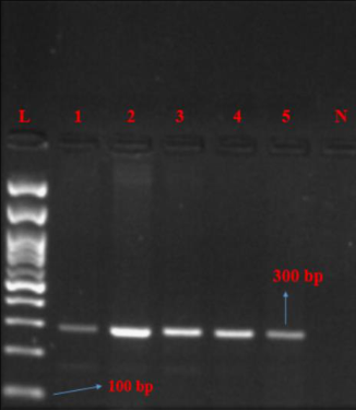 Figure 5
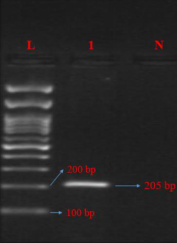 Figure 6
Introduction
Poultry are domesticated birds that are primarily raised for their meat, egg, and feathers. According to the most recent census, there are 23 billion eggs produced worldwide and 4.2 million tonnes of poultry meat are produced annually (Thopireddy, 2023) [13] . Compared to illnesses that affect other organs, respiratory tract illnesses constitute a major part of diseases that affect poultry and lead to significant economic losses for the industry globally (Glisson, 1998; Blackall and Soriano-Vargas, 2020) [5,2] . The respiratory organs of chicken include the lungs, syrinx, trachea, larynx, air sacs, nasal cavity, and nostrils. The respiratory illness is characterized by weakness, mucous discharge, gasping, dyspnea, mortality, sinus swelling, facial edema, tracheitis, exudative pneumonia, pump handled respiration, pleuritis, pericarditis, sinusitis, poor egg quality, and decrease in egg production (Zorman et al., 2000; Canal et al., 2005) [16,3] . Various bacterial pathogens affecting the respiratory system include Pasteurella multocida (P. multocida), Avibacterium paragallinarum (A.paragallinarum), Escherichia coli (E. coli), Bordetella avium (B. avium), Ornithobacterium rhinotracheale (O.rhinotracheale), Klebsiella pneumoniae (K. pneumoniae), Mycoplasma gallisepticum (M.gallisepticum), and Staphylococcus spp. (Popy et al., 2011) [9] . Molecular PCR methods that are more sensitive, specific, and effective could be more useful in the diagnosis of respiratory infections (Prabhu et al., 2021) [10] .
Materials and Methods
Materials and Methods
Sample Collection
Samples (lungs and trachea) were collected from 52 birds from commercial farms delivered to the department of Veterinary Pathology for the post-mortem examination. Samples were collected from 40 chicken flocks. The birds were of different ages and came from different regions of the Anand, Gujarat.
DNA extraction
The DNeasy® blood & tissue kit (50), Cat. No. 69504, Lot No. 172049243 was used to isolate bacterial genomic DNA directly from lung and trachea tissue samples of chickens, according to Kit's instructions. The negative control in the DNA extraction was the Tris buffer used for sample preparation. The quantity and quality of the DNA was determined using the Nanodrop 1000 system (Thermo Fisher Scientific, Waltham, MA, USA).
Molecular identification of bacteria by PCR
PCR was carried out in 25 µl volumes. The reaction mixture consisted of 2X PCR Master Mix (PCR Master Mix) 12.50 µl, Forward Primer (10pmol/µl) 1.0 µl, Reverse Primer (10pmol/µl) 1.0 µl, Template DNA 3.0 µl, and Nuclease Free Water 7.50 µl. PCR products were detected by running a 5 µl sample in 2% agarose gel containing ethidium bromide for 45 min at 80 V and visualization under UV light.
Results and Discussion
The present study was undertaken to study the presence of bacteria in respiratory tract infections from chickens. To find out the bacterial organisms from the respiratory tract of chickens using direct culture and molecular methods. In the present investigation samples were collected from the dead birds presented for post-mortem disease diagnosis at the Department of Veterinary Pathology, Veterinary College, Anand from December 2022 to March 2023. In the present study, 52 bird tissue samples (lung and trachea) were collected from chickens suffering from respiratory tract infections. Out of 52 tissue samples, 51 (98.07%) tissue samples were found positive for bacterial pathogen by PCR, while 01 (1.92%) tissue sample was negative for the presence of any pathogen. PCR-based detection of tissue samples revealed positivity for Escherichia coli 98.08% (51/52) followed by Staphylococcus spp. 59.61% (31/52), Mycoplasma gallisepticum 9.61% (05/52), Ornithobacterium rhinotracheale 7.69% (04/52), Avibacterium paragallinarum 3.85% (02/52), and Mycoplasma synoviae 1.92% (01/52). None of the samples found to be positive for Bordetella avium, Pasteurella multocida, and Pseudomonas aeruginosa through PCR.
Conclusion
Our study highlights the prevalence of bacterial pathogens in chickens suffering from respiratory tract infections. Utilizing molecular methods, notably PCR, we detected a high incidence of Escherichia coli and Staphylococcus spp. in lung and trachea tissue samples. This underscores the significance of these bacteria in poultry respiratory diseases. Importantly, the absence of Bordetella avium, Pasteurella multocida, and Pseudomonas aeruginosa suggests their limited involvement in the sampled cases. Our findings emphasize the necessity of thorough surveillance and targeted control measures against prevalent bacterial pathogens to mitigate the impact of respiratory infections in poultry populations. Future research could explore the molecular characteristics and virulence factors of the identified pathogens to enhance understanding and develop more effective control strategies.
Figures and tables from the paper
| Sr. No. | Name of targeted organism | Primer | Primer sequences (5’-3’) | — | size (bp) | References |
|---|---|---|---|---|---|---|
| 1. | Staphylococcus spp. | TStaG422 TStaG765 | F | GGCCGTGTTGAACGTGGTCAAATCA | 370 | Martineau et al. (2001) [8] |
| R | TTACCATTTCAGTACCTTCTGGTAA | |||||
| 2. | Pasteurella multocida | KMT1T7 KMT1SP6 | F | ATCCGCTATTTACCCAGTGG | 460 | Townsend et al. (1998) [14] |
| R | GCTGTAAACGAACTCGCCAC | |||||
| 3. | Escherichia coli | Eco 223 Eco 455 | F | ATCAACCGAGATTCCCCCAGT | 232 | Riffon et al. (2001) [11] |
| R | TCACTATCGGTCAGTCAGGAG | |||||
| 4. | Avibacterium paragallinarum | HPG-2 | F | TGAGGGTAGTCTTGCACGCGAAT | 500 | Anne et al. (2022) [1] |
| R | CAAGGTATCGATCGTCTCTCTACT | |||||
| 5. | Bordetella avium | N-avium C-avium | F | GGCGCCGTCAACACATACTCTTGAT | 520 | Savelkoul et al. (1993) [12] |
| R | AGGGAGGTCAGATAGCTCTAGAAT | |||||
| 6. | Ornithobacterium rhinotracheale | OR16S | F | GAGAATTAATTTACGGATTAAG | 784 | Van Emple and Hafez (1999) [15] |
| R | TTCGCTTGGTCTCCGAAGAT | |||||
| 7. | Pseudomonas spp. | algD | F | ATGCGAATCAGCATCTTTGGT | 1310 | Lanotte et al. (2004) [6] |
| R | CTACCAGCAGATGCCCTCGGC | |||||
| 8. | Mycoplasma gallisepticum | mgc2 | F | CGCAATTTGGTCCTAATCCCCAACA | 300 | Garcia et al. (2005) [4] |
| R | TAAACCCACCTCCAGCTTTATTTCC | |||||
| 9. | Mycoplasma synoviae | MS-16SrRNA | F | GAGAAGCAAAATAGTGATATCA | 205 | Lauerman (1998) [7] |
| R | CAGTCGTCTCCGAAGTTAAAA |
Table text extracted from the original PDF; superscript letters are shown inline. The original table image is below.
View original table image
| Name of targeted organism | Primers (Forward and Reverse) | Cycling conditions | — | — | — | — |
|---|---|---|---|---|---|---|
| Initial Denaturation | Denaturation | Annealing | Extension | Final Extension | ||
| Staphylococcus spp. | TStaG422 TStaG765 | 94 °C 5 minute | 94 °C 30 second | 52 °C 1 minute | 72 °C 1 minute | 72 °C 10 minute |
| Repeated for 40 cycles | ||||||
| Pasteurella multocida | KMT1(F) KMT1(R) | 95 °C 5 minute | 95 °C 1 minute | 58 °C 1 minute | 72 °C 1 minute | 72 °C 6 minute |
| Repeated for 30 cycles | ||||||
| E. coli | Eco 223 Eco 455 | 95 °C 5 minute | 94 °C 45 second | 64 °C 45 second | 72 °C 90 second | 72 °C 10 minute |
| Repeated for 35 cycles | ||||||
| Avibacterium paragallinarum | HPG-2 (F) HPG-2 (R) | 95 °C 5 minute | 94 °C 1 minute | 65 °C 1 minute | 72 °C 2 minute | 72 °C 10 minute |
| Repeated for 25 cycles | ||||||
| B. avium | N-avium (F) C-avium (R) | 95 °C 5 minute | 95 °C 30 second | 50 °C 30 second | 72 °C 30 second | 72 °C 5 minute |
| Repeated for 35 cycles | ||||||
| O. rhinotracheale | OR16S (F) OR16S (R) | 94 °C 7 minute | 94 °C 30 second | 53 °C 1 minute | 72 °C 2 minute | 72 °C 7 minute |
| Repeated for 30 cycles | ||||||
| Pseudomonas spp. | algD (F) algD (R) | 94 °C 5 minute | 94 °C 45 second | 62 °C 1 minute | 72 °C 1 minute | 72 °C 7 minute |
| Repeated for 30 cycles | ||||||
| M. synoviae | 16SrRNA (F) 16SrRNA (R) | 94 °C 5 minute | 94 °C 30 second | 55 °C 30 second | 72 °C 1 minute | 72 °C 5 minute |
| Repeated for 30 cycles | ||||||
| M. gallisepticum | mec 2 (F) mec 2 (R) | 93 °C 3 minute | 94 °C 30 second | 58 °C 30 second | 72 °C 1 minute | 72 °C 5 minute |
| Repeated for 30 cycles | ||||||
| Pseudomonas spp. | oprD (F) oprD (R) | 93 °C 3 minute | 93 °C 1 minute | 55 °C 1 minute | 72 °C 1 minute | 72 °C 7 minute |
| Repeated for 40 cycles | ||||||
| Staphylococcus spp. | mecA (F) mecA (R) | 94 °C 5 minute | 94 °C 30 second | 55 °C 30 second | 72 °C 1 minute | 72 °C 5 minute |
| Repeated for 40 cycles | ||||||
| Staphylococcus spp. | Coa (F) Coa (R) | 94 °C 45 second | 94 °C 20 second | 57 °C 15 second | 70 °C 15 second | 72 °C 2 minute |
| Repeated for 30 cycles |
Table text extracted from the original PDF; superscript letters are shown inline. The original table image is below.
View original table image
References
16 in the original paper- Anne NS, Malmarugan S, Prabhu M, Rajeswar JJ. Isolation and molecular serotyping of A. paragallinarum from desi birds. Indian Journal of Animal Health. 2022;61(1):78-83. DOI: https://doi.org/10.36062/ijah.2022.12021
- Blackall PJ, Soriano-Vargas E. Infectious coryza and related bacterial infections. Diseases of Poultry. 2020:890-906. DOI: https://doi.org/10.1002/9781119371199.ch20
- Canal CW, Leao JA, Rocha SLS, Macagnan M, LimaRosa CAV, Oliveira SD, Back A. Isolation and characterization of O. rhinotracheale from chickens in Brazil. Research in veterinary science. 2005;78(3):225230. DOI: https://doi.org/10.1016/j.rvsc.2004.10.003
- García M, Ikuta N, Levisohn S, Kleven SH. Evaluation and comparison of various PCR methods for detection of M. gallisepticum infection in chickens. Avian diseases. 2005;49(1):125-132. DOI: https://doi.org/10.1637/72610812204R1
- Glisson JR. Bacterial respiratory disease of poultry. Poultry Science. 1998;77(8):1139-1142. DOI: https://doi.org/10.1093/ps/77.8.1139
- Lanotte P, Watt S, Mereghetti L, Dartiguelongue N, Rastegar-Lari A, Goudeau A, et al. Genetic features of Pseudomonas aeruginosa isolates from cystic fibrosis patients compared with those of isolates from other origins. Journal of medical microbiology. 2004;53(1):7381. DOI: https://doi.org/10.1099/jmm.0.05324-0
- Lauerman LH, Hoerr FJ, Sharpton AR, Shah SM, Van Santen VL. Development and application of a polymerase chain reaction assay for M. synoviae. Avian Diseases. 1993:829-834. Doi: https://doi.org/10.2307/1592037
- Martineau F, Picard FJ, Ke D, Paradis S, Roy PH, Ouellette M, Bergeron MG. Development of a PCR assay for identification of staphylococci at genus and species levels. Journal of clinical microbiology. 2001;39(7):2541-2547.
- Popy N, Asaduzzaman M, Miah MS, Siddika A, Sufian MA. Pathological study on the upper respiratory tract infection of chickens and isolation, identification of causal bacteria. The Bangladesh Veterinarian. 2011;28:60-69. DOI: https://doi.org/10.3329/bvet.v28i2.10677
- Prabhu M, Malmarugan S, Sweetline Anne N, Parthiban S, Balakrishnan G, Johnson Rajeswar J. Detection of M. gallisepticum infection in turkey and chicken farms of Tamil Nadu, India. International Journal of Current Microbiology and Applied Sciences. 2021;10(01):31513158. DOI: https://doi.org/10.20546/ijcmas.2021.1001.367
- Riffon R, Sayasith K, Khalil H, Dubreuil P, Drolet M, Lagacé J. Development of a rapid and sensitive test for identification of major pathogens in bovine mastitis by PCR. Journal of clinical microbiology. 2001;39(7):25842589. DOI: https://doi.org/10.1128/jcm.39.7.25842589.2001
- Savelkoul PH, de Groot LE, Boersma C, Livey I, Duggleby CJ, Van der Zeijist BA, et al. Identification of Bordetella avium using the polymerase chain reaction. Microbial Pathogenesis. 1993;15(3):207-215. DOI: https://doi.org/10.1006/mpat.1993.1071
- Thopireddy NR. Isolation and Molecular Detection of E. coli from Common Respiratory Infections of Poultry in AP. 2023. DOI: 10.36349/easjvms.2023.v05i01.001
- Townsend KM, Frost AJ, Lee CW, Papadimitriou JM, Dawkins HJ. Development of PCR assays for speciesand type-specific identification of Pasteurella multocida isolates. Journal of Clinical Microbiology. 1998;36(4):1096-1100.
- Van Empel PCM, Hafez HM. Ornithobacterium rhinotracheale: a review. Avian pathology. 1999;28(3):217-227. DOI: https://doi.org/10.1080/03079459994704.
- Zorman-Rojs O, Zdovc I, Bencina D, Mrzel I. Infection of turkeys with O. rhinotracheale and M. synoviae. Avian Disease. 2000;44:1017-1022. DOI: https://doi.org/10.2307/1593082.
Cited by 1
From OpenAlex, OpenCitations and reference lists read by Scinovex
- Relevance of navel quality of day-old chick on organ development, initial performance, and bacterial presence ↗
Mariana Cristina Vieira, Henrique Carneiro Lobato, Laryssa Fernanda Bernardes, Clayton Barbosa Galvão, Júlia Macedo Fernandes Oliveira, Victória Veiga Alves et al. · Poultry Science · 2025
This study aimed to evaluate effects of day-old chick navel quality on the absorption of the residual yolk sac, visceral development, macroscopic organ alterations, yolk sac bacterial presence and performance of broilers up to 14 d of age. A total of 372 male Ross 308 day-old chicks were classified as first or second-grade based on navel quality. First-grade chicks were…
❝ 2PDF