
Research article · 2024 · Full text
International Journal of Advanced Biochemistry ResearchCanine anemia: Clinical assessment, prevalence and morphological classification
Volume 8(4S) · Pages 279–284 · DOI: 10.33545/26174693.2024.v8.i4sd.961 ↗
Research areasRabies epidemiology and controlVirus-based gene therapy researchBlood groups and transfusion
Abstract
Author-writtenThe present study entitled “Canine Anemia: Clinical assessment, Prevalence and Morphological classification” was conducted at Department of Veterinary Pathology, College of Veterinary and Animal Sciences, Parbhani. During study, total 146 dogs were screened from dog breeder farms, Veterinary dispensaries, and Veterinary Clinical Complex for detection of anemia. Out of that, 76 cases were considered anemic based on history, clinical signs, and laboratory tests. Suspected cases were examined for morphological classification of anemia. Clinical evaluation revealed that anemic dogs had rough skin and hair coats, pale visible mucus membranes, lethargy, anorexia, tachycardia and deep breathing as prominent clinical features. Hematology revealed significantly lower values of Hb, PCV, TEC, MCV, and MCHC, whereas, values of TLC, DLC and platelets were found within normal physiological range. Overall prevalence of anemia was 52.06%. It was found highest in adult dogs that to in males compared to females, in ND breed of dog and during monsoon season. Morphologically, there was marked variation in shape and size of erythrocytes. Morphologically, microcytic hypochromic anemia was recorded as a major type of anemia. Highlights 1. Dog plays most important role in human lives as a best companion. Due to their fundamental behavioral characters like obedience, loyalty, adoptability, sensitive olfaction, differentiation and investigating behavior, different dog breeds are utilized for various purposes viz., sporting, crime detection, explosive searching, etc. 2. Considering the various diseases of dogs, anemia is most common and frequently diagnosed complication caused due to many etiological factors, some of which are life-threatening in veterinary practice.
Summary
Source-checkedPlain-language reading of the full paper, prepared with AI and checked against the text. Each point links to the authors' own words.
Why it matters
- Anemia is one of the most common and frequently diagnosed complications in dogs, and some of its causes can be life-threatening. View evidence ↗
- Anemia is characterized by a reduction in the number of circulating red blood cells (erythrocytes) or a decrease in hemoglobin concentration. View evidence ↗
- The study was motivated by the critical role of red blood cells in oxygen transport and a lack of research on canine anemia in the Parbhani district of India. View evidence ↗
The question
- The study aimed to perform a clinical assessment, determine the prevalence, and establish the morphological classification of anemia in dogs. View evidence ↗
What they did
- A total of 146 dogs were screened for anemia from veterinary dispensaries, clinical complexes, and breeder farms. View evidence ↗
- Twelve apparently healthy dogs were included as a control group to compare hematological status with anemic dogs. View evidence ↗
- Anemia classification was based on 10 clinical parameters, including heart rate, respiration rate, and the condition of mucus membranes and skin. View evidence ↗
- Morphological classification was determined using blood smear examinations and erythrocytic indices, specifically Mean Corpuscular Volume (MCV) and Mean Corpuscular Hemoglobin Concentration (MCHC). View evidence ↗
What they found
- The overall prevalence of anemia among the screened dogs was 52.06%, with 76 out of 146 dogs identified as anemic. View evidence ↗
- Prevalence varied by age, peaking in adults (32.90%) and reaching its lowest point in the senile group (7.90%). View evidence ↗
- Anemia was more prevalent in males (55.26%) than in females (44.74%). View evidence ↗
- The Non-Descript (ND) breed showed the highest prevalence of anemia (32.90%), while several other breeds, including Pugs and Lhasa apsos, showed the lowest (1.32%). View evidence ↗
- Seasonal analysis showed that anemia was most frequent during the monsoon season (65.79%) and least frequent in winter (11.84%). View evidence ↗
- Prominent clinical signs in anemic dogs included pale mucus membranes, lethargy, anorexia, tachycardia, deep breathing, and rough skin/hair coats. View evidence ↗
- Microcytic hypochromic anemia was the most common morphological type (44.74%), followed by normocytic normochromic anemia (19.74%). View evidence ↗
- Blood smear analysis of anemic dogs revealed high rates of poikilocytosis (94.74%) and anisocytosis (78.95%), with 76.34% of cases exhibiting both. View evidence ↗
What it means
- The authors concluded that anemia has a 52.06% prevalence in the studied population, most frequently affecting adult males of the Non-Descript breed during the monsoon. View evidence ↗
- The authors identified microcytic hypochromic anemia as the primary morphological type of anemia in the studied dogs. View evidence ↗
Visual summary
1 visualsIllustrations explain the paper; they do not show study data.
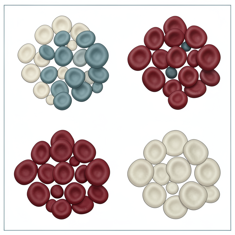
Morphological Classification of Canine Anemia
AI-generated conceptual illustration (canine red blood cells). It explains the idea and does not show study data.
View source evidence ↗Figures & tables
From the original paper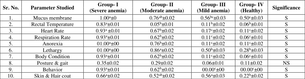 Table 1
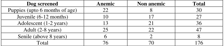 Table 2
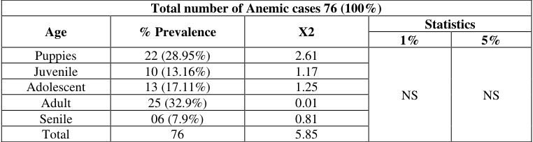 Table 3
 Table 4
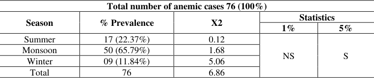 Table 5
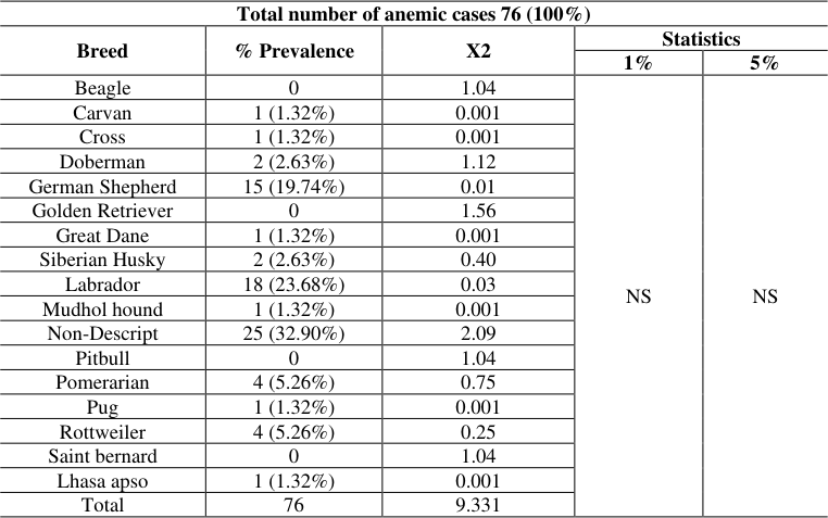 Table 6
Introduction
The dog (Canis familaris) is the oldest companion animal and is first domesticated species. The dog has fulfilled a variety of roles i.e., as a guard dog, hunting, companion and also guide. Hytönen, & Lohi, (2016) [11] stated that dog is a large animal model and is physiologically and clinically more similar to human than mouse. Lindblad et.al., (2005) [17] reported that dog genome is less divergent from the human genome than from the mouse genome. Therefore, more of the human genomic sequence can be syntenically aligned to the genome of the dog than that of the mouse. Dogs have approximately the same number of genes as humans, most of them being close Orthologues. Anemia is defined as a condition in which the body has a decreased number of circulating erythrocytes (RBCs), or decrease in hemoglobin concentration. It can also be defined as reduction in number of the erythrocytes in an animal for that particular age, species, breed, and geographic location (Vegad and Madhu Swamy, 2010) [29] . It can either be due to decreased production of erythrocytes or haemoglobin or increased destruction of red blood cells (RBCs). It is therefore associated with reduction in red cell count or volume of red cell. In a physiological way, anemia is defined as the decreased ability of blood to supply tissues with adequate oxygen for proper metabolic functions (Hoffbrand and Pettit, 1993) [12] . It is characterized by reduction in the hemoglobin (Hb), haematocrit (PCV) or total erythrocytic count (TEC) per unit volume of blood in a normally hydrated animal (Aiello, 1997) [2] . Different researchers viz. Moninder (2003) [21] , Furlanello et al. (2005) [8] , Singh (2012) [25] , Tandel et al. (2016) [28] and Meshram et al. (2019) [20] studied anemia in dogs at different places and showed that the prevalence of anemia ranged between 47-75%, which is quite higher and alarming condition. Wintrobe developed three erythrocytic indices viz. Mean corpuscular hemoglobin (MCH), Mean corpuscular volume (MCV) & mean corpuscular hemoglobin concentration (MCHC). The traditional morphological approach for classification of anemia is based on Mean cell volume (MCV) and mean cell hemoglobin concentration (MCHC). This classification is useful and diagnostically sound which classify anemia as normocytic-normochromic, macrocyticnormochromic, macrocytic-hypochromic and microcytichypochromic. Based on morphology, there are two more types such as normocytic-hypochromic and microcyticnormochromic. Also, one more addition to type based on aetiology is immunological haemolytic anemia (Chakrabarthi, 2007) [3] . The course and outcome of anemia is mainly dependent upon the degree and severity affecting the blood components and it could be life threatening or at other side it may be asymptomatic and even difficult to detect clinically. Therefore, one should give more attention towards predisposing causes, exciting causes, diagnosis and prevention of anemia rather than treating it. Considering the importance of RBCs and hemoglobin in transport of oxygen to vital organs and the high prevalence but very little research work on anemia in dogs in India, especially in and around the Parbhani district of Marathwada region, the present study has been conducted.
Materials and Methods
The study was conducted at College of Veterinary and Animal Sciences, Parbhani, Maharashtra. Samples (blood, serum, fecal and urine) for present study were taken from Veterinary Clinical Complex, Veterinary Polyclinics, Veterinary dispensaries and from dog breeder farms in and around Parbhani and screened for anemia.
Clinical assessment of anemia
Anemia was diagnosed on the basis of history, clinical manifestations and laboratory examination. Positive anemic dogs included in this study were selected from, dogs in study area and screened for hematological studies. The anemic dogs taken for the study were divided into five groups viz., up to 6 months of age (Puppies), 6 months -1year (Juvenile), 1-2 years (Adolescent), 2-8 years (Adult) and above 8 years of age i.e., Senile, (Harvey, 2021) [10] . For comparison of hematological status of anemic dog with that of healthy dog, a total of 12 apparently healthy dogs from well-maintained dog owners from Parbhani city were taken.
Inclusion criteria for assessment of anemia in dog
Following inclusion criteria was used along with clinical assessment of dogs for incorporation of anemic cases during study. [19] and (Assenmacher et al., 2019) [4] .
Collection of blood and serum samples
The blood samples were collected either from cephalic or saphenous vein of dogs suspected for anemia with all aseptic precautions, in vacutainers with anticoagulant and sterile vials containing Ethylene Diamine Tetra Acetate (EDTA) as an anticoagulant for hematological studies. Samples were collected from apparently healthy control animals in a similar pattern as that suspected for anemic dogs.
Classification of data
The data collected was analysed according to age, sex, breed of animals and also, season for prevalence study. For classification of anaemia, 10 clinical parameters were taken such as visible mucus membranes, rectal temperature, heart rate, respiration rate, anorexia, lethargy, body condition, posture and gait, behaviour, skin and hair coat. Scoring was made between 0-1 grade. Grading for each parameter was made as 0 if it was normal and 1 if it was deviated from normal i.e., abnormal. For example, normally the color of mucus membrane is pink, so it was graded as 0 whereas, other than pink like pale or congested or icteric was graded as 1. This was done for all 10 parameters and score card was made for each case out of 10 clinical parameters noted and after that groups were made according to grading as follows: 1. Group I (Severe anemia): The dogs with score 8-10 were categorized into this group.
Group II (Moderate anemia):
The dogs with score 5-7 were categorized into this group.
Group III (Mild anemia):
The dogs with score 1-4 were categorized into this group.
Group IV (Apparently healthy control): In this group
representative number of cases were taken from apparently healthy dog cases. (Kisielewicz et al., 2014) [16] .
Morphological classification of anemia
It was done on the basis of erythrocytic indices (MCV and MCHC) and blood smear examination during DLC.
Prevalence of anemia
Overall prevalence was calculated from number of dogs screened and out of which number of positive cases for anemia, which were selected for further studies. Prevalence of Clinical cases was further classified into Age-wise (Puppies, Juvenile group, Adult and Senile group, as described by Harvey, (2021) [10] and gender-wise (male and female), breed wise and season wise.
Statistical analysis
Data generated was analyzed statistically to find the level of significance by employing Chi-square test for analysis of prevalence of anemia (age, sex, breed and season wise) and Complete Randomized Design (CRD) for clinical assessment, hematological and biochemical studies in both anemic as well as healthy dog with standard statistical method described by Snedecor and Cochran, (1982) [26] . Prominent clinical signs and symptoms significant of anemia in dogs were reported by several researchers. From the reviewed literature, Joshi (2000) [13] , Furlanello et al.
Results and Discussion
(2005) [8] , Swann et al. (2013) [27] , Kisielewicz et al. (2014) [16] , Mahalingaiah et al. (2017) [19] , Assenmacher et al. (2019) [4] recorded similar clinical findings as recorded in present study. Skin & Hair coat 0.66 a ±0.02 0.52 ab ±0.02 0.56 a ±0.03 0.22 b ±0.02 S Where, Different superscripts in a row show significant difference. S-Significant, NS-Non-significant (Note: Values of all parameters of anemic dogs are depicted in 0-1 grade)
Prevalence of anemia
Overall prevalence of anemia in dogs was recorded as 52.06% i.e., out of total 146 dogs screened, 76 cases were found anemic. Different researchers viz. Moninder (2003) [21] , Furlanello et al. (2005) [8] , Singh (2012) [25] , Tandel et al. (2016) [28] and Meshram et al. (2019) [20] studied anemia in dogs at different places and showed that the prevalence of anemia was ranged between 47-75%. In present study we recorded 52.06% prevalence of anemia in dogs, which was found analogous to the findings of earlier workers.
Age wise prevalence
It was found highest (32.90%) in adults, followed by Puppies (28.95%), Adolescent (17.11%), Juvenile group (13.16%) and least (7.90%) in Senile group. Chervier et al. (2012) [7] and Tandel et al. (2016) [28] observed highest prevalence of anemia in old dogs, whereas, Singh (2012) [25] , Brahmbhatt et al. (2015) [6] , Meshram et al. (2019) [20] and Shah et al. (2020) [24] recorded higher incidence in young pups. The results found in present study were allied with the study conducted by Chervier et al. (2012) [7] , Tandel et al. (2016) [28] and Meshram et al. (2019) [20] .
Gender wise prevalence
It was found highest in males (42 cases out of total 76 cases of anemia i.e., 55.26%) than females (34 cases out of total 76 cases of anemia i.e., 44.74%). The highest prevalence of anemia in males observed during present study has been found akin with the studies conducted by Brahmbhatt et al. (2015) [6] , Tandel et al. (2016) [28] , Meshram et al. (2019) [20] and Shah et al. (2020) [24] .
Breed wise prevalence
It was found highest (32.90%) in ND breed of dog, followed by Labrador (23.68%), German shepherd (19.74%), Pomeranian (5.26%), Rottweiler (5.26%), Siberian Husky (2.63%), Doberman (2.63%) and least in Carvan, Crossbreds, Great Dane, Mudhol Hounds, Pug and Lhasa apso (Each were having 1.32% prevalence). The results obtained during present study regarding breed wise prevalence of anemia in dogs found congrous with the reports of earlier researchers, Singh et al. (2012) [25] ; Brahmbhatt et al. (2015) [6] ; Meshram et al. (2019) [20] and Shah et al. (2020) [24] .
Season wise prevalence
It was observed highest during monsoon season (65.79%), followed by summer (22.37%) and then winter (11.84%). The findings of present study about seasonal prevalence of anemia was found at par with the findings of Ahmad et al. (2007) [1] and Gadahi et al. (2008) [9] . [22] , Zygner et al. (2006) [30] , Balch & Mackin, (2007) [5] and Shah et al. (2009) [23] reported microcytosis, anisocytosis, poikilocytosis, hypochomasia, spherocytosis and agglutination of erythrocytes during their study of anemia in dogs. The findings on morphology of erythrocytes in the present study go well with the observations recorded by all these authors. Regarding morphological type of anemia, it was concluded that microcytic hypochromic anemia in 44.74% cases was a major type of anemia followed by normocytic normochromic anemia in 19.74% cases, microcytic normochromic anemia in 18.42% cases, macrocytic hypochromic anemia in 15.79% cases, normocytic hypochromic anemia in 6.58% cases, and macrocytic normochromic anemia in 1.32% cases out of 76 anemic dogs screened. Kim et al. (2006) [14] reported microcytic hypochromic anemia as a major type of anemia during their study, which very well go with findings of present study. Several other researchers like King et al. (1992) [15] , and Lucidi et al. (2017) [18] found normocytic normochromic anemia as major type of anemia during their research. This is also a co relating findings between their study and present study.
Conclusion
From the present study it can be concluded as 1. Pale visible mucus membrane, lethargy or reluctant to move, anorexia, tachycardia, deep breathing, rough and dehydrated skin & hair coat are the prominent clinical manifestations observed in anemic cases of dog. 2. Overall prevalence of anemia in dogs was 52.06%. It was found highest in adult dogs that to in males as compared to females and in ND breed of dog during monsoon season. 3. Microcytic hypochromic anemia was a major type of anemia followed by normocytic normochromic anemia then, microcytic normochromic anemia, macrocytic hypochromic anemia, normocytic hypochromic anemia and macrocytic normochromic anemia.
Acknowledgement
AcknowledgementAuthors wish to thank Associate Dean, College of Veterinary and Animal Sciences, Parbhani for conducting the experiment and providing facilities required for it.
Figures and tables from the paper
| Sr. No. | Parameter Studied | Group- I (Severe anemia) | Group- II (Moderate anemia) | Group- III (Mild anemia) | Group- IV (Healthy) | Significance |
|---|---|---|---|---|---|---|
| 1. | Mucus membrane | 1.00a±0 | 0.76ab±0.02 | 0.56bc±0.03 | 0.50c±0.03 | S |
| 2. | Rectal Temperature | 0.83a±0.01 | 0.05b±0.01 | 0.11b±0.02 | 0.06b±0.01 | S |
| 3. | Heart Rate | 0.93a ±0.01 | 0.67b±0.02 | 0.17c±0.02 | 0.11c±0.02 | S |
| 4. | Respiration Rate | 0.93a±0.01 | 0.62b±0.02 | 0.11c±0.02 | 0.06c±0.01 | S |
| 5. | Anorexia | 01.00a±00 | 0.76b±0.02 | 0.11c±0.02 | 0.11c±0.02 | S |
| 6. | Lethargy | 01.00a±00 | 0.86a±0.02 | 0.50b±0.03 | 0.28b±0.03 | S |
| 7. | Body Condition | 0.93a±0.01 | 0.62b±0.02 | 0.11c±0.02 | 0.06c±0.01 | S |
| 8. | Posture & gait | 0.35±0.02 | 0.29±0.02 | 0.06±0.01 | 0.11±0.02 | NS |
| 9. | Behavior | 0.93a±0.01 | 0.62b±0.02 | 00.00c±00 | 00.00c±00 | S |
| 10. | Skin & Hair coat | 0.66a±0.02 | 0.52ab±0.02 | 0.56a±0.03 | 0.22b±0.02 | S |
Table text extracted from the original PDF; superscript letters are shown inline. The original table image is below.
View original table image
| Dog screened | Anemic | Non anemic | Total |
|---|---|---|---|
| Puppies (upto 6 months of age) | 22 | 8 | 30 |
| Juvenile (6-12 months) | 10 | 17 | 27 |
| Adolescent (1-2 years) | 13 | 21 | 36 |
| Adult (2-8 years) | 25 | 22 | 47 |
| Senile (above 8 years) | 6 | 2 | 8 |
| Total | 76 | 70 | 176 |
Table text extracted from the original PDF; superscript letters are shown inline. The original table image is below.
View original table image
| Total number of Anemic cases 76 (100%) | — | — | — | — |
|---|---|---|---|---|
| Age | % Prevalence | X2 | Statistics | |
| 1% | 5% | |||
| Puppies | 22 (28.95%) | 2.61 | NS | NS |
| Juvenile | 10 (13.16%) | 1.17 | ||
| Adolescent | 13 (17.11%) | 1.25 | ||
| Adult | 25 (32.9%) | 0.01 | ||
| Senile | 06 (7.9%) | 0.81 | ||
| Total | 76 | 5.85 |
Table text extracted from the original PDF; superscript letters are shown inline. The original table image is below.
View original table image
| Total number of Anemic cases 76 (100%) | — | — | — |
|---|---|---|---|
| Sex | % Prevalence | X2 | Statistics |
| Male | 42 (55.26%) | 0.78 | NS |
| Female | 34 (44.74%) | 0.85 | |
| Total | 76 | 1.63 |
Table text extracted from the original PDF; superscript letters are shown inline. The original table image is below.
View original table image
| Total number of anemic cases 76 (100%) | — | — | — | — |
|---|---|---|---|---|
| Season | % Prevalence | X2 | Statistics | |
| 1% | 5% | |||
| Summer | 17 (22.37%) | 0.12 | NS | S |
| Monsoon | 50 (65.79%) | 1.68 | ||
| Winter | 09 (11.84%) | 5.06 | ||
| Total | 76 | 6.86 |
Table text extracted from the original PDF; superscript letters are shown inline. The original table image is below.
View original table image
| Total number of anemic cases 76 (100%) | — | — | — | — |
|---|---|---|---|---|
| Breed | % Prevalence | X2 | Statistics | |
| 1% | 5% | |||
| Beagle | 0 | 1.04 | NS | NS |
| Carvan | 1 (1.32%) | 0.001 | ||
| Cross | 1 (1.32%) | 0.001 | ||
| Doberman | 2 (2.63%) | 1.12 | ||
| German Shepherd | 15 (19.74%) | 0.01 | ||
| Golden Retriever | 0 | 1.56 | ||
| Great Dane | 1 (1.32%) | 0.001 | ||
| Siberian Husky | 2 (2.63%) | 0.40 | ||
| Labrador | 18 (23.68%) | 0.03 | ||
| Mudhol hound | 1 (1.32%) | 0.001 | ||
| Non-Descript | 25 (32.90%) | 2.09 | ||
| Pitbull | 0 | 1.04 | ||
| Pomerarian | 4 (5.26%) | 0.75 | ||
| Pug | 1 (1.32%) | 0.001 | ||
| Rottweiler | 4 (5.26%) | 0.25 | ||
| Saint bernard | 0 | 1.04 | ||
| Lhasa apso | 1 (1.32%) | 0.001 | ||
| Total | 76 | 9.331 |
Table text extracted from the original PDF; superscript letters are shown inline. The original table image is below.
View original table image
References
22 in the original paper- Ahmad SS, Khan MS, Khan MA. Prevalence of canine babesiosis in Lahore, Pakistan. J Anim Plant Sci. 2007;17:11-13.
- Assenmacher TD, Jutkowitz LA, Koenigshof AM, Lucidi CdA, Scott MA. Clinical features of precursortargeted immune-mediated anemia in dogs: 66 cases (2004-2013). J Am Vet Med Assoc. 2019;3:255.
- Balch A, Mackin A. Canine immune-mediated hemolytic anemia: Pathophysiology, clinical signs, and diagnosis. Compend. 2007;29(4):217-225.
- Brahmbhatt NN, Patel PV, Hasnani JJ, Pandya SS, Joshi BP. Study on prevalence of ancylostomosis in dogs at Anand district, Gujarat, India. Vet World. 2015;8(12):1405.
- Chervier C, Cadoré JL, Rodriguez-Piñeiro MI, Deputte BL, Chabanne L. Causes of anaemia other than acute blood loss and their clinical significance in dogs. Vet J. 2012;53(4):223-227.
- Furlanello T, Fiorio F, Caldin M, Lubas G, SolanoGallego L. Clinicopathological findings in naturally occurring cases of babesiosis caused by large form Babesia from dogs of northeastern Italy. Vet Parasitol. 2005;134(1-2):77-85.
- Gadahi JA, Arijo AG, Abubakar M, Javaid SB, Arshed MJ. Prevalence of Blood parasites in stray and pet Dogs in Hyderabad Area: Comparative sensitivity of different Diagnostic techniques for the detection of microfilaria. Vet World. 2008;1(8):229-232.
- Harvey ND. How old Is my dog? identification of rational age groupings in pet dogs based upon normative age-linked processes. Front Vet Sci. 2021;8:643085.
- Kim HJ, Park C, Jung DI, Kang BT, Kim JW, Lim CY, et al. A case of protein losing nephropathy in a dog infected with canine Babesia gibsoni. Korean J Vet Res. 2006;46(1):77-81.
- King LG, Giger U, Diserens D, Nagode LA. Anemia of Chronic Renal Failure in Dogs. Vet Intern Med. 1992;6(5):264-270. doi:10.1111/j.1939-1676.1992.tb00350.x.
- Kisielewicz C, Self I, Bell R. Assessment of Clinical and Laboratory Variables as a Guide to Packed Red Blood Cell Transfusion of Euvolemic Anemic Dogs. J Vet Intern. 2014;28:576-582.
- Lindblad-Toh K, Wade CM, Mikkelsen TS, et al. Genome sequence, comparative analysis and haplotype structure of the domestic dog. Nature. 2005;438:803819.
- Lucidi CdA, de Rezende CL, Jutkowitz LA, Scott MA. Histologic and cytologic bone marrow findings in dogs with suspected precursor-targeted immune-mediated anemia and associated phagocytosis of erythroid precursors. Vet Clin Pathol. 2017;46(3):401-415. doi:10.1111/vcp.12502.
- Mahalingaiah MKC, Asoor M, Thimmaiah RP, Narayanaswamy HD, Mukartal SY, Elattuvalappil AM, et al. Prevalence of canine babesiosis in different breeds of dogs in and around Bengaluru. Adv. Anim. Vet Sci. 2017;5(3):140-144.
- Meshram PV, Bembde NS, Deshpande RR, Bomble PB, Shetye MV, Dhule RP, et al. Analysis of Haematological Data for Canine Samples with Special Reference to Anaemia. Chem Sci. Rev Lett. 2019;8(30):221-225.
- Nassiri SM, Shirani D, Khazrainia P, Hajmohammadali A, Sharifi H. The investigation of the prevalence of immune-mediated hemolytic anemia (IMHA) in anemic dogs referred to the Veterinary Teaching Hospital of the University of Tehran. Comp Clin Pathol. 2005;14(3):121-124. DOI:10.1007/s00580-005-0578-y.
- Shah SA, Sood NK, Uppal SK, Gupta K. Studies on canine secondary immune mediated hemolytic anemia. J Immunol Immunopathol. 2009;11(2):67-70.
- Shah SA, Sood NK, Singh A, Gupta K, Wani BM, Shafi M, et al. Evaluation of Etiopathology of Canine Anemia in Ludhiana, India. Vet Res. 2020;8(04):388391.
- Singh R, Ravjoyt S, Beigh SA, Bhardwaj RK. Incidence of Anaemia in Dogs from Jammu Region. Ind. J Canine Pract. 2012;4(1):1-3.
- Swann JW, Skelly BJ. Systematic Review of Evidence Relating to the Treatment of Immune-Mediated Hemolytic Anemia in Dogs. J Vet Intern. 2013;28:576582.
- Tandel FB, Jani RG, Rao N, Bilwal AK, Raval SR. Epidemiological Status of Anemia in Dogs of in and Around Anand Region, Gujarat. Adv. Life Sci. 2012;5(6):2180-2182.
- Zygner W, Gójska O, Rapacka G, Jaros D, Wędrychowicz H. Hematological changes during the course of canine babesiosis caused by large Babesia in domestic dogs in Warsaw (Poland). Vet Parasitol. 2007;145(1-2):146-151.