
Research article · 2024 · Full text
International Journal of Veterinary Sciences and Animal HusbandryPrevalence of theileriosis in donkeys (Equus asinus) Based on parasitological and molecular technique
Volume 9(2) · Pages 770–772 · DOI: 10.22271/veterinary.2024.v9.i2k.1289 ↗
Research areasHelminth infection and controlVector-borne infectious diseasesCoccidia and coccidiosis research
Abstract
Author-writtenEquine theileriosis, being an OIE listed disease mainly requires frequent disease screening and monitoring. The aim of the present study was to monitor the prevalence of theileriosis in donkeys by using both parasitological method and molecular diagnostic technique based on Equine Merozoite Antigen-5 and 6. In the present study, samples from 216 donkeys were collected randomly. Positive samples detected by using Giemsa stained blood smear technique (GSBT) were used as the positive control samples for screening randomly collected blood samples by using Polymerase Chain Reaction technique (PCR) based on Equine Merozoite Antigen-5 and 6. Out of 216 donkeys samples screened for Theileria equi DNA, 67 were positive indicating an overall prevalence rate of 31.02% by universal PCR whereas GSBT revealed only 8.80% prevalence. This study emphasized the usefulness of universal PCR technique as an efficient diagnostic tool for determination of T. equi in field samples rather than Giemsa stained blood smear technique.
Summary
Source-checkedPlain-language reading of the full paper, prepared with AI and checked against the text. Each point links to the authors' own words.
Why it matters
- Equine theileriosis is a tick-borne disease caused by Theileria equi that has significant global socio-economic effects on the equine industry and is listed by the OIE. View evidence ↗
- The disease is difficult to manage because parasitemia levels are often very low, allowing infected animals to act as lifelong carriers that spread the pathogen via ticks. View evidence ↗
- In donkeys, piroplasmosis is an economically important problem that can lead to decreased working capacity and loss of appetite. View evidence ↗
What they did
- Blood samples were collected from 216 donkeys via the jugular vein using both anticoagulant (K2EDTA) and non-anticoagulant tubes. View evidence ↗
- The parasitological method involved preparing thin blood smears, fixing them in methanol, and staining them with 10% Giemsa stain for 40 minutes. View evidence ↗
- PCR products were analyzed using 1.5% agarose gel electrophoresis and visualized with a UV transilluminator or gel documentation system. View evidence ↗
What they found
- The Giemsa stained blood smear technique detected the parasite in 19 out of 216 donkeys, resulting in a prevalence rate of 8.80%. View evidence ↗
- The universal PCR technique identified 67 positive samples out of 216, showing a significantly higher prevalence rate of 31.02%. View evidence ↗
- The study found that the Giemsa stained blood smear technique has low sensitivity for detecting carrier status or low parasitemia compared to PCR. View evidence ↗
What it means
- The authors conclude that the universal PCR technique is a more efficient diagnostic tool for detecting T. equi in field samples than the Giemsa stained blood smear technique. View evidence ↗
- The authors recommend prioritizing the development and validation of diagnostic tools that are simple, specific, and highly sensitive to distinguish between infected and non-infected donkeys. View evidence ↗
Visual summary
1 visualsIllustrations explain the paper; they do not show study data.
Comparison of Diagnostic Sensitivity
AI-generated conceptual illustration (donkeys (Equus asinus)). It explains the idea and does not show study data.
View source evidence ↗Figures & tables
From the original paper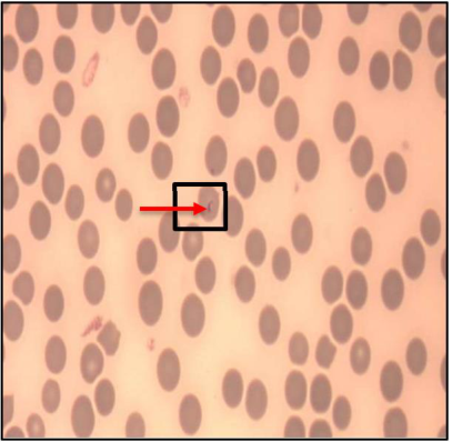 Figure 1
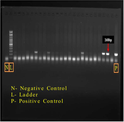 Figure 2
Introduction
Since the beginning of time, equines have made a significant contribution to the advancement of the human species, and India is blessed with valuable equine genetic material. According to the 2019 census of animals, India has roughly 0.54 million equines. The donkey, mule, horse, and pony populations, which make up the majority of equids, support rural and semi-urban communities. There are three registered breeds of donkeys, including Spiti, Halari, and Kachchhi. Therefore, it is crucial to protect and conserve horse and donkey breeds both in-situ and ex-situ, especially in light of their declining numbers (Devi A., 2017) [5] . The total population of donkeys in India is 1.24 lakhs in 2019, which was decreased by 61.2% over the previous census (3.19 lakhs). Amongst India, the major donkey population is in the states of Rajasthan (20,935), Uttar Pradesh (12,590), Maharashtra (13,345), Bihar (10,622), and Gujarat (11,286). Gujarat ranked at the 5 th position and contributed 8.1% in India. 50% donkey population lies in districts (Anand, Kheda, and Panchmahal) of middle Gujarat, and remaining 50% population is sporadically spread in different districts of Gujarat (Anonymous, 2019) [3] Interest in the welfare and diseases of donkeys is constantly increasing in several countries, mostly due to the recent rediscovery of donkey milk as an alternative food source for milkintolerant children. Clinical research on donkeys needs to be in continual development since they have different reactions compared to donkeys in many conditions (resistance to pain in case of colic), including infectious diseases, making it more difficult to recognize the symptoms normally observed in horses. Equine theileriosis caused by Theileria equi is an OIE listed tick transmitted hemoprotozoan disease with worldwide socio-economic impact on equine industry. Theileria equi (Previously known as Babesia equi, later reclassified as Theileria equi (Ibrahim et al., 2011; Mehlhorn and Schein, 1998) [6,8] is the causative agent of equine theileriosis which occurs in tropical and subtropical regions of the world. Being an endemic disease in Asia, Southern Europe, Latin America and Africa (Ristic, 1988) [9] , long-term monitoring of the prevalence of infection is very important in these areas. The disease has been reported by more than 20 countries of the world and the prevalence study results are found to be varying depending upon the study design, sample size and diagnostic techniques used. Equine piroplasmosis is among the listed diseases of the World Organization for Animal health (OIE), notifiable within 72 hours. Long-term monitoring of the prevalence of infection therefore is very important in areas where the disease has been reported, given the international importance of the disease and the potential impact on the horse industry. So knowledge of the prevalence of infection is essential to set up efficient control measures. Equine theileriosis is a challenge as the parasitemia often remains very low and the infected horses may act as the lifelong nucleus for spreading the disease through vector ticks, thereafter the outbreak or re-emergence of the disease (Wise et al., 2013) [11] . The latently infected horses may exhibit poor performance following physical, immunological or mental stress and may predispose horses to the clinical manifestation of the disease and death. But the clinical signs of equine theileriosis are variable and non-specific making the diagnosis difficult (Chhabra et al., 2012) [4] . So it is important to identify infected and non-infected horses based on more sensitive and specific diagnostic tool, thus to prevent the spread of the disease among the susceptible equine population by appropriate preventive and control measures. The aim of the present study was PCR based unravelling of prevalence of equine theileriosis by targeting the equine merozoite antigen (EMA-5 and EMA-6) which may contribute well to the future diagnostic as well as therapeutic approaches in cases of equine theileriosis. In the present study, Polymerase chain reaction as well as microscopic methods was compared in which PCR, being molecular diagnostic technique determines the actual presence of parasitic DNA for confirmation of horses as a latent carrier of T. equi infection. Piroplasmosis in donkeys has been recognized as a serious problem of major economic importance, since the affected animals manifest loss of appetite and decreased working capacity. Donkeys usually show an asymptomatic form of the disease, with lower T. equi parasitemia when compared to infection in donkeys. Donkeys usually remain asymptomatic carriers with positive antibody titres throughout life. The present work is aimed at detecting infection of donkeys to Theileria equi using molecular approach and at recording the hemato-biochemical alterations in affected donkeys.
Materials and Methods
Materials and Methods
Sample Collection
A total of 216 blood samples from donkeys were collected via jugular vein using anti-coagulant (K 2 EDTA) and without anticoagulant sterile vacutainers tubes. The sex, age and breed of the donkeys were also recorded during this study. The study was carried out for a period of nine months and blood samples were collected for blood smear examination as well as for PCR. The blood samples were collected as per the standard procedure without any stress or harm to the donkeys.
Examination of Blood smears
Thin blood smears were prepared from blood samples in EDTA tubes. The smears were air dried, fixed in absolute methanol for 10 minutes, stained with 10% Giemsa stain for 40 minutes, washed with distilled water and allowed to dry. The stained blood smears were examined under the high power and oil immersion lens (100x) of the microscope.
DNA extraction
The whole genomic DNA was extracted from the blood samples of donkeys by using commercial DNA Extraction kit (QIAamp-DNA extraction blood mini kit, Quigen, USA) according to the manufacturer's instructions. The obtained DNA was stored at -20 °C until further use.
Primers
DNA of Theileria equi was amplified by using PCR where the primer sequences used were Forward (EMA-5) 5'-TCGACTTCCAGTTGGAGTCC-3' and Reverse (EMA-6) 5'-AGCTCGACCCACTTATCAC-3' (Vidhyalakshmi et al., 2015) [10] yielding a 268bp product.
PCR protocol
All PCR reactions were performed in 25 µl of reaction mixture consisting of 12.5 µl Master mix, 2 µl of each forward and reverse primer, 5.5 µl of Millique water and 5 µl of extracted DNA. PCR reactions also included a negative control, consisting of the 19.5 µl of reaction mix and 5.5 µl of Millique water. The amplification conditions for T. equi included 40 cycles with enzyme activation at 95 °C for 10 min, denaturation at 94 °C for 1 min, primer annealing at 60 °C for 1 min and amplification at 72 °C for 1 min followed by final extension at 72 °C for 5 min. The final PCR products were subjected to electrophoresis in a 1.5% agarose gel with TBE buffer and DNA bands were visualized by using UV transilluminator or Gel documentation System. Positive samples for T. equi detected by using Giemsa stained blood smear technique were used as positive control.
Agarose gel electrophoresis
Preparation of 2% agarose gel was done by taking 1 gm of agarose powder into a 250 ml flask and added 50ml of 0.5x TBE buffer. The solution was heated in a microwave until agarose was completely dissolved. Gel casting tray was used for casting and appropriate number of comb was placed in gel tray and added 2.5 μl of ethidium bromide to the gel. Gel was poured into tray and allowed to cool for 15-30 minutes at room temperature. Later the comb was removed from gel tray and placed in electrophoresis chamber and covered with TBE buffer. Thereafter 1 μl of 1oading dye was added to a 6 μl PCR product and mixed well and loaded. About 2-3 μl of standard (Ladder) or 3 μl positive control was loaded in another well and electrophoresis was done at 80 V for 30 minutes. Finally DNA bands were visualized using UV transilluminator or Gel documentation System. For molecular detection, Theileria equi oligo-primers specifically amplify at 268 bp, 18s rRNA gene of Theileria equi (Vidhyalakshmi et al., 2015) [10] .
Results and Discussion
Results and Discussion
Parasitological method
Examination of the stained blood smears revealed few parasitized red blood cells with T. equi and shown in Figure 1. Out of 216 blood smears examined, 19 were found to be positive indicating a prevalence rate of 8.80% indicating a low sensitivity when compared to PCR.
Polymerase chain reaction
Out of 216 donkey samples screened for T. equi DNA by using universal PCR technique and gel electrophoresis, 67 were found to be positive indicating an overall prevalence rate of 31.02%. Out of 67 donkeys positive for T. equi as detected by PCR, only 8 donkeys have shown varying clinical symptoms whereas rest of 59 donkeys positive for theileriosis were found to be apparently healthy. DNA extracted from 216 blood samples were amplified and shown in Figure 2. The present study revealed low sensitivity of Giemsa stained blood smear examination technique to detect low level of parasitemia or subclinical or carrier status when compared to the PCR technique therefore supporting observations of other studies. ( [1,7,2] . Abedi et al., (2015) [1] observations consists of prevalence of T. equi as 3.77% positive by Giemsa stained blood smear examination technique, whereas 50.94% by PCR technique (n=106). Kurra H. M. & Nageib B. R., (2017) [7] shows the prevalence of T. equi as 14%(7/50) positive by Giemsa stained blood smear examination technique, whereas 38% (19/50) by PCR. Ahedor et al., (2022) [2] observations consists of prevalence of T. equi as 57.7% (64/111) positive by Giemsa stained blood smear examination technique, whereas 85.6% (95/111) by PCR technique (n=106).
In the present study, PCR technique is used to detect T. equi DNA based on Equine merozite antigen-5 and 6 and found to be more sensitive than microscopic methods. and EMA-6 antigen of T. equi by using universal molecular technique and the same is found to be more dependable diagnostic tool than microscopic method as the former detects circulating parasitic DNA. Thus present study revealed the efficiency of molecular technique for monitoring Theileriosis among donkeys. Therefore universal PCR technique may be helpful to develop better strategies to minimise the likelihood of clinical cases or outbreaks of theileriosis in donkeys of endemic areas under the circumstances of existing challenge in complete elimination of theileriosis. Priority should be given for the development and validation of highly sensitive, simple and specific diagnostic tool to distinguish infected and noninfected donkeys.
References
9 in the original paper- Abedi V, Razmi G, Seifi H, Naghibi A. Molecular detection of equine piroplasms in donkeys (Equus asinus) in North Khorasan province, Iran. Iranian journal of Veterinary Research. 2015;16(2):202.
- Ahedor B, Kothalawala H, Kanagaratnam R, Vimalakumar SC, Otgonsuren D, Tuvshintulga B, et al. First detection of Theileria equi in free-roaming donkeys (Equus africanus asinus) in Sri Lanka. Infection, Genetics and Evolution. 2022;99:105244.
- Chhabra S, Ranjan R, Uppal SK, Singla LD. Transplacental transmission of Babesia equi (Theileria equi) from carrier mares to foals. Journal of Parasitic Disease. 2012;36:31-33.
- Devi A. Claasico-Molecular Studies and Characterization of RoTat 1.2 VSG of Trypanosoma evansi in equines. M. V. Sc. Thesis. U. P. Pandit Deen Dayal Upadhyaya Pashu-Chikitsa Vigyan Vishwavidyalaya Evam Go Anusandhan Sansthan, Mathura, India, 2017.
- Ibrahim AK, Gamil IS, Abd-El baky AA, Hussein MM, Tohamy AA. Comparative Molecular and Conventional Detection Methods of Babesia equi in Egyptian Equine. Global Veterinaria. 2011;7:201-210.
- Kurra HM, Nageib BR. Parasitological and molecular detection of Theileria equi in donkeys in Assiut governorate. Egyptian Journal of Agricultural Research. 2017;95(1):271-284.
- Mehlhorn H, Schein E. Redescription of Babesia equi (Laveran, 1901) as Theileria equi. Parasitology Research. 1998;84:467-475.
- Vidhyalakshmi TM, Sunant Raval K, Bharat Bhanderi B, Amit Kanani K. Prevalence of Theileria equi among Horses Based on Parasitological and Universal Molecular Technique. International Journal of Current Microbiology and Applied Science. 2018;7(10):318-323.
- Wise LN, Kappmeyer LS, Mealey RH, Knowles DP. Review of Equine Piroplasmosis. J Vet Intern Med. 2013;27(6):1334-1346.