
Research article · 2024 · Full text
International Journal of Veterinary Sciences and Animal HusbandryIncidence of fungal dermatitis in dogs
Volume 9(2) · Pages 679–686 · DOI: 10.22271/veterinary.2024.v9.i2j.1281 ↗
Research areasNail Diseases and TreatmentsAntifungal resistance and susceptibilityPoxvirus research and outbreaks
Abstract
Author-writtenThe present research work entitled “Incidence of fungal dermatitis in dogs” was carried out at Department of Veterinary Medicine, Junagadh. A total 817 dogs were brought at small animal medicine OPD, Veterinary Clinical Complex, Kamdhenu University, Junagadh during October, 2022 - March, 2023 and out of them 123 (15.06%) were having different dermatological disorders. Among them a total 39 (4.77%) dogs were positive with canine fungal dermatitis. Canine fungal dermatitis was more common in
Summary
Source-checkedPlain-language reading of the full paper, prepared with AI and checked against the text. Each point links to the authors' own words.
Why it matters
- Skin disorders are among the most frequent health issues in companion animals, with dogs being susceptible to various causes including allergies, bacteria, parasites, and fungi. View evidence ↗
- Among the various skin conditions dogs face, fungal dermatitis is noted as one of the conditions that causes the most suffering. View evidence ↗
- Global occurrences of fungal infections affecting the nails, hair, and skin have increased dramatically over the last 20 years. View evidence ↗
The question
- The study aimed to determine the incidence of fungal dermatitis in dogs at the Department of Veterinary Medicine in Junagadh. View evidence ↗
What they did
- The study examined 817 dogs brought to the small animal medicine OPD at Kamdhenu University, Junagadh, between October 2022 and March 2023. View evidence ↗
- Researchers collected hair samples and skin scrapings, which were then cultured on Sabourad’s dextrose agar (SDA) and dermatophyte test medium (DTM), with confirmation via staining. View evidence ↗
- The study recorded epidemiological data including the dog's breed, sex, age, clinical sign history, and the type and distribution of skin lesions. View evidence ↗
What they found
- Out of 817 dogs, 123 (15.06%) had dermatological disorders, and 39 (4.77%) of the total population were positive for fungal dermatitis. View evidence ↗
- Fungal dermatitis was most prevalent in dogs under 1 year old (53.85%; 21/39 cases), with incidence decreasing as age increased. View evidence ↗
- Male dogs were more frequently affected by fungal dermatitis than females, accounting for 66.67% (26/39) of the positive cases. View evidence ↗
- The Labrador retriever was the most affected breed (30.77%; 12/39 cases), followed by non-descript and German Shepherd dogs. View evidence ↗
- The most common clinical signs in affected dogs were itching (94.87%), erythema (82.05%), and alopecia or mixed lesions (both 79.49%). View evidence ↗
- Lesions were most frequently located on the inner thighs (74.36%) and abdomen (53.85%). View evidence ↗
What it means
- The authors concluded that fungal dermatitis occurred in 4.77% of the studied dogs, with a higher predisposition in male dogs, dogs under one year of age, and Labrador retrievers. View evidence ↗
Visual summary
2 visualsIllustrations explain the paper; charts are replotted from the paper's own table values.
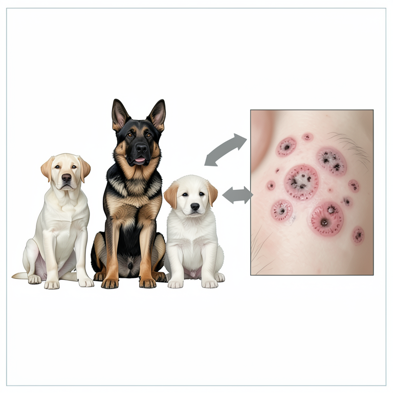
Risk Factors for Canine Fungal Dermatitis
AI-generated conceptual illustration (Labrador Retriever and German Shepherd dogs). It explains the idea and does not show study data.
View source evidence ↗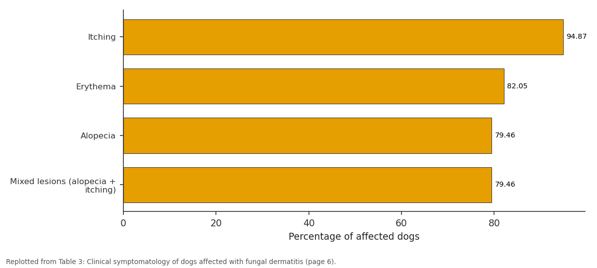
Percentage of affected dogs: Itching vs Erythema vs Alopecia vs Mixed lesions (alopecia...
Replotted from Table 3 (page 6) of the original paper. Every bar is the paper's own table value.
View source evidence ↗Figures & tables
From the original paper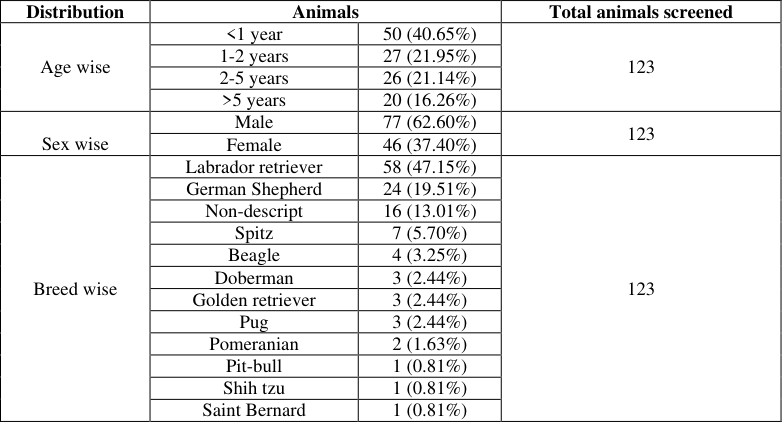 Table 1
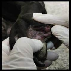 Figure 1
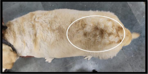 Figure 1
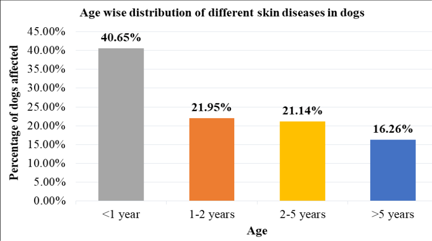 Figure 1
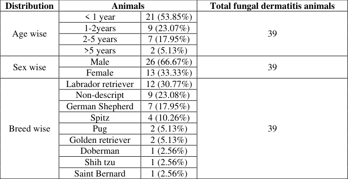 Table 2
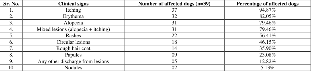 Table 3
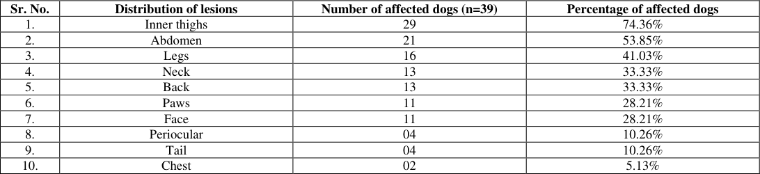 Table 4
Introduction
Dogs are one of the first domesticated animals. The association of dogs and humans have promoted co-evolution of many pathogens. The body's largest organ and outermost integument, skin serves as the first line of defence for an individual, making it vulnerable to environmental hazards. Skin problems are one of the most common disorders in companion animals. Dogs are prone to a wide range of skin conditions including parasitic, fungal, bacterial or allergies of various origins. One of these conditions that produces the most suffering is fungal dermatitis. Fungal infections of the skin, hair and nails are widespread throughout the world and their occurrence has been dramatically rising over the past 20 years. The majority of mycotic skin infections are cutaneous and those involving dermatophytes are referred to as "dermatophytosis", while those involving non-dermatophyte fungi are known as "dermatomycosis". The seven genera Microsporum, Trichophyton, Epidermophyton, Nannizzia, Arthroderma, Paraphyton and Lophophyton belong to the group of keratinolytic fungus known as dermatophytes (Bagra et al., 2022) [1] . One of the most prevalent mycotic disorders with a significant public health impact is dermatophytosis (Ivaskiene et al., 2016) [6] . Dermatophytosis is a keratinized layers of the epidermis and superficial fungal infection of hair and is caused by keratinophilic and keratinolytic genera such as Microsporum, Trichophyton and Epidermophyton. Microsporum canis, M. gypseum and Trichophyton mentagrophytes are the pathogens that are most frequently isolated (Scott et al., 2001) [17] . Skin, hair and nails are typically infected by Trichophyton species. Skin and hair are infected by Microsporum spp., while skin and nails are infected by Epidermophyton spp. (Nasimuddin et al., 2014) [12] .
Materials and Methods
The present research work was conducted at Department of Veterinary Medicine, College of Veterinary science and Animal Husbandry, Kamdhenu University, Junagadh from October, 2022 to March, 2023.
A total of 123 dogs presented to the TVCC with dermatological problems were screened for dermatophytosis. Skin scrapings and hair samples were collected and cultured using dermatophyte test medium (DTM) and Sabourad's dextrose agar (SDA). Dermatophytes were confirmed by staining of fungal isolates. Detailed epidemiological data such as age, sex, breed, a complete history of the development of clinical signs, distribution of skin lesions and its type, presence of itching, alopecia or skin lesions were recorded.
Results and Discussion
A total 817 dogs were brought at small animal medicine OPD, Veterinary Clinical Complex, Kamdhenu University, Junagadh during October, 2022 -March, 2023 and out of them 123 (15.06%) dogs were having different dermatological disorders and out of them 39 (4.77%) dogs were found positive for fungal dermatitis. Similar level of occurrence was reported by Debnath et al. (2016) [3] , Sever et al. (2017) [18] and Tarra (2021) [22] , had reported 20.93, 29. 6 [21] , Khurana et al. (2016) [9] and Thapa and Sarkar (2018) [24] . Highest number of cases of canine fungal dermatitis (39 cases) was found in dogs with <1 year age group (53.85%; 21 cases) followed by 1-2 years (23.07%; 9 cases), 2-5 years (17.95%; 7 cases) and >5 years (5.13%: 2 cases) age group in the present study. Table . 2 and Graph. 4. Similar findings were noticed by Katiraee et al. (2021) [8] , Prakash et al. (2022) [13] and Reddy et al. (2023) [14] . This might be due to an undeveloped immune system and lack of the fungistatic linoleic acid in young animals. (Ghadeer et al., 1997) [5] . Higher number of cases of different canine dermatological disorders (123 cases) were found in males (62.60%; 77 cases) as compare to female dogs (37.40%; 46 cases). [16] and Thapa and Sarkar (2018) [24] . Highest number of cases of canine fungal dermatitis (39 cases) was found in males (66.67%; 26 cases) as compare to female dogs (33.33%; 13 cases). Table . 2 and Graph. 5. Similar findings were reported by Saleem (2021) [15] , Siam et al. (2022) [19] and Tarra et al. (2022) [23] . Highest number of cases of different skin conditions (123 cases) were found in Labrador retriever (47.15%; 58 cases) followed by German Shepherd (19.51%; 24 cases), Non-descript (13.01%; 16 cases), Spitz (5.70%; 7 cases), Beagle (3.25%; 4 cases), Doberman (2.44%; 3 cases), Golden retriever (2.44%; 3 cases), Pug (2.44%; 3 cases), Pomeranian (1.63%; 2 cases), Pit bull (0.81%; 1 case), Shih tzu (0.81%; 1 case) and Saint Bernard (0.81%; 1 case). [16] and Thapa and Sarkar (2018) [24] . Highest number of cases of canine fungal dermatitis (39 cases) was found in Labrador retriever (30.77%; 12 cases) followed by nondescript (23.08%; 9 cases), German Shepherd (17.95%; 7 cases), Spitz (10.26%; 4 cases), Pug (5.13%; 2 cases), Golden retriever (5.13%; 2 cases), Doberman (2.56%; 1 case), Shih tzu (2.56%; 1 case) and Saint Bernard (2.56%; 1 case). Table . 2 and Graph. 6. Comparable findings were reported by Jena et al. (2019) [7] , Saleem (2021) [15] and Prakash et al. (2022) [13] . Labrador retrievers and German shepherds have the highest incidence due to their long hair. German Shepherds may have a higher incidence because of their long hair coats and increase humidity levels, which play a significant role in the easy growth and spread of fungal components. A total 39 dogs affected with fungal dermatitis showed different clinical symptoms like itching (94.87%; 37 cases), erythema ( [4] , Da Cunha et al. (2019) [2] , Minnat and Khalaf (2019) [10] and Prakash et al. (2022) [13] . Singathia (2014) [20] , Moriello et al. (2017) [11] and Da Cunha et al. (2019) [2] explained that lesions of dermatophytosis could range from mild to severe, depending on the variety of factors such as the infecting organisms, virulence factors, location of infection, secondary infections and environmental circumstances. This may be caused by variations in local skin factors, such as saturated long chain fatty acids, anatomical and functional sebaceous gland involution and unidentified immunologic factors.
Conclusion
The present study investigated on different epidemiological factors which predisposes dogs to fungal dermatitis. The incidence of different dermatological disorder in dogs was observed to be 15.06%. Different dermatological disorders were found to be higher in dogs of <1 year age group and in male dogs. In breed wise prevalence Labrador retriever (47.15%) was more affected. The incidence of fungal dermatitis was 4.77% in dogs. It was found high in male dogs (66.67%) and in dogs with less than 1 year of age group. Labrador retriever dogs were highly affected with canine fungal dermatitis followed by non-descript and German Shepherd dogs during the study period. The most common clinical signs showed by the dogs with fungal dermatitis were itching, erythema, alopecia, mixed lesions (alopecia + itching), rashes, circular lesions, rough hair coat, papules, any other discharge from lesions and nodules.
Acknowledgement
AcknowledgementThe authors thank the Dean, College of Veterinary Sciences and Animal Husbandry, Kamdhenu University, Junagadh for providing facilities for conduct of the study.
Figures and tables from the paper
| Distribution | Animals | — | Total animals screened |
|---|---|---|---|
| Age wise | <1 year | 50 (40.65%) | 123 |
| 1-2 years | 27 (21.95%) | ||
| 2-5 years | 26 (21.14%) | ||
| >5 years | 20 (16.26%) | ||
| Sex wise | Male | 77 (62.60%) | 123 |
| Female | 46 (37.40%) | ||
| Breed wise | Labrador retriever | 58 (47.15%) | 123 |
| German Shepherd | 24 (19.51%) | ||
| Non-descript | 16 (13.01%) | ||
| Spitz | 7 (5.70%) | ||
| Beagle | 4 (3.25%) | ||
| Doberman | 3 (2.44%) | ||
| Golden retriever | 3 (2.44%) | ||
| Pug | 3 (2.44%) | ||
| Pomeranian | 2 (1.63%) | ||
| Pit-bull | 1 (0.81%) | ||
| Shih tzu | 1 (0.81%) | ||
| Saint Bernard | 1 (0.81%) |
Table text extracted from the original PDF; superscript letters are shown inline. The original table image is below.
View original table image
| Distribution | Animals | — | Total fungal dermatitis animals |
|---|---|---|---|
| Age wise | < 1 year | 21 (53.85%) | 39 |
| 1-2years | 9 (23.07%) | ||
| 2-5 years | 7 (17.95%) | ||
| >5 years | 2 (5.13%) | ||
| Sex wise | Male | 26 (66.67%) | 39 |
| Female | 13 (33.33%) | ||
| Breed wise | Labrador retriever | 12 (30.77%) | 39 |
| Non-descript | 9 (23.08%) | ||
| German Shepherd | 7 (17.95%) | ||
| Spitz | 4 (10.26%) | ||
| Pug | 2 (5.13%) | ||
| Golden retriever | 2 (5.13%) | ||
| Doberman | 1 (2.56%) | ||
| Shih tzu | 1 (2.56%) | ||
| Saint Bernard | 1 (2.56%) |
Table text extracted from the original PDF; superscript letters are shown inline. The original table image is below.
View original table image
| Sr. No. | Clinical signs | Number of affected dogs (n=39) | Percentage of affected dogs |
|---|---|---|---|
| 1. | Itching | 37 | 94.87% |
| 2. | Erythema | 32 | 82.05% |
| 3. | Alopecia | 31 | 79.46% |
| 4. | Mixed lesions (alopecia + itching) | 31 | 79.46% |
| 5. | Rashes | 22 | 56.41% |
| 6. | Circular lesions | 18 | 46.15% |
| 7. | Rough hair coat | 14 | 35.90% |
| 8. | Papules | 09 | 23.08% |
| 9. | Any other discharge from lesions | 05 | 12.82% |
| 10. | Nodules | 02 | 5.13% |
Table text extracted from the original PDF; superscript letters are shown inline. The original table image is below.
View original table image
| Sr. No. | Distribution of lesions | Number of affected dogs (n=39) | Percentage of affected dogs |
|---|---|---|---|
| 1. | Inner thighs | 29 | 74.36% |
| 2. | Abdomen | 21 | 53.85% |
| 3. | Legs | 16 | 41.03% |
| 4. | Neck | 13 | 33.33% |
| 5. | Back | 13 | 33.33% |
| 6. | Paws | 11 | 28.21% |
| 7. | Face | 11 | 28.21% |
| 8. | Periocular | 04 | 10.26% |
| 9. | Tail | 04 | 10.26% |
| 10. | Chest | 02 | 5.13% |
Table text extracted from the original PDF; superscript letters are shown inline. The original table image is below.
View original table image
References
22 in the original paper- Bagra J, Abhishek, Nair SS, Athira V, Kumar M, Thomas P, et al. Dermatophytosis in Canines and Felines. The Science World a Monthly E Magazine. 2022 Jun;2(6):661-669.
- Da Cunha MM, Capote-Bonato F, Capoci I, Bonato DV, Ghizzi LG, Paiva-Lima P, et al. Epidemiological investigation and molecular typing of dermatophytosis caused by Microsporum canis in dogs and cats. Prev Vet Med. 2019;167:39-45.
- Debnath C, Mitra T, Kumar A, Samanta I. Detection of dermatophytes in healthy companion dogs and cats in eastern India. Iran J Vet Res. 2016;17(1):20-24.
- Gangil R, Dutta P, Tripathi R, Singathia R, Lakhotia RL. Incidence of dermatophytosis in canine cases presented at Apollo Veterinary College, Rajashtan, India. Vet World. 2012;5(11):682-684.
- Ghadeer AA, Rashad MN, Walid AB. Distribution and prevalence of superficial fungal infections in Amman and other Jordan cities. Dirasat. Med Biol Sci. 1997;24(2):219-227.
- Ivaskiene M, Matusevicius AP, Grigonis A, Zamokas G, Babickaite L. Efficacy of Topical Therapy with Newly Developed Terbinafine and Econazole Formulations in the Treatment of Dermatophytosis in Cats. Pol J Vet Sci. 2016;19(3):535-543.
- Jena S, Mishra R, Patra R, Das DP, Behera SS. Analysis of Skin Microbiota from Canine Dermatitis in Odisha, India. Int J Curr Microbiol Appl Sci. 2019;8(8):14891494.
- Katiraee F, Kosari YK, Soltani M, Shokri H, Minooieanhaghighi MH. Molecular identification and antifungal susceptibility patterns of dermatophytes isolated from companion animals with clinical symptoms of dermatophytosis. J Vet Res. 2021;65(2):175-182.
- Khurana R, Kumar T, Agnihotri D, Sindhu NEELESH. Dermatological disorders in canines-a detailed epidemiological study. Haryana Vet. 2016;55(1):97-99.
- Minnat TR, Khalaf JM. Epidemiological, clinical and laboratory study of canine dermatophytosis in Baghdad Governorate, Iraq. Iraqi J Vet Med. 2019;43(1):183-196.
- Moriello KA, Coyner K, Paterson S, Mignon B. Diagnosis and treatment of dermatophytosis in dogs and cats. Clinical Consensus Guidelines of the World Association for Veterinary Dermatology. Vet Dermatol. 2017;28(3):266-268.
- Nasimuddin S, Appalaraju B, Surendran P, Srinivas CR. Isolation, Identification and comparatative analysis of SDA and DTM for dermatophytes from clinical samples in a tertiary care hospital. IOSR J Dent Med Sci. 2014;13(11):68-73.
- Prakash VA, Rathish RL, Deepa PM, Bipin KC, John L. Host and environmental risk factors of canine dermatophytosis. Pharma Innovation J. 2022;11(10):936939.
- Reddy BL, Mishra R, Rath PK, Patra BK, Mishra BP, Soren N, et al. Microbial prevalence of dermatitis in dogs in Odisha. Pharma Innovation J. 2023;12(1):497-501.
- Sarma K, Mondal DB, Sarvanan M, Kumar M, Vijaykumar H. Incidence of dermatological disorders and its therapeutic management in canines. Intas Polivet. 2013;14(2):186-192.
- Scott DW, Miller Jr. WH, Griffin CE. Dermatoses of pet rodents, rabbits, and ferrets: Muller & Kirk's Small Animal Dermatology. Philadelphia: W. B. Saunders; 2001.
- Sever NK, Ustun T, Omerovic M, Mustafa ONOL, Zahiri AK, Dogan B, et al. Prevalence of dermatophytes isolated from domestic animals in Ankara within a threeyear period (2014-2017). Vet J Mehmet Akif Ersoy Univ. 2017;6(1):1-7.
- Siam MO, El-Sheikh AEK, Attia NE. Alopecia in dogs: causes, incidence and clinical signs with a special reference to nutritional alopecia. Zagazig Vet J. 2022;50(2):151-160.
- Singathia R, Gupta SR, Yadav R, Gupta Y, Lakhotla R. Prevalence of canine dermatophytosis in semi-arid Jaipur, Rajasthan. Haryana Vet. 2014;53(1):43-45.
- Singh R, Beigh SA, Soodan JS, Tikoo A, Tantaray H. Clinico-epidemiological studies in canine dermatitis. Indian J Canine Pract. 2012;4(2):96-99.
- Tarra M, Davis KJ, Vinodkumar K, Vijayakumar K, Menon VK. Occurrence of dermatophytosis in dogs from Thrissur, Kerala. J Vet Anim Sci. 2022;53(2):322-327.
- Thapa G, Sarkar S. Occurrence of canine skin disorder and its haematobiochemical alterations. Int J Curr Microbiol Appl Sci. 2018;7(12):184-195.
Cited by 2
From OpenAlex, OpenCitations and reference lists read by Scinovex
- Phenotypic characterization of non-dermatophyte moulds from companion animals with mycotic dermatitis
Padala Harini, Deepthi Balam, Anand Kumar P, Bindu Kiranmayi Ch · International Journal of Advanced Biochemistry Research · 2026
Cutaneous mycoses represent a significant proportion of dermatological disorders in companion animals, adversely affecting animal welfare and necessitating accurate laboratory diagnosis for effective management. Although dermatophytes are traditionally regarded as the principal causative agents of fungal skin infections, non-dermatophyte moulds have increasingly been implicated as opportunistic pathogens and potential contributors to dermatological lesions. The present study was undertaken to phenotypically characterize non-dermatophyte moulds recovered from dogs and cats exhibiting clinical m
- Epidemiological pattern of canine fungal dermatitis in Tarai area of Kumaon, Uttarakhand
R S Negi, Satish Kumar, Deepali Bhandari, P Bhatt · International Journal of Advanced Biochemistry Research · 2026
Canine dermatology is multidisciplinary aspects incorporates knowledge from diverse disciplines including bacteriology, mycology, parasitological, immunology, endocrinology and oncology. A detailed clinical history, physical examination and assessment of hematological and biochemical alterations are important components of the diagnostic process. Further diagnostic confirmation may involve skin scrapings and microbiological culture using selective and enrichment media. The prevalence of cutaneous fungal infections in dogs varies considerably according to geographical and climatic conditions, w