
Research article · 2024 · Full text
International Journal of Advanced Biochemistry ResearchUltrasonographical diagnosis of pregnancy in sheep at an organized farm
Volume 8(3S) · Pages 522–525 · DOI: 10.33545/26174693.2024.v8.i3sg.804 ↗
Research areasReproductive Physiology in Livestock
Abstract
Author-writtenThe present study was aimed to assess pregnancy and embryonic mortality in ewes. Transrectal real-time B-mode ultrasonography (USG) was carried out on day 28 post-mating and pregnancy was confirmed by the presence of amniotic vesicle, or fetal image and embryonic heartbeat. Further, the USG was repeated on day 45 post-mating to estimate the embryonic mortalities and overall pregnancy rate. Out of 210 ewes on 28th day, 181 (86.19%) ewes were diagnosed as pregnant. On day 45th post-mating, The overall pregnancy rate was 82.85%, whereas the embryonic mortality was 3.86%. The transrectal real-time B- mode ultrasonography appeared to be safe, quick and convenient mean of pregnancy diagnosis in sheep.
Summary
Source-checkedPlain-language reading of the full paper, prepared with AI and checked against the text. Each point links to the authors' own words.
Why it matters
- Early pregnancy diagnosis in sheep is economically valuable for the industry. View evidence ↗
- Detecting pregnancy early allows for better nutritional management of ewes, which can improve lamb birth weights, weaning weights, and survival rates. View evidence ↗
- Early detection helps flock management by allowing quick re-insemination of non-pregnant ewes, informing culling decisions, reducing expenses for infertile animals, and improving the scheduling of vaccinations and marketing. View evidence ↗
The question
- The study aimed to assess pregnancy rates and embryonic mortality in ewes. View evidence ↗
What they did
- The research was conducted at the Central Sheep Breeding Farm in Hisar, Haryana, India. View evidence ↗
- The study involved 210 ewes in estrus that were naturally mated with proven rams. View evidence ↗
- Pregnancy was diagnosed using a portable real-time B-mode ultrasonography machine with a 5-7 MHz trans-rectal multi-frequency transducer. View evidence ↗
- Ultrasonography was performed on day 28 post-mating to confirm pregnancy and repeated on day 45 to determine the overall pregnancy rate and embryonic mortality. View evidence ↗
- Ewes were placed in dorsal recumbency during the imaging process. View evidence ↗
What they found
- On day 28 post-mating, 181 out of 210 ewes (86.19%) were diagnosed as pregnant. View evidence ↗
- By day 45 post-mating, the overall pregnancy rate was 82.85%, with 174 ewes reconfirmed as pregnant. View evidence ↗
- The embryonic mortality rate was found to be 3.86%. View evidence ↗
- Pregnancy diagnosis was rapid, taking approximately 2-3 minutes per ewe based on the observation of uterine fluid, the embryo/fetus, and concave placentomes. View evidence ↗
What it means
- The authors concluded that real-time B-mode ultrasonography is the fastest, safest, most accurate, and most economical method for diagnosing pregnancy in sheep at the farm level. View evidence ↗
- The authors state that transrectal ultrasonography can be used to assess late embryonic deaths that occur after the mother has recognized the pregnancy. View evidence ↗
Visual summary
1 visualsIllustrations explain the paper; they do not show study data.
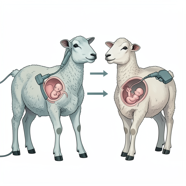
Study Timeline
AI-generated conceptual illustration (pregnant ewes). It explains the idea and does not show study data.
View source evidence ↗Figures & tables
From the original paper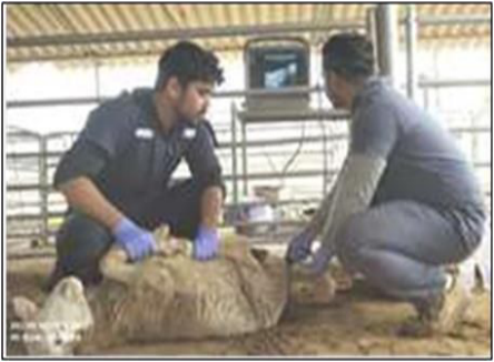 Figure 1
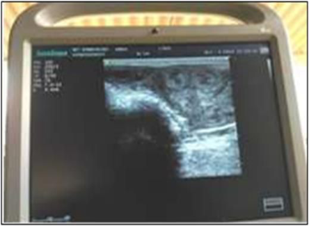 Figure 2
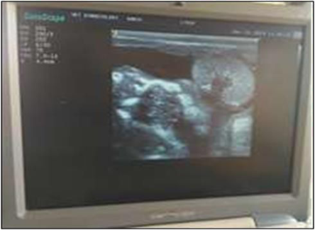 Figure 3
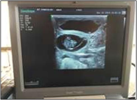 Figure 4
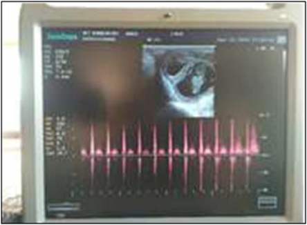 Figure 5
Introduction
Ewes are seasonally polyestrous animals, and a fertile ewe is desired to be pregnant as early as possible during the limited breeding season. Early diagnosis of pregnancy is of considerable economic value to the sheep industry. Improved control over the nutrition of ewes in late gestation would optimize birth weights, weaning weights and survivability of lambs (Scott, 1986) [6] . Early pregnancy detection may provide several important advantages in sheep flock management: (i) non-pregnant ewes can be quickly re-inseminated; (ii) it facilitates culling or selling decisions; (iii) it decreases feeding and managing expenses by detecting infertile ewes in flocks; (iv) feeding, vaccination, weaning, and marketing can be programmed more effectively; and (v) it provides an opportunity for assisted reproductive techniques such as laparoscopic artificial insemination and embryo transfer in valuable commercial sheep flocks (Ishwar, 1995) [3] . Ultrasound imaging is one of the most widely used powerful tools in animal diagnostics and research, which allows the real-time visualization of internal organs, tissues, structures or fluid accumulations and the investigation of blood flow without any radiation risk. It depends on the computerized analysis of reflected ultrasound waves. Due to its non-invasive character and apparent high safety, it has become one of the most rapidly advancing technologies in the clinical fields. An ideal pregnancy diagnosis method should be non-invasive, cheap, reliable, practical, repeatable, and easily applied during early pregnancy (Ishwar, 1995) [3] . Currently, the preferred method in field conditions is real-time transrectal/transabdominal ultrasonography. The main advantage of transrectal USG over transabdominal USG is earlier detection of pregnancy (Ishwar, 1995) [3] . After this first sign, the reliability of the ultrasound based diagnosis reaches 100% with observation of the embryo and its heart beat in the next few days (20 days after mating) (Schrick and Inskeep, 1993) [5] .
Materials and Methods
Place of study: This study was conducted at Central Sheep Breeding Farm, Hisar, (Haryana) India. The institute is located at latitude 29⁰ N and longitude 75⁰ E with average elevation of 215 m from the sea level. The institute is located at the place where mainly continental climatic conditions are present with a significant annual variation in the temperature during summers and winters. A total of 210 ewes in estrus detected by ram parading were subjected to natural mating with proven rams. Transrectal real-time B-mode ultrasonography (USG) was carried out on day 28 post-mating and pregnancy was confirmed by the presence of amniotic vesicle, or embryo/fetal image and embryonic/heart-beat. Further, the USG was repeated on day 45 post-mating to estimate the embryonic mortalities and overall pregnancy rate.
Ultrasonographic scanning:
For the ultrasonographic study of reproductive status and early pregnancy diagnosis, a real time B-mode portable Ultrasonography machine (SONPSCAPE S6/S6Pro/S6BW Portable Digital Color Doppler Ultrasound System) equipped with a linear array trans-rectal multi-frequency transducer using frequency of 5-7 MHz was used. The urinary bladder was the point of reference during trans-rectal ultrasonography.
Technique for Imaging
The ewes were restrained in dorsal recumbency. Corboxy gel (coupling gel) was applied on active portion of probe to minimize the air gap and also for lubrication. The rectal probe was inserted in to the rectum, and firstly, the urinary bladder was located that served as a point of reference for scanning of reproductive tract. The probe was rotated in clockwise and anti-clockwise direction to examine the both uterine horns and ovaries. Uterine horns were observed just at cranial aspect of urinary bladder. After proper imaging of scanned structures, the images were freezed and the pictures were taken using mobile phone. Hardly 2-3 minutes were taken per ewe for detailed scanning (Fig. 1).
Pregnancy rate (%) was calculated by calculating the number of pregnant animals divided by total number of animals multiplied by hundred (Paula et al., 2020) [7].
Embryonic mortality:
The embryonic mortality (%) was calculated as the difference in number of ewes that were diagnosed as pregnant on day 28 post-mating and nonpregnant on day 45 th post-mating, multiplied by 100 and divided by the number of ewes pregnant on day 28 th postmating.
Results and Discussion
Out of 210 was on 28 th day post-mating, 181 (86.19%) ewes were diagnosed as pregnant. Subsequently on day 45 th postmating, 174 (82.85%) ewes were reconfirmed pregnant and embryonic mortality rate was 3.86%. Pregnancy diagnosis at first schedule of examination (day 28 th post-mating) was done by observation of circular anechoic areas in the uterine lumen and embryo proper (Fig. 4). At second schedule of examination (45 days), the fetuses were identified easily (Fig. 5, Table . 1). The diagnosis of pregnancy was very fast (about 2-3 minutes) by observation of fluid in the uterine lumen, embryo proper/fetus and concave placentomes The ovine pregnancy could be diagnosed at an earliest day of 17-19 of gestation based on presence of anechoic intrauterine fluid by trans-rectal USG (5 MHz) while the embryo proper could be imaged between days 21 to 34 of gestation, with placentomes visualization on day 26-28 (Garcia et al., 1993) [9] . The heart beat can be detected as early as the 18 th or 19 th day of pregnancy, heart beat may not be observed in all pregnant sheep until as late as day 23 (Martinez et al., 1998) [8] . The position of the female will affect the distance between the uterus and the transducer, therefore, affecting the quality and accuracy of the scanned images. Compared with transabdominal application, transrectal application allows for earlier pregnancy diagnosis, due to the shorter distance between the uterus and probe. A transducer with high frequency generates a better image resolution, but has a shallower depth of penetration. Therefore, close proximity of the uterus to the rectal wall during transrectal ultrasonography will improve the visualization and scanning efficiency (Griffin and Ginther, 1992) [2] . It is established that embryos may be observed from day 28 (Picazo et al., 1991) [10] and absence of embryonic vesicle, amniotic fluid and absence of detectable heart rate in fetuses on day 45 post-mating are diagnostic features for late embryonic mortality (Yadav et al., 2020) [11] . Results of this study indicated that transrectal ultrasonographic examination in dorsal recumbency is a reliable method for pregnancy diagnosis in ewes, and give high accuracy reaching to 100% especially after 45 days of natural mating (Buckrell et al., 1986) [1] . The accuracy of transrectal ultrasonography is higher than those of transabdominal ultrasonography (Kahn, 1992) [4] . Although factors causing early embryonic mortality in sheep are not well established, there is some evidence suggesting the involvement of luteal inadequacy, resulting from environmental factors such as heat stress or nutrition, has been shown to be a major cause of embryonic loss in sheep (Binelli et al., 2001) [12] . The findings of the present study indicated that the early pregnancy diagnosis through transrectal ultrasonography could be a mean for assessment of late embryonic deaths occurring post maternal recognition of pregnancy.
Conclusion
The study concluded that real-time B-mode ultrasonography is the earliest, most accurate, safest, faster and economical method of pregnancy diagnosis in sheep at farm level. The embryonic heart beat was first detected via transrectal scanning on day 28 th and reconfirmation was attempted on day 45 th post-mating. The characteristic cotyledonary placentation of ruminants was easily recognizable when examining sheep transrectally facilitated a rapid, accurate determination of pregnancy status.
Figures and tables from the paper
References
12 in the original paper- Buckrell BC, Bonnett BN, Johnson WH. The use of real-time ultrasound rectally for early pregnancy diagnosis in sheep. Theriogenology. 1986;25(5):665673.
- Griffin PG, Ginther OJ. Research applications of ultrasonic imaging in reproductive biology. J Anim Sci. 1992;70:953-972.
- Ishwar AK. Pregnancy diagnosis in sheep and goats: a review. Small Ruminant Research. 1995;17(1):37-44.
- Kahn W. Ultrasonography as a diagnostic tool in female animal reproduction. Anim Reprod Sci. 1992;28:110.
- Schrick FN, Inskeep EK. Determination of early pregnancy in ewes utilizing transrectal ultrasonography. Theriogenology. 1993;40(2):295-306.
- Scott GE. The sheepman's production handbook. Sheep Industry Development, Denver, Colorado. 1983;80206:4-26.
- Paula CJ, Fabjan JMG, Gonçalves JD, Dias JH, Souza GN, Oliveira MEF, et al. Effect of a 12-h increment in the short-term treatment regimen on ovarian status, estrus synchrony, and pregnancy rate in artificially inseminated dairy goats. Anim Reprod Sci. 2020;221:106571.
- Martinez MF, Bosch P, Bosch RA. Determination of early pregnancy and embryonic growth in goats by transrectal ultrasound scanning. Theriogenology. 1998;49(8):1555-1565.
- Garcia A, Neary MK, Kelly GR, Pierson RA. Accuracy of ultrasonography in early pregnancy diagnosis in the ewe. Theriogenology. 1993;39(4):847-861.
- Picazo RA, Barragan ML, Valenciano M, Sebastian AL. Evolución de la imagen ecográficadurante la gestación de la oveja. Med Vet. 1991;8:300-315.
- Yadav V, Chandolia RK, Dutt R, Bisla A, Saini G, Singh G. Effect of estrus synchronization using AVIKESIL-S® with eCG on the reproductive efficiency in crossbred ewes. Int J Livest Res. 2020;10(3):49-59.
- Binelli M, Thatcher WW, Mattos R, Baruselli PS. Antiluteolytic strategies to improve fertility in cattle. Theriogenology. 2001;56(9):1451-1463.
Cited by 1
From OpenAlex, OpenCitations and reference lists read by Scinovex
- Ultrasonographic detection of fetal sexing in nanny goat ↗
Eman H. Lazim, Barra D. Al-Watar · Al-mağallaẗ al-ʻirāqiyyaẗ li-l-ʻulūm al-bayṭariyyaẗ/Iraqi journal of veterinary sciences · 2025
We aimed to evaluate the accuracy of early fetal sexing by sonography in nanny goats during three periods of gestation and their correlation with the number of fetuses. A total of 50 pregnant nanny goats were included in this study. The B mode of real-time scanner with 3.5-5 MHz transabdominal transducer was used to identify the fetal external genital organs…
❝ 1PDF