
Research article · 2024 · Full text
International Journal of Advanced Biochemistry ResearchThe prevalence of E. coli in raw milk and their susceptibility to antibiotics
Volume 8(3) · Pages 406–410 · DOI: 10.33545/26174693.2024.v8.i3e.758 ↗
Research areasMilk Quality and Mastitis in Dairy CowsEnterobacteriaceae and Cronobacter Research
Abstract
Author-writtenEscherichia coli, sometimes known as E. coli, is a beneficial member of the natural symbiotic bacterial flora found in the stomach. One of the main causes of bovine mastitis, which can range from a serious systemic illness to a subclinical mammary gland infection, is E. coli. Most commensals are benign, but some have developed virulence features that can spread through contaminated milk and infect otherwise healthy people. Antibiotic-resistant E. coli found in raw cow's milk may be the cause of resistance illnesses in people. The study looks into the antibiotic susceptibility of E. coli and how often it is in raw milk samples from dairy farms in Haryana. For this cross-sectional investigation, 40 raw cow milk samples were gathered from different areas in Haryana. Following the presumed isolation of E. coli from raw milk, a two-stage NDRI assay was conducted to confirm the isolates of E. coli utilising quick procedures. The IMViC assays were used to biochemically characterise isolates of E. coli samples. Using the Kirby-Bauer method, antibiotic susceptibility was assessed for third- and fourth-generation cephalosporins. The NDRI test verified that 15% of the 40 raw milk samples had E. coli germs in them. Every E. coli sample with phenotypic confirmation was susceptible to carbapenem and broad spectrum cephalosporin drugs. 15% of raw milk from Haryana's dairy had E. coli, although the bacteria did not show signs of drug resistance to cephalosporins.
Summary
Source-checkedPlain-language reading of the full paper, prepared with AI and checked against the text. Each point links to the authors' own words.
Why it matters
- E. coli is a primary cause of bovine mastitis, a condition that can vary from subclinical mammary gland infections to severe systemic illness. View evidence ↗
- The presence of antibiotic-resistant E. coli in raw cow's milk is a concern because it may contribute to drug-resistant infections in humans. View evidence ↗
- The use of third and fourth-generation cephalosporins is particularly concerning due to their link to the development of antimicrobial resistance. View evidence ↗
The question
- The study aimed to determine the prevalence of E. coli in raw milk samples from dairy farms in Haryana and evaluate their susceptibility to antibiotics. View evidence ↗
What they did
- Isolates were identified using IMViC biochemical tests and further confirmed using a two-stage NDRI assay. View evidence ↗
- Antibiotic susceptibility testing (AST) was performed using the Kirby-Bauer disk diffusion method to detect resistance to ESBL and Carbapenems. View evidence ↗
What they found
- All six E. coli isolates tested positive for indole and methyl red, and negative for Voges-Proskauer and citrate utilization, matching the positive control. View evidence ↗
- All six confirmed E. coli isolates were susceptible to broad-spectrum cephalosporins and carbapenem antibiotics. View evidence ↗
What it means
- The authors concluded that none of the E. coli isolates in this study exhibited multidrug-resistant characteristics. View evidence ↗
- The authors suggest that the lack of multidrug-resistant isolates may be due to the small number of milk samples included in the study. View evidence ↗
Visual summary
1 visualsIllustrations explain the paper; they do not show study data.
Study Workflow
AI-generated conceptual illustration (raw cow milk and E. coli bacteria). It explains the idea and does not show study data.
View source evidence ↗Figures & tables
From the original paper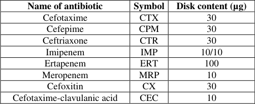 Table 1
 Table 3
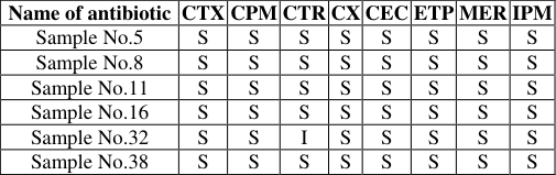 Table 4
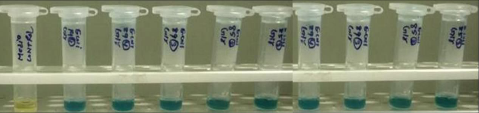 Figure 1
Introduction
Escherichia coli (E. coli) is a rod-shaped, facultatively anaerobic, Gram-negative bacteria that is classified as a member of the family Enterobacteriaceae within the Gammaproteo bacteria class (Aidara -Kane, 2012) [1] . Existing independently in nature it is also a part of normal bacterial flora in humans and animals. They benefit their hosts by producing vitamin K2 and prevent colonization of the intestine with pathogenic bacteria, having a symbiotic relationship (Cabrera et al., 2020) [2] . These bacteria live in the intestines of many animals and are usually transmitted to people when they eat foods contaminated with the bacteria (Clark et al., 1957) [3] . The majority of E. coli are harmless commensals of the mammalian gastrointestinal tract, yet some strains have deviated from this ancestral niche, adapting specific virulence traits that enable them to cause disease in otherwise healthy individuals (Duan et al., 2006) [4] . The resulting clinical syndromes may include gastroenteritis, urinary tract infection, septicemia, and meningitis. Escherichia coli infection may itself be caused by at least six distinct E. coli pathotypes: enterotoxigenic (ETEC), enteropathogenic (EPEC), enterohemorrhagic (EHEC), enteroinvasive (EIEC), diffusely adherent (DAEC) and enteroaggregative E. coli (EAEC). These pathotypes exhibit distinct clinical, epidemiologic, and pathogenic profiles. Strains within each pathotype are characterized by shared virulence traits and can typically be further distinguished by O (lipopolysaccharide) and H (flagellar) antigens (Jafari et al., 2012) [10] . Since its discovery, an increasing number of studies have implicated E. coli in causing persistent diarrhea, and traveler diarrhea among neonates and patients with weak immune systems. E. coli is the most frequent cause of community and hospital-acquired urinary tract infections (including infections of the kidney), bloodstream infection, meningitis and intraabdominal infections such as peritonitis (Gebremedhinm et al., 2018) [7] . E. coli is also one of the major causative agents of bovine mastitis ranging from being a subclinical infection of the mammary gland to a severe systemic disease (Suojala et al., 2013) [17] . Broad-spectrum antimicrobials are commonly used to treat E. coli mastitis (Erskine et al., 2003) [5] . Most of these antibiotics have been classified as "critically important" by the World Health Organization (WHO). The use of third and fourth-generation cephalosporins is of particular concern because they have been attributed to the development of antimicrobial resistance in certain bacteria (Erskine et al., 2003; Martinson and Walk, 2020) [5,14] . Studies have been conducted in India in which antimicrobial-resistant E. coli were isolated from raw milk, mastitis milk, and unpasteurized milk (Ghatak et al., 2013; Kakkar et al., 2017) [8,12] . It has been found that these bacterial species have characteristics of both multi-drug as well as carbapenem resistance. The major cause of the prevalence of ESBL and carbapenem-resistant bacteria is the indiscriminate use β-lactam antibiotics, horizontal transfer of genes by the mastitis milk isolate from human or environmental sources. Also among dairy animal's absence of antimicrobial treatment records, lack of written plans, and failure to complete an antimicrobial treatment course are the major factors that could lead to the inappropriate use of antimicrobials and the emergence of antibiotic-resistant bacteria (Sawant et al., 2007) [15] . Further European Food Safety Authority (EFSA) in 2013 stated that the β-lactam resistant bacteria are now seen as a new and potentially emerging problem in dairy animals because of the specific use of cephalosporins and β-lactam antibiotics for mastitis treatment (Kucukbasmaci et al., 2008) [13] . Isolates of E. coli, which are resistant to 3rd or 4th generation cephalosporin should be screened for ESBL and Carbapenem resistance. According to the National Action Plan (2017) on AMR, surveillance of AMR in humans, foods, animals and environment sectors focus on strengthening laboratories is one of the prioritized strategies for the containment of AMR in India. The present study investigated the Isolation and Identification of E. coli from Raw Milk Samples and the Antibiotic Susceptibility test for E. coli isolates.
Materials and Methods
Over the course of three months, the Department of Microbiology carried out this cross-sectional investigation. Forty raw cow milk samples were obtained from different regions in Haryana to be included in the study. Initially, 70% alcohol was used to sanitise the milking man's hands in preparation for collecting milk samples. Tap water was used to wash the cow's urine. The first few milk streams were thrown away, and the remaining streams were sampled right into the sample collection container. After being labelled, all of the sample bottles were quickly brought with ice packs to the laboratory.
Isolation and identification of E. coli in raw milk samples
Isolation of E. coli was performed as per the procedure laid down in the IS 5887; Methods for detection of bacteria responsible for food poisoning-isolation, identification and enumeration of E. coli. All milk samples were processed for the primary isolation of bacteria. Initially, 1 ml of milk samples was transferred into 10 ml of MacConkey broth tube in the bio-safety cabinet under aseptic conditions. Inoculated test tubes were incubated at 37 °C for 24 h.
Positive isolates from enriched MacConkey broth tubes which showed a change in colour from purple to yellow were streaked on MacConkey agar and EMB agar plates and incubated at 37 °C for 24 hours. Gram staining was used to differentiate two large groups of bacteria based on their different cell wall constituents. It distinguishes between gram-positive and gram-negative groups. The Gramnegative short rods were presumptively considered as E. coli species and were subjected to biochemical tests.
Biochemical characterization of E. coli
The IMViC tests are a group of individual tests used in microbiology labs to identify an organism in the coliform group. The term "IMViC" is an acronym for each of these tests. "I" is for the indole test; "M" is for the methyl red test; "V" is for the Voges-Proskauer test, and "C" is for the citrate test. Isolates having characteristic pink smooth colonies on MacConkey agar plates and greenish metallic sheen colonies on EMB agar plates were further evaluated for confirmation using the IMViC test.
Indole test
Prepared tryptone broth medium was inoculated with overnight grown culture and it was incubated at 37 °C for 24 h. Point five ml of Kovacs reagents was added, which was prepared by dissolving 10 g p-dimethyl-amino benzaldehyde in 150 ml amyl alcohol or iso-amyl alcohol and to which 50 ml of concentrated hydrochloric acid was slowly added. After adding the Kovacs reagent, the appearance of a distinct red color in the upper layer was an indication of the presence of indole.
Methyl red test
Prepared glucose peptone medium was inoculated with overnight grown culture and it was incubated at 37 °C for 48 h. After incubation, tubes were added with 2-3 drops of methyl red indicator solution into the medium. The appearance of red color was an indication of a positive reaction.
Voges-Proskauer reaction test
Prepared glucose peptone medium was inoculated with overnight grown culture and it was incubated at 37 °C for 48 h. After incubation, tubes were added with 0.6 ml of alphanaphthol solution prepared as 5 percent solution in ethanol and 0.2 ml of 40 percent aqueous solution of potassium hydroxide. The positive reaction was indicated by the appearance of a rose-red color ring on the top of the tube was an indication of positive reaction.
Citrate utilization test
Prepared Simmons citrate agar slant was inoculated with overnight grown culture and slant tubes were incubated at 37 °C for 48 h. The change in color of the slant from green to blue indicated a positive reaction. The negative reaction was indicated by no color change.
Confirmation of E. coli isolates using rapid techniques by NDRI two-stage assay of Presumptive of E. coli isolates
Added 0.9 ml of sterile distilled water (pH 7.0) into the tube containing lyophilized ECSM. Tubes were inoculated with 0.1 mL of milk and were incubated at 37 °C for 12±1.0 h. A change in color was observed from milky white to bluish-green. Tube was centrifuged at 5,000 rpm for 10.0 min. The cell pellet was washed twice with 1.0 ml of Sodium phosphate buffer (pH-6.8). After draining the supernatant, the sides of the tube were wiped with the sterile cotton swab to absorb the residual buffer without disturbing the pellet. Took stage-2 tube containing pre-lyophilized ESM and reconstituted with 200 µl of distilled water (pH 7.0). Transferred the contents of stage -2 tube to washed cell pellet and finally incubated at 37±1 °C for 3.00±0.15 h. Colour change was observed from yellow to blue which confirmed the presence of E. coli.
Antibiotic susceptibility test for E. coli
All the E. coli isolates obtained were subjected to an in vitro antibiotic susceptibility test (AST) as per the Kirby-Bauer method. This method is usually used for antimicrobial susceptibility testing and is recommended by the Clinical and Laboratory Standards Institute (CLSI). Mueller Hinton Agar Plates were prepared as per the manufacturer's instruction. A sterile non-toxic cotton swab on a wooden applicator was dipped into the standardized inoculum and was rotated. The soaked swab was firmly pressed against the upper inside wall of the tube to remove excess fluid. This inoculum was streaked the entire agar surface of the plate with the swab three times, turning the plate at a 60° angle between each streaking. Antimicrobial disks (Table 1) were placed on the surface of the prepared Muller Hinton agar plate which was inoculated with the test culture with the help of sterile forceps. Plates were transferred to the incubator and incubated at 35-37 °C for 16-18 hours. The detailed AST for E. coli isolates was carried out using disk diffusion test by using antibiotics disk given in (Table 1) for detection of ESBL/Carbapenem-resistant E. coli. For preparation of inoculum isolates of E. coli were incubated in BHI broth at 37 °C for 24 h. overnight, grown cultures of E. coli were added into sterile saline until its turbidity reached equivalent to 0.5 McFarland standards (HI media Lab. Pvt. Ltd.).
| Name of antibiotic | Symbol | Disk content (µg) |
|---|---|---|
| Cefotaxime | CTX | 30 |
| Cefepime | CPM | 30 |
| Ceftriaxone | CTR | 30 |
| Imipenem | IMP | 10/10 |
| Ertapenem | ERT | 100 |
| Meropenem | MRP | 10 |
| Cefoxitin | CX | 30 |
| Cefotaxime-clavulanic acid | CEC | 10 |
Table text extracted from the original PDF; superscript letters are shown inline. The original table image is below.
View original table image
Results
Forty raw cow milk samples were examined for the presence and isolation of E. coli variants in the current study. Based on the enrichment of milk samples in McDonald's broth, six samples of cow's milk were found to be positive for E. coli bacteria, or 15% of the samples overall. EMB or MacConkey plates were used for streaking in order to identify E. coli colonies inferentially. On EMB Agar, all six samples displayed a green metallic Sheen colour, while on MacConkey, they displayed a smooth pink colony. The positive strain of Escherichia coli was verified using the ATCC2599 strain. Gram staining was used to categorise the six E. coli isolates before microscopic inspection. Under a microscope, the E. coli was observed to have a rod-shaped pink colour and had lost its crystal violet colour.
On biochemical characterization by IMViC test, all 6 isolates of E coli were found to have the presence of indole using the indole test and was also positive on methyl red testing indicator. All samples of E. coli were observed to be negative on the Vogus Proskauer test and citrate utilization by the Simmons Citrate Agar slant test. The results were similar to isolate ATCC25922 (positive control) which was taken as positive controls (Table 3) All the 6 cow milk samples which were biochemically identified were further confirmed for presence of E. coli using another rapid technique of NDRI 2 stage enzyme assay. The result showed that all six samples were also confirmed for the presence of E. coli strain. Similar to the conventional method. 4) [16,9] conducted comparable studies in which they tested 380 raw milk samples for the presence and isolation of Escherichia coli.
| Species | Indole Test | Methyl Red Test | Voges-Proskauer Test | Citrate Test |
|---|---|---|---|---|
| ATCC 25922 | Positive | Positive | Negative | Negative |
| Escherichia coli Isolates | Positive | Positive | Negative | Negative |
Table text extracted from the original PDF; superscript letters are shown inline. The original table image is below.
View original table image
Results were favourable for 129 (33%) of the samples. It was found that cefoxitin, cefepime, streptomycin, and norfloxacin were effective against several isolates. In another study, out of 300 raw milk samples, 75 (or 25%) had positive E. coli tests. There have been reports of high susceptibility to imipenem, gentamicin, ertapenem, and kanamycine. Duan et al. (2006) [4] reported a 3.1% prevalence rate of ESBL producers among E. coli isolates from cattle (Suojala et al., 2013; Fairbrother and Nadeau, 2006) [17,6] . In a Turkish study, a 2.1% prevalence of ESBL among Enterobacteriaceae isolates was found in cattle (Suojala et al., 2013; Jang et al., 2017) [17,11] . However, none of the E. coli isolates showed multidrug-resistant characteristics in our study. This may be due to less number of milk samples included in this study.
Conclusion
Every E. coli isolate shown susceptibility to cephalosporins of the third and fourth generations. The isolates of E. coli were also sensitive to cefoxitin and cefotaxime-clavulanic acid. Against carbapenem, imipenem, meropenem, and ertapenem, none of the isolates demonstrated susceptibility.
Figures and tables from the paper
| Name of antibiotic | CTX | CPM | CTR | CX | CEC | ETP | MER | IPM |
|---|---|---|---|---|---|---|---|---|
| Sample No.5 | S | S | S | S | S | S | S | S |
| Sample No.8 | S | S | S | S | S | S | S | S |
| Sample No.11 | S | S | S | S | S | S | S | S |
| Sample No.16 | S | S | S | S | S | S | S | S |
| Sample No.32 | S | S | I | S | S | S | S | S |
| Sample No.38 | S | S | S | S | S | S | S | S |
Table text extracted from the original PDF; superscript letters are shown inline. The original table image is below.
View original table image
References
17 in the original paper- Aidara-Kane A. Containment of antimicrobial resistance due to use of antimicrobial agents in animals intended for food: WHO perspective. Revue Scientifique et Technique. 2012;31(1):277-87.
- Cabrera-Sosa L, Ochoa TJ. Escherichia coli diarrhea. InHunter's Tropical Medicine and Emerging Infectious Diseases, 2020, 481:485
- Clark HF, Geldreich EE, Kabler PW, Bordner RH, Huff CB. The Coliform Group: I. The Boric Acid Lactose Broth Reaction of Coliform IMViC Types. Applied Microbiology. 1957;5(6):396-400.
- Duan RS, Sit TH, Wong SS, Wong RC, Chow KH, Mak GC, et al. Escherichia coli producing CTX-M βlactamases in food animals in Hong Kong. Microbial drug resistance. 2006;12(2):145-8.
- Erskine RJ, Wagner S, DeGraves FJ. Mastitis therapy and pharmacology. Veterinary Clinics: Food Animal Practice. 2003;19(1):109-38.
- Fairbrother JM, Nadeau E. Escherichia coli: on-farm contamination of animals. Revue Scientifique et Technique. 2006 25(2):555-69.
- Gebremedhinm Yohannes Isolation, identification and antimicrobial susecptibilyi testing of E coli isolated from selected dairy farms in and around mekelle, Ethiopia Dairy Veterinary and Animal Research. 2018;7(6):287-291
- Ghatak S, Singha A, Sen A, Guha C, Ahuja A, Bhattacharjee U, et al. Detection of N ew D elhi Metallo-beta-Lactamase and Extended-Spectrum beta-Lactamase Genes in E scherichia coli Isolated from Mastitic Milk Samples. Transboundary and emerging diseases. 2013;60(5):385-389.
- Harrington SM, Dudley EG, Nataro JP. Pathogenesis of enteroaggregative Escherichia coli infection. FEMS microbiology letters. 2006;254(1):12-8.
- Jafari A, Aslani MM, Bouzari S. Escherichia coli: a brief review of diarrheagenic pathotypes and their role in diarrheal diseases in Iran. Iranian journal of microbiology. 2012;4(3):102.
- Jang J, Hur HG, Sadowsky MJ, Byappanahalli MN, Yan T, Ishii S. Environmental Escherichia coli: ecology and public health implications: A review. Journal of applied microbiology. 2017;123(3):570-81.
- Kakkar M, Walia K, Vong S, Chatterjee P, Sharma A. Antibiotic resistance and its containment in India. BMJ. 2017;5:358.
- Kucukbasmaci O, Ciftcioglu G, Midilli K, Issa G. Detection of extended spectrum β-lactamase producing Enterobacteriaceae from food animals In Turkey. Revue Médicale Vétérinaire. 2008;159:586-92.
- Martinson JN, Walk ST. Escherichia coli residency in the gut of healthy human adults. EcoSal Plus. 2020;9(1):10-128.
- Sawant AA, Hegde NV, Straley BA, Donaldson SC, Love BC, Knabel SJ, Jayarao BM. Antimicrobialresistant enteric bacteria from dairy cattle. Applied and environmental microbiology. 2007;73(1):156-63.
- Seiffert SN, Hilty M, Perreten V, Endimiani A. Extended-spectrum cephalosporin-resistant Gramnegative organisms in livestock: an emerging problem for human health. Drug Resistance Updates. 2013;16(12):22-45.
- Suojala L, Kaartinen L, Pyörälä S. Treatment for bovine E scherichia coli mastitis-an evidence-based approach. Journal of veterinary pharmacology and therapeutics. 2013;36(6):521-31.
Cited by 1
From OpenAlex, OpenCitations and reference lists read by Scinovex
- Environmental monitoring of microbiological contamination in raw cow milk from dairy farms in Western Turkey ↗
Gözde Türköz Bakırcı, Ulaş Baysan · Environmental Monitoring and Assessment · 2026
❝ 0