
Research article · 2024 · Full text
International Journal of Veterinary Sciences and Animal HusbandrySuccessful management of dystocia due to Schistosoma reflexes in a Gir cow: A case report
Volume 9(2) · Pages 166–168 · DOI: 10.22271/veterinary.2024.v9.i2c.1193 ↗
Research areasGlobal Socioeconomic and Political Dynamics
Abstract
Author-writtenA pluriparus Gir cow in its 5th parity with completed gestation and a history of straining for the last 10-12 hours without advancement in calving as well as fails to make any progress towards calving. The condition was diagnosed as dystocia due to Schistosoma reflexes fetus in a Gir cow by gynaeco-clinical examination. The Schistosoma reflexes fetus was extracted successfully through cesarean operation at the left flank approach by using the standard operating procedure under local and low caudal epidural anesthesia. The case was successfully managed through a caesarian section.
Summary
Source-checkedPlain-language reading of the full paper, prepared with AI and checked against the text. Each point links to the authors' own words.
Why it matters
- Dystocia is defined as an abnormal birth process or trouble during calving that necessitates human intervention to complete. View evidence ↗
- Dystocia is treated as an emergency because failure to intervene immediately can lead to fetal death, fetal emphysema, and maternal septicemia. View evidence ↗
- Schistosoma Reflexus (SR) is a congenital fetal malformation characterized by spinal inversion and exposed thoracic or abdominal organs, which causes delivery disorders. View evidence ↗
The question
- The objective was to manage and resolve a case of dystocia in a Gir cow caused by a fetus with Schistosoma reflexus. View evidence ↗
What they did
- The subject was a Gir cow in her 5th parity who had completed gestation and had been straining for 10-12 hours without progress in delivery. View evidence ↗
- Diagnosis was made via gynaeco-clinical examination, which identified an open birth canal, ventral vertebral curvature, and palpable visceral organs through an incomplete ventral body wall. View evidence ↗
- The fetus was extracted using a cesarean operation via a left flank approach under local and low caudal epidural anesthesia. View evidence ↗
What they found
- The fetus with Schistosoma reflexus was successfully delivered through the surgical intervention. View evidence ↗
- Standard obstetrical mutation and fetotomy were deemed too difficult for this case due to birth canal constriction caused by severe inflammation and edema. View evidence ↗
- Forced traction for vaginal delivery was avoided because fetal bone protrusions could cause uterine rupture or birth canal injury. View evidence ↗
What it means
- The authors conclude that an emergency cesarean section was the necessary and successful method for delivering the fetus in this specific case. View evidence ↗
- The authors state that while the exact cause of this type of dystocia is unclear, genetic and teratogenic abnormalities are likely factors. View evidence ↗
Visual summary
1 visualsIllustrations explain the paper; they do not show study data.
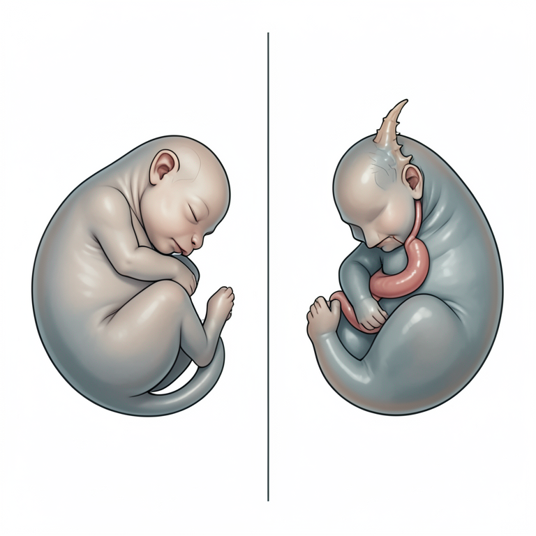
Anatomical Features of Schistosoma Reflexus
AI-generated conceptual illustration (Gir cow fetus with Schistosoma reflexus). It explains the idea and does not show study data.
View source evidence ↗Figures & tables
From the original paper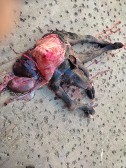 Figure 1
Introduction
Dystocia refers to trouble in calving or an abnormal birth process. It requires human intervention to finish the process (Blood et al., 2011) [3] . The factors responsible for difficulty in delivery may be both maternal and fetal. The fetal factors include oversized fetus, malpresentation, malposition, postural defects, and congenital abnormalities (Aitken, 2008) [1] and the maternal factors consist of overfeeding of the dam in the course of pregnancy, uterine inertia, and small diameter of the pelvic brim (Pugh et al., 2012) [10] . The dystocia is taken into considered an emergency circumstance and it requires immediate intervention otherwise it will result in the death of the fetus with various consequences like emphysematous of the fetus, and septicemia of the dam. It can be dealt with medically, with various mutation operations, fetotomy, or surgically. The numerous medical controls and mutations are counseled when a dam is strong and properly dilated beginning canal to allow our hand to control the fetus (Noakes 2009) [9] . Obstructive dystocia due to systemic illness, uterine inertia, prolonged active hard work, or failure in medicinal control requires a cesarean segment (Majeed et al. 1993) [8] or fetotomy. The fetal monstrosity because of dystocia can be due to progressive anomalies of the ovum, embryo, or fetus that can cause brilliant distortion of the animal, commonly resulting in dystocia (Vegad, 2007) [18] . Schistosoma Reflexus (SR) is a delivery disorder due to the malformation of the fetus. This fetal congenital syndrome is characterized by the presence of exposed abdominal and sometimes thoracic viscera (Schistosomes) and marked spinal inversion producing a unique ventral convex curvature (Roberts, 1998) [15] . It's far visible most typically in farm animals as abnormalities of the trunk of the fetus convert dystocia in livestock (Roberts, 1971) [13] . It typically includes the intermediate mesoderm and occurs in the early gastrulation embryo but, the exposed viscera and inversion of the backbone are taken into consideration as genuine schistosomus reflexus (Laughton, 2005) [7] . Various medical management and mutations are advised when a dam is stable and properly dilated birth canal to allow our hand to manipulate the fetus (Noakes, 2009) [9] . Obstructive dystocia due to systemic illness, fatigue in the uterus, prolonged straining, or failure in medicinal treatment requires a cesarean section (Majeed et al., 1993) [8] or fetotomy. The fetal monstrosity caused by dystocia may be due to developmental anomalies of the ovum, embryo, or fetus that can cause great distortion of the individual, generally resulting in dystocia (Vegad, 2007) [18] . Schistosoma Reflexus (SR) is a birth defect resulting in the malformation of the entire body. This fetal congenital syndrome is characterized by the presence of exposed abdominal and sometimes thoracic viscera (Schistosomes) and marked spinal inversion producing a distinctive ventral convex curvature (Roberts, 1998) [15] . It is seen most commonly in cattle as anomalies of the trunk of the fetus causing dystocia in cattle (Roberts, 1971) [13] . It generally involves the intermediate mesoderm and occurs in the early post-gastrulation embryo. However, the exposed viscera and inversion of the spine are considered true schistosomus reflexus (Laughton, 2005) [7] . The various environmental pollutions surrounding the animal's habitat have become a major cause of failure in conception and adversely affect prenatal development. The hereditary factors may be responsible for prenatal development defects in the embryo and fetus. The internetwork of multiple genes is a frequent and most important genetic mechanism for the occurrence of such extensive defects (Jana and Ghosh, 2001) [5] . The combination of multiple abnormal genes is responsible for such a condition. The viable normal twin calves were reported in the Schistosoma reflexes (Knight, 1996) [6] . The twin fetuses with freemartin (Cavalieri and Farin, 1999) [4] , were also associated with dystocia. The incidence of SR is between 0.01% (Sloss and Johnston, 1967) [16] to 1.3% (Knight, 1996) [6] with the highest incidence seen in cattle (Roberts, 1986) [14] . The fetus with this condition was not delivered normally but it requires a caesarian operation or fetotomy. Its typical characteristics involve spinal inversion, exposure of the abdominal viscera because of a fissure of the ventral abdominal wall, limb ankylosis, positioning of the limbs adjacent to the skull, and, lung and diaphragm hypoplasia.
Case history and Clinical findings
A Gir cow in its 5 th parity with completed gestation was brought to the Veterinary Clinical Complex, Junagadh, Gujarat with severe straining for the last 10-12 hours and no any progress towards delivery. The gynaeco-clinical examination revealed an open birth canal, ventral curvature of the vertebral column, and the head, both hind limbs and tail were in the birth canal. The outside visceral organs were palpable through an incompletely closed ventral body wall. So, it was diagnosed as a case of Schistosoma reflexus. The successful delivery of the Schistosoma reflexus fetus (Figure 1) was carried out through the left flank by following appropriate procedures under local low caudal epidural anesthesia.
Conclusions
The monstrosity of Schistosoma reflexes is generally seen in cattle (Srivastava et al., 1998) [11] . The etiology of such type dystocia is still not clear but teratogenic and genetic abnormalities cannot be ignored. The occurrence of such abnormalities is mostly due to a combination of abnormal multiple genes and their genetic mechanism (Jana and Ghosh, 2001) [5] . The dystocia due to such a condition can be resolved by using obstetrical mutation, fetotomy, or cesarean. In the present case, mutation and fetotomy were very difficult to perform to relieve dystocia because the development of severe edema and inflammation leads to constriction of the birth canal. The literature consulted also suggests delivery of Schistosoma reflexes through either obstetrical mutation (Selvaraju et al., 2013) [12] or cesarean operation (Azawi et al., 2012) [2] . The per vaginally normal calving of such a fetus is very difficult using forced traction because a prominent bone of the fetus may lead to a rupture of the uterus and tearing/injury of the birth canal which may further cause postpartum calving related problems therefore, in the present case emergency caesarian section was performed to deliver fetus successfully.
References
14 in the original paper- Aitken RJ, Iuliis GND, McLachlan RI. Biological and clinical significance of DNA damage in the male germ line. International Journal of Andrology. 2008;32(1):4656.
- Azawi OI, Ahmed OS, Abass SF. Schistosomus reflexus fetus in cross breed Iraqi cow: a case report. Iraqi Journal of Veterinary Science. 2012;26(2):103-104.
- Cavalieri J, Farin PW. Birth of a Holstein freemartin calf co-twinned to Schistosomus fetus. Theriogenology. 1999;52:815-826.
- Jana D, Ghosh M. Dystocia due to foetal monster with Schistosomus reflexus in ectobic viscera-a case report. Indian Vet. J. 2001;78:333-334.
- Knight RP. The occurrence of Schistosomus reflexus in bovine dystocia. Australian Veterinary Journal. 1996;73:105-107.
- Laughton KW, Fisher KR, Halina WG, Partlow GD. Schistosomus reflexus syndrome: a heritable defect in ruminants. Anat Histol Embryol. 2005;34:312-318.
- Majeed AF, Taha MB. Azawi OI Cesarean section in Iraqi Awassi ewes: A case study. Theriogenology. 1993;40:435-439
- Noakes DE. Arthur's veterinary reproduction and obstetrics. Saunders WB. 2009, 404-405.
- Pugh DG, Baird AN. Sheep and goat medicine. Elsevier Health Sciences, 2012.
- Srivastava KK, Sharma AK, Ahlawat SPS, Maithy SK. Schistosomus reflexus perosomus elumbis in Holstein Friesian cow. Indian J Anim Reprod. 1998;19(1):75.
- Silvaraju M, Kumaresan A, Ravikumar K, Sivaraman A, Manokaran S, Prakash S. Schistosomus reflexus fetus in a cow case report. Shanlax International Journal of Veterinary Science. 2013;(1):28-29.
- Roberts SJ. Veterinary Obstetrics and Genital Diseases (Theriogenology) (2ndedn) New Delhi, CBS Publishers and Distributors. 1998, p. 180-183.
- Sloss VE, Johnston DE. The cause and treatment of dystocia in beef cattle in western Victoria. Aust. Vet. J. 1967;43:13-21.
- Knight RP. The occurrence of Schistosomus reflexus in bovine dystocia. Aust. Vet. J. 1996;73:105-107.