
Research article · 2024 · Full text
International Journal of Veterinary Sciences and Animal HusbandrySurgical management of unilateral horn cancer in cattle
Volume 9(2) · Pages 97–99 · DOI: 10.22271/veterinary.2024.v9.i2b.1180 ↗
Research areasVeterinary Oncology ResearchMicrobial infections and disease research
Abstract
Author-writtenThe report describes a case of unilateral (left) horn cancer in a 15 year old cattle. The horn was soft at the base and bent downward and backward. The affected horn was surgically amputated under inj. Xylazine HCl sedation and cornual nerve block and ring block around base of the horn with 2% Lignocaine HCl. The cattle recovered uneventfully. Histopathology confirmed the condition was squamous cell carcinoma of horn.
Summary
Source-checkedPlain-language reading of the full paper, prepared with AI and checked against the text. Each point links to the authors' own words.
Why it matters
- Horn cancer frequently occurs in bulls with large horns, typically affecting one side in animals aged 5 to 10 years. View evidence ↗
- Several factors are associated with the development of horn cancer, including solar radiation, genetic predisposition, sex hormones, horn dye, and irritation. View evidence ↗
- Common symptoms of horn cancer in animals include nasal bleeding, head shaking, and bending at the impact angle. View evidence ↗
The question
- The objective of this report is to describe the successful surgical treatment of a case of unilateral horn cancer in cattle. View evidence ↗
What they did
- The subject was a 15-year-old cattle that presented with a two-month history of left horn bending and discharge from the horn base. View evidence ↗
- Sedation was achieved using intramuscular Xylazine HCl at 0.03 mg/kg, combined with a cornual nerve block and a 2% Lignocaine HCl ring block for analgesia. View evidence ↗
- The surgical procedure involved ligating the cornual artery with chromic catgut No.1 and removing part of the skull bone at the base using a hexablade. View evidence ↗
- Tissue growth and pus were cleared from the frontal sinus via gauze, suction, and normal saline flushing, followed by skin suturing with Nylon No.1. View evidence ↗
What they found
- Histopathology of the removed tissue confirmed the diagnosis of squamous cell carcinoma, characterized by keratinized squamous cells and epithelial pearls. View evidence ↗
- The animal recovered without complications, and no recurrence of the cancer was observed following the surgery. View evidence ↗
- The surgical site healed sufficiently to allow for the removal of skin sutures on the 22nd post-operative day. View evidence ↗
What it means
- The authors conclude that surgical dehorning under Xylazine HCl sedation and Lignocaine local analgesia provides a satisfactory outcome for cattle with horn cancer. View evidence ↗
- The authors state that early diagnosis and treatment are essential for achieving a good prognosis. View evidence ↗
- Chemotherapy was not administered in this case due to the owner's economic constraints. View evidence ↗
Visual summary
1 visualsIllustrations explain the paper; they do not show study data.
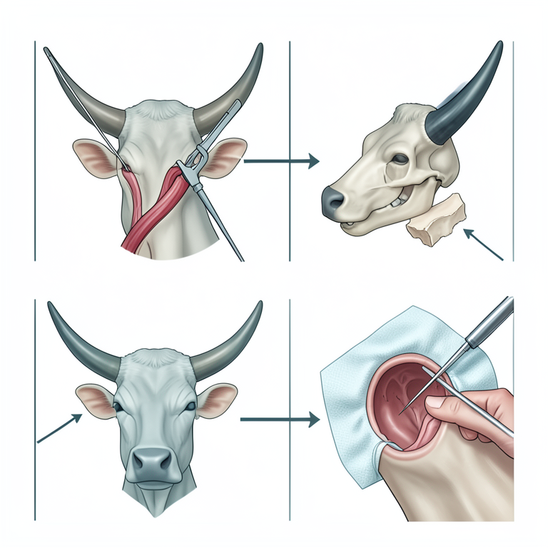
Surgical Workflow for Horn Amputation
AI-generated conceptual illustration (cattle head and horn). It explains the idea and does not show study data.
View source evidence ↗Figures & tables
From the original paper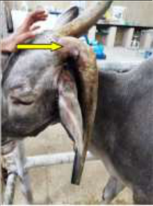 Figure 1
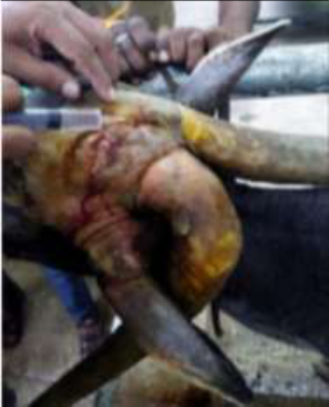 Figure 2
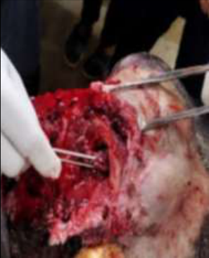 Figure 2
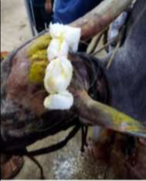 Figure 2
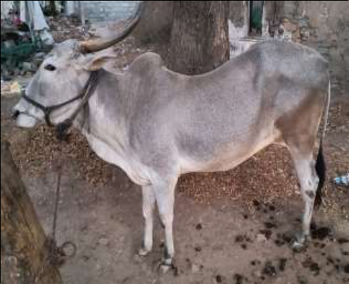 Figure 3
Introduction
Horn cancer is common in bulls with big horns and usually affects one-sidedness in the 5-10 age groups (Giri et al., 2011; Veena et al., 2011; Tyagi and Singh, 2006) [2,20,18] . This pain is caused by chronic pain at the base of the horns as a result of animals rubbing against the skin. Many predisposing factors such as irritation, horn dye, solar radiation, genetic predisposition, and sex hormones are associated with it (Sahoo et al., 2022) [3] . Head shaking, bending at impact, bending at impact angle, and nasal bleeding are common symptoms in animals (Joshi et al., 2009; Sodhi and Sangwan, 2019) [6,17] . Amputation is recommended to treat horn cancer. However, chemotherapy and amputation were also reported by Udharwar et al. (2008) [19] , Kalim et al. (2021) [7] and Goode et al. (2022) [3] . This document describes the successful treatment of unilateral horn cancer in cattle.
Case details
15 year old cattle presented with history of bending of the horn of left horn along with discharge from horn base since 2 months. Clinical examination showed unstable horn and foul smelled discharge from the left horn base. Based on history and clinical examination a tentative diagnosis of horn cancer and animal was scheduled for surgery. Anaesthesia and surgical procedure Inj. Xylazine HCl @ 0.03 mg/ kg was administered intramuscularly to sedate the cattle and Cornual nerve block was performed parallel to the frontal crest to desensitize the nerve and additional ring block around the base of the horn (Figure 2A) with 2% Ligocaine HCl achieve complete analgesia. The surgical site was prepared aseptically by shaving hairs around base of the horn and scrubbing using Chlorhexidine solution followed by surgical spirit and povidone iodine. An elliptical skin incision was given at the base, saving maximum possible skin. The skin was undermined for the loosening and exposure of skull bone.
Following skin incision the cornual artery were located and ligated by chromic catgut No.1. The bleeding was controlled using artery forceps and with gauze pieces. The part of skull bone at the base was evenly removed using hexablade to make space for skin suturing (Figure 2B). The tissue growth and the thick pus were removed from the frontal sinus using gauzes and were flushed with normal saline solution and suction. The skin was sutured with Nylon No.1 in vertical mattress suture pattern. Antiseptic bandaging was done to prevent surgical site (Figure 2C). The sample of the tissue removed from the sinus was sent for histopathology.
Post-operative care
Post-operative care included antibiotics inj. Streptopencillin @ 10.000 IU/kg, for 7 days, and inj. Tolfenamic acid @ 2 mg/kg and inj. Chlorpheniramine maleate @ 0.3mg/kg intramuscularly for 3 days. Dressing of the suture site with 5% Povidone iodine solution and fly repellent spray. The skin sutures were removed on 22 nd post-operative day.
Results and Discussion
Horn cancer is a sporadic, malignant disease affecting the horn core epithelium and predominantly seen in aged bovines.
In the present report 15 year old cattle affected with horn cancer. Clinical signs include downward bending of affected horn, soft at the base and shaky with loss of rigid orientation. Similar findings were recorded by Kumar et al. (2013) [9] , Prasad et al. (2016) [13] , Reddy, et al. (2017) [14] and Sodhi and Sangwan (2019) [17] . Dehorning is a delicate procedure because poor technique and management of wounds may result in ample complications that will lead to septicemia and delayed wound healing (Jesse et al., 2016) [5] . In the present case dehorning of affected horn was performed under inj. Xylazine HCl sedation and cornual nerve block with 2% inj. Lignocaine HCl. Similar anaesthesia protocol recorded by Kumar et al. (2013) [9] , Reddy et al. (2017) [14] , Sodhi and Sangwan (2019) [17] and Mulatu et al. (2021) [10] . The chemotherapy treatment was not undertaken due to economic considerations of the owner. However, Udharwar et al. (2008) [19] and Kumar et al. (2013) [9] reported successful use of intravenous injection of Vincristine sulphate @ 0.025 mg/kg thrice at interval of seven days. Cattle recovered uneventfully and there was no recurrence after surgery. Similar observations were reported by Giri et al. (2011) [2] , Jaiswal et al. (2014) [4] , Sharma and Singh, (2014) [16] , Pitlawar et al.
(2016) [16] and Behera et al. (2016) [1] . Histological examination of collected sample was suggestive of squamous cell carcinoma with typical keratinized squamous cell with characteristic epithelial pearls. Similar finding were reported by Kumar et al., 2013 [9] , Jaiswal et al. (2014) [4] , Reddy et al.
(2017) [14] and Sodhi and Sangwan (2019) [17] .
Acknowledgement
AcknowledgmentThe authors thankful to department of Veterinary Pathology and Veterinary Physiology and Biochemistry, Veterinary College, Gadag.
Figures and tables from the paper
References
20 in the original paper- Behera SS, Nayak S, Behera M. Surgical Management of Squamous Cell Carcinoma of Horn in a Cow. Intas Polivet. 2016;17(2):241-242.
- Giri DK, Kashyap DK, Dewangan G, Tiwari SK, Ghosh RC, Sinha B. Squamous cell carcinoma of horn and its surgical management -a report of three cases. International Journal of Livestock Research. 2011;1:5558.
- Goud A, Sahoo AK, Killi L, Patra R, Behera HS, Umar MD. Successful surgical management of squamous cell carcinoma of horn in cattle: Case report. Communications. 2022;46(3):711-717.
- Jaiswal S, Singh HN, Kumar S, Jadon NS. Horn Cancer - A clinical insight into its Diagnosis and Management in 10 cattle. Intas Polivet. 2014;15(1):3-6.
- Jesse FA, Abba Y, Sadiq MA, Umer M, Chung ELT, Bitrus AA, et al. A suspected case of suppurative frontal sinusitis in a Friesian heifer: Clinical management. International Journal of Livestock Research. 2016;6(8):50-54.
- Joshi BP, Soni PB, Ferar DT, Ghodasara DJ, Prajapati KS. Epidemiological and pathological aspects of horn cancer in cattle of Gujarat. Indian Journal of Field Veterinarians. 2009;5:15-18.
- Kalim MO, Tiwari SK, Sharda R, Shakya S, Sannat C, Praveen K, et al. Studies on prevalence, predisposing factors and microscopic evaluation of bovine horn cancer. Pharma Innovation Journal. 2021;10(2):268-270.
- Kumar A. Veterinary Surgical Techniques. 2nd ed. New Delhi: Vikas Publishing House Pvt. Ltd; 2005. p. 217219.
- Kumar V, Mathew DD, Kumar N, Arya RS. Horn cancer in bovines and its management. Intas Polivet. 2013;14(1):56-57.
- Mulatu Z, Dugassa J, Telila C. Unilateral Surgical Amputation of Horn Due to Different causes in Cattle and Goat: Case Report. Stechnolock Veterinary Sciences. 2021;1:1-8.
- Nicholas HB, Mc Donald LE. Jones Veterinary Pharmacology and Therapeutics. 6th ed. New Delhi: Kaltani Printings; 2001. p. 874.
- Pitlawar SS, Gahlot BM, Akhare SB, Upadhye SV, Jadhao PT. Surgical Management of Horn Affections -A Report of 2 Bullocks. Intas Polivet. 2016;17(2):239-240.
- Prasad VD, Kumar PR, Harikrishna NVV, Sreenu M, Vidyasagar P, Mahesh R, et al. A comprehensive study on surgical affections of horns in buffaloes (Bubalus bubalis). Journal of Livestock Science. 2016;7:280-287.
- Reddy KJM, Kumar VG, Ganesh S, Raghavender KBP. Unilateral horn cancer in cow and its surgical management-A case report. International Journal of Current Microbiology and Applied Sciences. 2017;6(8):3349-3352.
- Sastry GA. Veterinary Pathology. 7th ed. New Delhi: CBS Publisher and Distributors; 2001. p. 205-249.
- Sharma N, Singh D. Unilateral Horn Cancer in a Bullock and its Management. Intas Polivet. 2014;15(1):7-8.
- Sodhi HS, Sangwan V. Successful Horn Amputation for the Treatment of Horn Cancer in an 18-Year-Old Humped Bullock. International Journal of Current Microbiology and Applied Sciences. 2019;8(08):537541.
- Tyagi RPS, Singh J. Ruminant Surgery. 1st ed. New Delhi: CBS Publisher and Distributors; 2006. p. 415-416.
- Udharwar SV, Aher VD, Yadav GU, Bhikane AU, Dandge BP. Study on incidence, predisposing factors, symptomatology and treatment of horn cancer in bovine with special reference to surgery and chemotherapy. Veterinary World. 2008;1:7-9.
- Veena P, Kumar RVS, Sankar P, Dhanalakshmi N, Kokila S. Squamous cell carcinoma of horn in a bullockA case report. Indian Journal of Animal Research. 2011;45:226-227.