
Research article · 2024 · Full text
International Journal of Veterinary Sciences and Animal HusbandrySuccessful management of cataracts in dogs by phacoemulsification with IOL implantation: Review of two cases
Volume 9(2) · Pages 100–103 · DOI: 10.22271/veterinary.2024.v9.i2b.1181 ↗
Research areasIntraocular Surgery and LensesConnexins and lens biologyOcular Surface and Contact Lens
Abstract
Author-writtenThe retina macula appeared as strip-like and pale in colour located dorsal to the optic disc in buffaloes. Retina macula contained large RPE cells with tightly packed melanin pigment, a relatively larger number of cone cells, cells of inner nuclear, inner plexiform and nerve fibre layers showed less cystoid degeneration when compared to the peripheral retina. The total thickness (µm) of retinal macula in groups I, II and III was 229.05±3.56, 230.55±3.28 and 231.72±2.87 respectively. There were few degenerating changes observed in the macula when compared to the peripheral retina with the advancement of age in buffaloes.
Summary
Source-checkedPlain-language reading of the full paper, prepared with AI and checked against the text. Each point links to the authors' own words.
Why it matters
- Cataracts are common in dogs and typically require surgical removal of the lens and the implantation of an artificial lens for effective treatment. View evidence ↗
- Cataracts are a major global health issue, accounting for 47.8% of all blindness cases. View evidence ↗
- Using an intraocular lens (IOL) improves vision in eyes lacking a natural lens and reduces the risk of posterior capsular opacity after surgery. View evidence ↗
The question
- The study aimed to evaluate the surgical outcomes of using phacoemulsification combined with a flexible acrylic IOL in dogs with cataracts. View evidence ↗
What they did
- Anesthesia was induced with propofol (4 mg/kg), maintained with isoflurane, and Atracurium (0.2 mg/kg) was used as a muscle relaxant. View evidence ↗
- The surgical approach involved making a 2.8-mm corneal incision located 2 mm from the corneal limbus edge. View evidence ↗
- The cataract was removed using phacoemulsification with a 'divide and conquer' technique for the nucleus. View evidence ↗
- A hydrophobic foldable acrylic IOL was implanted into the capsular bag through the existing incision. View evidence ↗
What they found
- Both dogs experienced an uneventful recovery and regained their vision. View evidence ↗
- Following phacoemulsification, the treated eyes regained vision, allowing the dogs to perform daily activities without visual discomfort. View evidence ↗
What it means
- The authors conclude that phacoemulsification is a vital procedure for restoring eyesight in dogs with cataracts. View evidence ↗
Visual summary
3 visualsIllustrations explain the paper; charts are replotted from the paper's own table values.
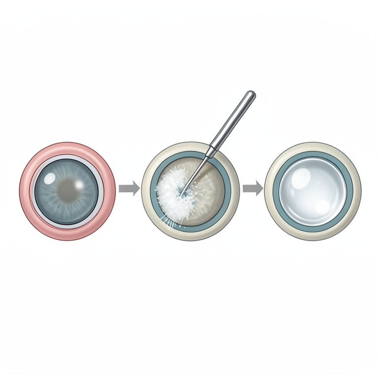
Phacoemulsification and IOL Process
AI-generated conceptual illustration (canine eye). It explains the idea and does not show study data.
View source evidence ↗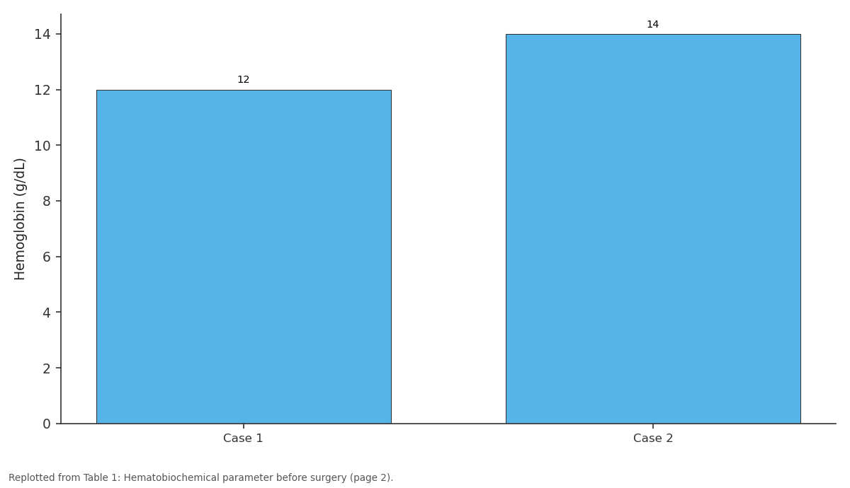
Hemoglobin (g/dL): Case 1 vs Case 2
Replotted from Table 1 (page 2) of the original paper. Every bar is the paper's own table value.
View source evidence ↗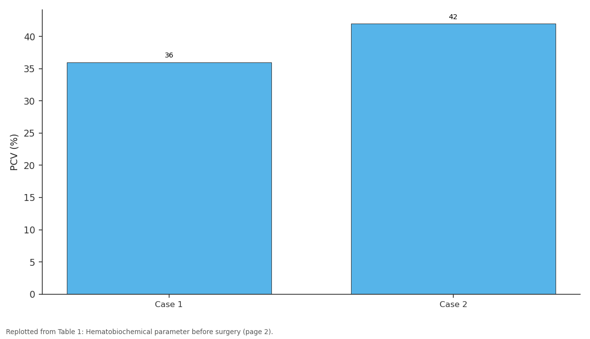
PCV (%): Case 1 vs Case 2
Replotted from Table 1 (page 2) of the original paper. Every bar is the paper's own table value.
View source evidence ↗Figures & tables
From the original paper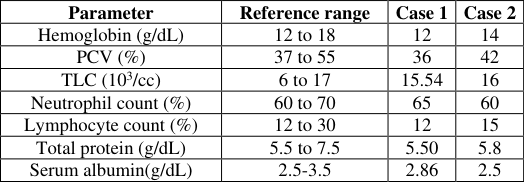 Table 1
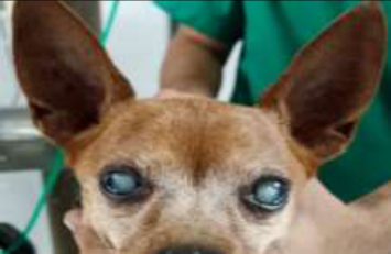 Figure 1
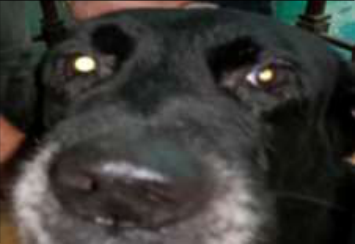 Figure 1
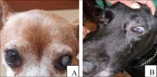 Figure 4
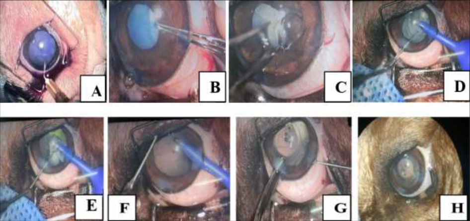 Figure 3
Introduction
In dogs, cataracts are a prevalent eye disease that can be effectively treated only with surgical extraction followed by intraocular lens implantation (Bhutto et al., 2021) [1] . Congenital and acquired cataracts are the two types of cataracts. According to Liu et al. (2017) [9] , cataracts constitute the leading cause of global blindness, contributing to 47.8% of all blindness cases worldwide. The most prevalent risk factor for cataracts is advanced age. Physical risk factors are not present in cases of senile cataracts. In veterinary medicine, cataract extraction is a frequently performed ocular procedure that uses a variety of surgical procedures. At present, the recommended treatment approach involves phacoemulsification combined with the implantation of an intraocular lens (IOL). The use of an IOL enhances visual function in eyes without a natural lens and diminishes the likelihood of developing posterior capsular opacity (PCO) post-surgery. IOL displacement, however, may lead to altered vision and increased risks following surgery (Bhutto et al., 2021; Nochez et al., 2010) [1] . The IOL material that is most frequently used in veterinary practice is polymethyl methacrylate, or PMMA (Liu et al., 2017) [9] . But researchers have also looked into substitute materials including silicone and hydroxyethyl methacrylate (HEMA) optic IOLs (Davidson et al., 1990; Kim and Oh 2018) [4,7] . When compared to alternative lens materials, Utilizing an acrylic intraocular lens (IOL) with a squared edge has been linked to a reduced occurrence of posterior capsular opacification (PCO), as indicated by studies conducted by Christine et al. (2011) [3] and Montes-Mico and Cervino (2009) [11] . The reduced incidence of posterior capsular opacification (PCO) associated with acrylic lenses is attributed to their firm attachment to the posterior capsule and their distinctive optical design, which hinders the migration of lens epithelial cells into the optic region, as highlighted in the research by Christine et al. (2011) [3] . While flexible acrylic intraocular lenses (IOLs) are commonly used in dogs, the prevention of PCO is generally not a primary consideration in their optical design, as indicated by Harathi et al. (2020) [5] . Our study aimed to investigate the surgical results linked to the combination of phacoemulsification and the placement of a pliable acrylic intraocular lens (IOL) in dogs diagnosed with cataracts.
Case history and observation Case 1
A 10-year-old male miniature pinscher, weighing 10kg, was presented to the surgical department at Mumbai Veterinary College, Parel, with the primary complaint of vision impairment. Upon ophthalmic examination, cloudy and white lenses were observed in both eyes, indicative of cataract symptoms. According to the dog owner, the onset of cataract symptoms began several months ago in the right eye and one year ago in the left eye (refer to Fig. 1). Cataract symptoms typically progress gradually over an extended period. Consequently, even though object images appear blurred and unclear, and there may be some visual discomfort, patients often fail to recognize these symptoms. The extent of visual impairment varies based on the degree of lens opacity and lesion location. In cases of partial opacity, object images may overlap, even in the vision of one eye, as noted by Mahbubul et al. (2019) [10] . The Schirmer tear test results indicated a rate of 18 mm/min for the right eye and 19 mm/min for the left eye. Additionally, intraocular pressure (IOP) measurements using TonopenVet fell within the normal range, registering at 17 mmHg for the right eye and 22 mmHg for the left eye. While the patient exhibited no responses in the menace response test, dazzle reflex test, and maze test, both palpebral reflexes and corneal reflexes were present. These findings align with similar observations reported by Myeong-Gon Kang et al. (2022) [6] .
Case 2
An eight-year-old non-descript male dog weighing 15.5 kg was presented with a history of blindness, hitting the wall while walking, and whiteness inside both eyes. On ophthalmologic examination, the dog did not exhibit a menace reflex, showed a positive response to the pupillary light, and the fundus was not visible in either eye. A bilateral mature cataract was confirmed (Fig. 2). Cornea, conjunctiva, and sclera were clear. Intra-ocular pressure was measured using TonopenVet and was 15 mmHg for both eyes. The animal was healthy. Hematological and serum biochemistry results were normal as shown in (Table 1). [8] and You et al. (2020) [14] . The cataractous lens in one eye was removed using phacoemulsification.
| Parameter | Reference range | Case 1 | Case 2 |
|---|---|---|---|
| Hemoglobin (g/dL) | 12 to 18 | 12 | 14 |
| PCV (%) | 37 to 55 | 36 | 42 |
| TLC (103/cc) | 6 to 17 | 15.54 | 16 |
| Neutrophil count (%) | 60 to 70 | 65 | 60 |
| Lymphocyte count (%) | 12 to 30 | 12 | 15 |
| Total protein (g/dL) | 5.5 to 7.5 | 5.50 | 5.8 |
| Serum albumin(g/dL) | 2.5-3.5 | 2.86 | 2.5 |
Table text extracted from the original PDF; superscript letters are shown inline. The original table image is below.
View original table image
Preoperative Preparation for Cataract Surgery:
In preparation for cataract surgery, topical treatments included the use of broad-spectrum ophthalmic antibiotics, Moxifloxacin HCL 0.5%, administered 6 hours for 24-48 hours before the procedure, along with topical corticosteroids for 48-72 hours preoperatively. On the day of surgery, mydriasis was induced using 1% atropine sulfate three times at 30-minute intervals starting 2 hours before surgery. Topical non-steroidal anti-inflammatory agents were applied every 30 minutes, beginning 1-3 hours before the procedure. Additionally, systemic non-steroidal anti-inflammatory agents and antibiotics were administered immediately before surgery.
Treatment and Discussion
Following standard aseptic surgery preparation, the canine patient received premedication with atropine sulfate (0.04 mg/kg SC) and intramuscular triflupromazine hydrochloride (1 mg/kg IV). General anesthesia was induced with propofol (4 mg/kg slow IV), and isoflurane gas was used for maintenance. To prevent pupil movement during surgery, Atracurium (0.2 mg/kg IV), a muscle relaxant, was administered. The patient was positioned supine, and a 2.8mm corneal incision was made at the corneal limbus edge, 2 mm away. Trypan blue dye was instilled for improved visualization of the anterior capsule. A continuous curvilinear capsulorhexis was performed with Utrata capsule forceps, and visco-elastic agents (hydroxypropyl methylcellulose) were injected into the anterior chamber for stability. Hydrodissection was carried out using lactated Ringer's solution to loosen the outer cortex from the lens capsule. Phacoemulsification commenced in the anterior lens cortex and nucleus, employing the divide and conquer technique for nucleus removal. The remaining cortex and nucleus were fragmented through parallel sculpting. Careful removal of cataractous material was conducted to avoid posterior lens capsule tearing. An irrigation-aspirating handpiece facilitated the removal of residual cataract material. If an intraocular lens (IOL) was implanted, the anterior capsulectomy diameter was adjusted to be 1 mm less than the IOL optic diameter. A hydrophobic foldable acrylic lens was used, and inserted into the capsular bag without enlarging the corneal incision. To ensure physiological pressure in the anterior chamber, a balanced salt solution was injected in the final step, estimating intraocular pressure (IOP) through digital palpation instead of a tonometer. Postoperative care for cataract surgery Animals were post-operatively followed for 30 days along with an infusion of topical antibiotics (Moxifloxacin HCL 0.5%), artificial tears (Carboxymethyl Cellulose 0.5%) and anti-inflammatory Eye Drops Fluorometholone 0.1% were given 3-4 times daily. Broad-spectrum systemic antibiotics and anti-inflammatory drugs for BID for 7 days were given.
Advised for the Elizabethan collar on at all times for 4 to 8 weeks after surgery, to keep him from hurting himself.
Conclusion
The patient regained vision in the treated eye following cataract surgery with PHACO and presently engages in daily activities without experiencing visual discomfort. In the context of canine cataract surgery, PHACO emerges as a vital therapeutic procedure essential for restoring eyesight.
Acknowledgement
AcknowledgmentThe author extends their support to the Department of Veterinary Pathology.
Figures and tables from the paper
References
14 in the original paper- Bhutto IA, Memon MN, Ali I, Mirani AH. Comparison of Complications between Manual Small Incision Cataract Surgery and Phacoemulsification. The Pakistan Journal of Ophthalmology. 2021;37(4):384-387.
- Biros DJ, Brooks DE. Development of glaucoma after cataract surgery in dogs: 200 cases (1987-1998). Journal of American Veterinary Medical Association. 2000;216:1780-1786.
- Christine CL, Shannan CB, Cheryl LW, and Bruce HG. Cataracts in 44 dogs (77 eyes): A comparison of outcomes for no treatment, topical medical management, or phacoemulsification with intraocular lens implantation. Canadian Veterinary Journal. 2011;52:283288.
- Davidson MG, Nasisse MP, Rusnak IM, English RV. Success rates of unilateral vs bilateral cataract extraction in dogs. Veterinary Surgery. 1990;19:232-236.
- Harathi K, Scott H, Ellison B. Comparison of outcomes in cataracts eyes of dogs undergoing phacoemulsification versus eyes not undergoing surgery. Veterinary Ophthalmology. 2020;23:286-291.
- Kang MG, Han DH, Han SM, Jung EG, Kim GM, Cho JH, et al. A case of cataract surgery using phacoemulsification in a 12-year-old poodle. The Korean Journal of Veterinary Service. 2022;45(3): 221-228.
- Kim MH, Oh JH. A Case of Suprachoroidal Hemorrhage after Pars Plana Vitrectomy for Rhegmatogenous Retinal Detachment. The Journal of the Korean Ophthalmological Society. 2018;59(11):1082-1086.
- Linebarger EJ, Hardten DR, Lindstrom RL. Phacoemulsification and modern cataract. Survey of ophthalmology. 1999;44:123-147.
- Liu YC, Wilkins M, Kim T, Mehta JS. Cataracts. The Lancet. 2017;390(10094):600-612.
- Mahbubul HS, Melinda KD. Cataract surgeon viewpoints on the need for novel preventative anti-inflammatory and anti-posterior capsular opacification therapies. Curr Med Res Opin. 2019;35(11):1971-1981.
- Montés-Micó R, Cerviño A. Analysis of the possible benefits of aspheric intraocular lenses: review of the literature. Journal of Cataract and Refractive Surgery. 2009;35:172-81.
- Nochez Y, Favard A, Pisella PJ. Measurement of corneal aberrations for customization of intraocular lens asphericity: impact on quality of vision after microincision cataract surgery. British Journal of Ophthalmology. 2010;94:440-4.
- Sigle KJ, Nasisse MP. Long-term complications after phacoemulsification for cataract removal in dogs: 172 cases (1995-2002). Journal of American Veterinary Medical Association. 2006;228:74-79.
- You JI, Bang SK, Jin KH. Visual Prognosis and Satisfaction of Advanced Cataract Patients Unable to be Evaluated by Fundus Imaging. The Journal of the Korean Ophthalmological Society. 2020;61(3):235-242.
Cited by 2
From OpenAlex, OpenCitations and reference lists read by Scinovex
- Development of Postoperative Ocular Hypertension After Phacoemulsification for Removal of Cataracts in Dogs ↗
Myeong-Gon Kang, Chung-Hui Kim, Shin-Ho Lee, Jae‐Hyeon Cho · Animals · 2025
A cataract is a disease in which the lens of the eye becomes clouded, causing a partial or complete loss of vision. Phacoemulsification (PHACO) is a modern surgical technique used in cataract surgery. Study findings: This study observed changes in intraocular pressure (IOP) after surgery in 31 dogs (48 eyes) with cataracts that visited a veterinary hospital. The procedure involved…
❝ 1PDF - Cataract in dogs: A comprehensive review
Monika Rani, Santoshmani D Tripathi, G.S. Khandekar, S.V. Gaikwad, Dishant Saini, Manika Jadhav · International Journal of Advanced Biochemistry Research · 2024
Cataracts are a leading cause of vision impairment in dogs, characterized by the opacity of the lens that can significantly affect a dog's quality of life. Advanced age is the main risk factor for developing cataracts. This comprehensive review explores the etiology, classification, and current treatment options, emphasizing surgical interventions such as phacoemulsification. The study concludes that Phacoemulsification is currently the preferred approach due to its smaller incision requirement compared to intracapsular cataract extraction (ICCE) or extracapsular cataract extraction (ECCE), le