
Research article · 2024 · Full text
International Journal of Veterinary Sciences and Animal HusbandryOccurrence of Schistosoma spindale infection in bovine of Puducherry
Volume 9(2) · Pages 90–91 · DOI: 10.22271/veterinary.2024.v9.i2b.1170 ↗
Research areasHelminth infection and controlParasites and Host Interactions
Abstract
Author-writtenA five years old female cross bred Jersey cow was presented with the history of blood mixed diarrhoea with fetid odour since 15 days and lateral recumbency. Upon fecal examination, the ova of Schistosoma spindale was revealed. The present case deals with occurrence of S. spindale infection in a Crossbred Jersey breeds of cattle of Union territory of Puducherry. Eggs of Schistosoma spindale was noticed in fecal sample of affected animals and identified as per standard taxonomical keys (Soulsby, 1982).
Summary
Source-checkedPlain-language reading of the full paper, prepared with AI and checked against the text. Each point links to the authors' own words.
Why it matters
- Visceral schistosomiasis in cattle is caused by Schistosoma spindale, a type of snail-borne digenetic trematode. View evidence ↗
- This infection is characterized as a chronic wasting disease that leads to significant economic losses. View evidence ↗
- The disease causes severe intestinal pathology, including mucosal thickening, ulcers, and hemorrhage. View evidence ↗
The question
- The study aims to report the occurrence of S. spindale infection in a crossbred Jersey cow located in Puducherry, India. View evidence ↗
What they did
- The subject was a five-year-old crossbred Jersey cow presenting with lateral recumbency and fetid, blood-mixed diarrhea for 15 days. View evidence ↗
- Direct fecal examination involved mixing a small amount of feces with water on a slide and observing it under a 10X objective lens. View evidence ↗
- The sedimentation technique involved centrifuging the sample at 2000 rpm for 3 minutes before examining the pellet under a 10X lens. View evidence ↗
- The flotation technique used a saturated salt solution and a 15-minute incubation period to isolate eggs on a slide. View evidence ↗
What they found
- Microscopic analysis of the cow's fecal samples revealed spindle-shaped eggs with a terminal spine and a developed miracidium, identifying the parasite as S. spindale. View evidence ↗
- The identification of the eggs as S. spindale was confirmed using standard taxonomical keys from Soulsby (1982). View evidence ↗
- The authors note that previous research by Banerjee et al. (1988) recorded a 41.2% infection rate in cattle. View evidence ↗
What it means
- Based on clinical history and fecal examination, the authors conclude that the crossbred Jersey cow was affected by schistosomiasis caused by S. spindale. View evidence ↗
- The authors state that fecal examination remains a valid diagnostic method for confirming cases and determining treatment. View evidence ↗
- The authors suggest that further research into the epizootiology in farm animals and cercarial bionomics in snails is needed to better understand the disease dynamics. View evidence ↗
Visual summary
1 visualsIllustrations explain the paper; they do not show study data.
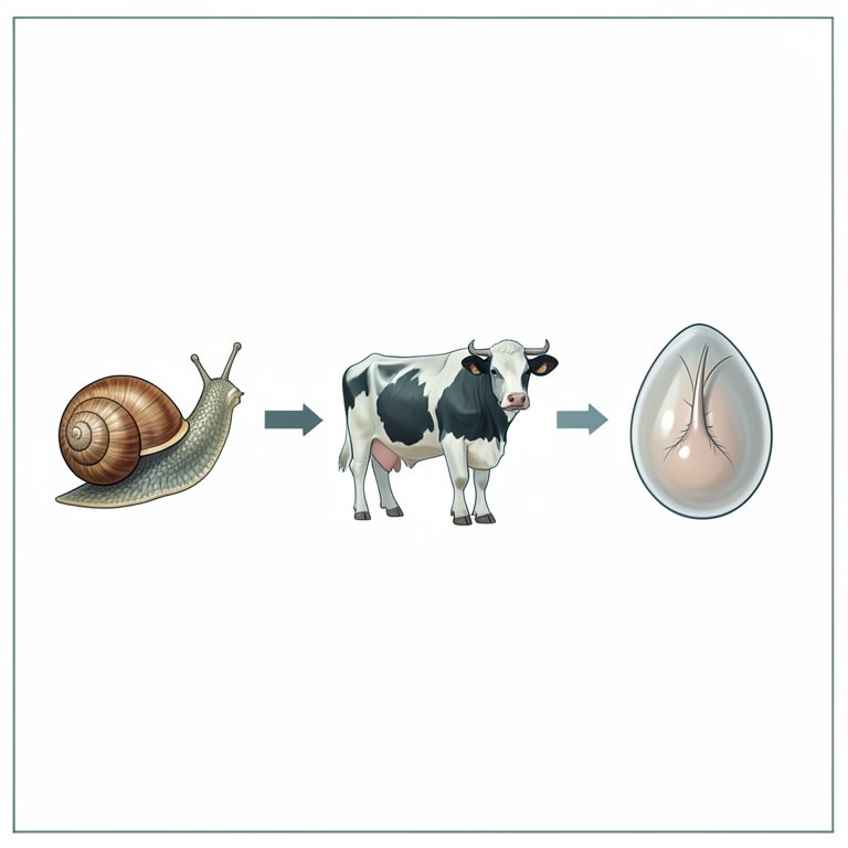
Life Cycle and Identification
AI-generated conceptual illustration (Bovine cattle, freshwater snails, and Schistosoma spindale eggs). It explains the idea and does not show study data.
View source evidence ↗Figures & tables
From the original paper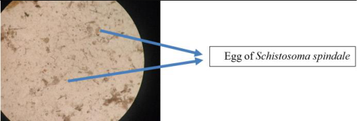 Figure 1
Introduction
Visceral schistosomiasis caused by Schistosoma spindale is a snail borne digenetic trematode infection of cattle. It was first identified in 1906 by Montgomery from cattle in Mukteswar, India (Montgomery, 1906) [6] . It affects domestic animals, man and wild animals in different parts of Asia and Africa (Latchuminkanthan et al., 2014) [4] . The species which commonly occur in India are Schistosoma nasale and S. spindale in cattle, S. indicum in equines and sheep and S. incognitum in pig. It resides in a mesenteric vein of cattle, Buffalo, Horse, Pig (Taylor, 2016) [12] . The freshwater snail Indoplanorbis exustus acts as an intermediate host. It is a chronic wasting disease contributing substantial economical loss (Bulbul et al., 2021) [2] . Visceral schistosomiasis were reported from Tamil Nadu (Jeyathilakan et al., 2008) [9] , Kerala (Ravindran et al., 2007) [10] , Assam (Bulbul et al., 2021) [2] . The pathology of infection and clinical signs are severe intestinal haemorrhage in intestinal mucosa, thickening of intestinal mucosa and ulcers covered with blood stained mucosa. The present communication reports the presence of S. spindale infection in Crossbred Jersey cow of Puducherry, a southern Union Territory of India
Materials and methods 2.1 Clinical history
A five years old Cross bred Jersey cow was presented with a history of blood mixed diarrhoea with fetid odour since 15 days and with history of lateral recumbency, diarrhoea since 2 days.
Sample collection
The fresh dung sample was examined grossly for the presence of helminthes and approximately two hundred grams of dung sample was collected in an air tight container/ polythene cover. The sample was subjected for direct and concentration techniques like floatation and sedimentation in the department.
Microscopic examination of dung sample
The direct fecal examination was done by taking a small quantity of faeces is placed on a slide, mixed with some droplets of water and a cover slip is placed on the fluid and examined under 10X objective lens. Further, the faecal samples were subjected to sedimentation and floatation techniques.
Sedimentation technique
A 1-2g of dung sample is kept in a mortar and with the help of pestle grind the sample properly. Add 10-15 ml of water and grind it till homogenous suspension is formed. Filter the contents through sieve and filtrate is transferred to the centrifuge tube. Centrifuged for 2000 rpm for 3 min. Discard the supernatant and reconstitute the pellet. The sediment is then examined under low power objective lens (10X).
Floatation technique
A 1-2g of dung sample is kept in a mortar and with the help of pestle grind the sample properly. Add saturated salt solution and grind it till homogenous suspension is Filter the contents through sieve and filtrate is transferred to the flotation tube, place a slide over the top of it. Incubated for 15 min and take a slide in one stroke. The slide is examined under low power objective lens (10X).
Results and Discussion
The microscopic examination of faecal samples of Cross bred Jersey revealed the presence of spindle shaped egg with terminal spine and fully developed miracidium inside (Schistosoma spindale) (Fig. 1). The eggs belong to S. spindale as per the standard taxonomical keys given by Soulsby (1982) [11] . The presence of spindle shaped eggs in fecal sample of cattle was also reported by Vimalraj and Latchumikanthan (2015) [13] , Jeyathilakan et al., (2008) [9] in Chennai, Shiyad et al. (2004) [8] in Kerala. The similar observations have been made by Banerjee et al., 1988 [1] where he has recorded 41.2% infection rate in cattle. While there may be minimal or no apparent clinical indications in the short term, the elevated occurrence of prolonged schistosome infections ultimately leads to substantial detriments to the herd. These detriments manifest through less easily discernible impacts on animal growth and productivity, as well as an increased vulnerability to other parasitic and bacterial diseases, as documented by Dargie (1980) [3] , Pitchford and Visser (1982) [7] and McCauley et al. (1984) [5] . Additional investigations into the epizootiology of S. spindale in farm animals and the cercarial bionomics in snails are necessary to enhance our comprehension of S. spindale and the dynamics of the associated diseases.
Conclusion
Considering the history of the case, clinical observation and faecal examination of the crossbred Jersey cow it is evident that schistosomiasis caused by Schistosoma spindale affected the cattle and faecal examination is still a diagnostic method for the confirmation of the case and further treatment.
References
11 in the original paper- Bulbul KH, Das M, Islam S, Sarmah PC, Tamuly S, Borah P, et al. Molecular epidemiology of visceral schistosomosis caused by Schistosoma spindale infection in cattle of Assam, India. Biological Rhythm Research. 2021;52(7):1101-1108.
- Dargie JD. The pathogenesis of Schistosoma bovis infection in Sudanese cattle. Trans R Soc Trop Med Hyg. 1980;74:560-2.
- Latchumikanthan A, Pothiappan P, Ilayabharathi D, Das SS, Kumar D, Ilangovan C. Occurrence of Schistosoma nasale infection in bullocks of Puducherry. Journal of parasitic diseases. 2014;38(2):238-240.
- Mc Cauley EH, Majid AA, Tayeb A. Economic evaluation of the production impact of bovine schistosomiasis and vaccination in Sudan. Preventive Veterinary Medicine. 1984;2:735-54.
- Pitchford RJ, Visser PS. Schistosoma mattheei Veglia &Le Roux, 1929, egg output from cattle in a highly endemic area in Eastern Transvaal. Ondersteport Journal of Veterinary Research. 1982;49:233-5.
- Shiyad MA, Jayakumar PK, Padmaraj K, Devada Chandrababu E. Schistosomosis in a goat -a case report. Journal of Veterinary and Animal Science. 2004;35:9193.
- Jeyathilakan N, Latha BR, Basith SA. Seasonal prevalence of Schistosoma spindale in ruminants at Chennai. Tamil Nadu Journal of Veterinary and Animal Science. 2008;4(4):135-138.
- Ravindran R, Lakshmanan B, Ravishankar C, Subramanian H. Research note. Visceral schistosomiasis among domestic ruminants slaughtered in Wayanad, South India. Southeast Asian journal of tropical medicine and public health. 2007;38(6):1008.
- Soulsby EJL. Helminths, arthropods and protozoa of domesticated animals. 7th ed. Bailliere Tindall. London, 1982, p. 1-778.
- Taylor MA, Coop RL, Wall RL. Veterinary Parasitology. 4 th Ed. Wiley Blackwell. 2016, p. 1-1029.
- Vimalraj PG, Latchumikanthan A. Schistosoma spindale infection in a captive jackal (Canis aureus). Journal of parasitic diseases. 2015;39:120-121.