
Research article · 2024 · Full text
International Journal of Veterinary Sciences and Animal HusbandryChronic ruminal impaction in goats and its surgical management: A two case studies
Volume 9(1) · Pages 1388–1390 · DOI: 10.22271/veterinary.2024.v9.i1s.1143 ↗
Research areasForensic Entomology and Diptera StudiesBacillus and Francisella bacterial research
Abstract
Author-writtenA four-year-old female non-descriptive Goat and a two-year-old male white African Boer was presented to the Veterinary clinical complex, College of Veterinary Animal Sciences, Udgir with a history of anorexia and weight loss in eight days. Clinically, all physiological parameters were in normal range, however abdominal palpation revealed hard ruminal consistency indicates the chronic impaction. Lateral radiographic examination of reticulo-thorax of both animals showed presence of non non-penetrating foreign body (NPFB) in the reticulum. On basis of history, physical and radiographic examination, both the goats were operated for laparo-rumenotomy for removal of NPFB under regional and local infiltration anaesthesia. To conclude, both the animals with non penetrating reticular foreign bodies causing chronic ruminal impaction leading to loss of body condition and it was successfully removed via Laparo-rumenotomy without complications and they assumed normal appetite.
Summary
Source-checkedPlain-language reading of the full paper, prepared with AI and checked against the text. Each point links to the authors' own words.
Why it matters
- The accumulation of foreign bodies in the rumen is economically significant because it prevents the absorption of volatile fatty acids, which reduces animal production. View evidence ↗
- Goats may ingest unwanted materials like polythene bags due to urbanization, lack of grazing areas, or a condition called pica. View evidence ↗
- The buildup of indigestible material in the rumen can create a hard mass leading to impaction, gas buildup, and potential death. View evidence ↗
The question
- The study aims to report rare cases of chronic ruminal impaction in two goats caused by non-penetrating reticular foreign bodies diagnosed through radiography. View evidence ↗
What they did
- The study involved two goats: a four-year-old non-descriptive female and a two-year-old male African Boer. View evidence ↗
- Physical examination of the left paralumbar fossa was used to identify rumen distention and impaction. View evidence ↗
- Lateral radiographic examination of the reticulo-thorax was performed to identify radio-opaque foreign bodies in the reticulum. View evidence ↗
- Both animals underwent exploratory rumino-laparotomy to remove the identified reticular foreign bodies. View evidence ↗
What they found
- An oval-shaped foreign body was manually removed from the female goat, while a polythene bag was disintegrated and removed from the male Boer goat. View evidence ↗
- Palpation of the rumino-reticular fold, esophageal orifice, and omasal orifice revealed no abnormalities. View evidence ↗
- Both animals successfully recovered their normal appetite after the foreign bodies were removed via laparo-rumenotomy without complications. View evidence ↗
What it means
- The authors conclude that non-penetrating ruminal foreign bodies causing loss of appetite can be successfully managed through rumenotomy. View evidence ↗
Visual summary
1 visualsIllustrations explain the paper; they do not show study data.
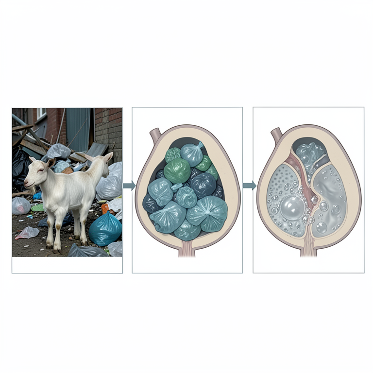
Path to Ruminal Impaction
AI-generated conceptual illustration (goat kids). It explains the idea and does not show study data.
View source evidence ↗Figures & tables
From the original paper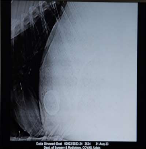 Figure 1
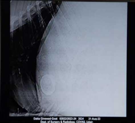 Figure 2
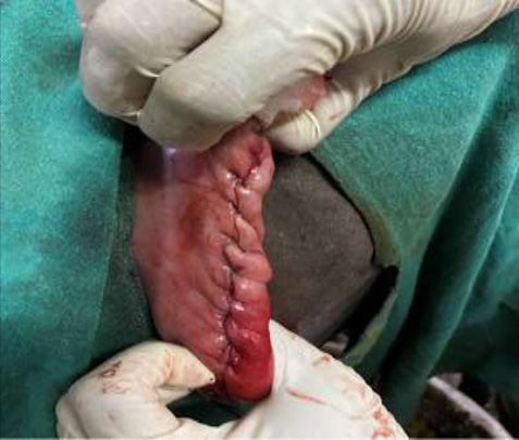 Figure 3
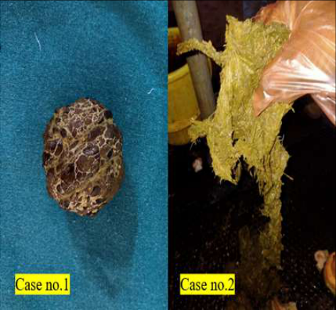 Figure 4
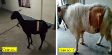 Figure 5
Introduction
Ruminants have an unusual digestive system that is divided into four sections, the rumen, reticulum, omasum, and abomasum. The abomasum alone produces digestive enzymes; hence it is in responsible for breaking down food (Phillipson, 1999) [1] . Due to the production of volatile fatty acids (VFAs) by the microorganisms found in the rumen (mostly ciliated protozoa and bacteria, but also yeasts), ruminants can digest fibrous plant material. VFAs are absorbed through the ruminal wall or later in the omasum and abomasum (Barcroft et al., 1944) [2] , and they are derived from complex carbohydrates and other products (such as proteins and B vitamins) (Ducharme, 1990) [3] . The ruminal volume of small ruminants is estimated to be 5.3 litres, accounting for around 13% of their body weight (Owens and Goetsch, 1993) [4] . Other authors estimate the ruminal content in sheep to be around 4-6 kg (Phillipson, 1999) [1] , however these figures may vary depending on nutrition and transit through the digestive tract. Foreign body accumulation in the rumen inhibits the absorption of volatile fatty acids, which lowers the animal's production. As a result, this has a significant economic impact (Igbokwe et al., 2003) [5] . The present paper represents the rare cases of chronic ruminal impaction in two goats leading to loss of body condition due to presence of non penetrating reticular foreign bodies which was diagnosed via radiography.
Case History
A Four-year-old female Non-descriptive (Case no.1) and A 2 year old male African Boer Goat (Case no. 2) were presented to the Department of Veterinary Surgery and Radiology, College of Veterinary Animal Sciences, Udgir, Maharashtra at different intervals of time with a same history of anorexia since 6 and 4 days respectively accompanied with ruminal impaction and absence of rumination. Both the animals were treated symptomatically by local veterinarian; however there was no relief from the ruminal impaction. The heart rate, temperature, and respiration rate were all in the normal range. The blood picture showed leucocytosis (14,000 cells/cmm), neutrophilia (94%) mild anaemic in case no.1 and mild leucocytosis (12,000 cells/cmm), and neutrophilia (87%) in case no. 2 respectively. A thorough physical examination of the left paralumbar fossa revealed distention and impaction of rumen.
Lateral radiographic examination of reticulo-thorax revealed a radio-opaque oval-shaped non-penetrating foreign body in the reticulum in case no.1 (Fig. 1) and a radio-opaque large structure in the reticulum (might be polythene bag) in case no. 2 (Fig. 2). On the basis of history, physical and radiographic examination, both the animals were subjected for exploratory rumino-laparotomy for removal of reticular foreign bodies. Following premedication with Inj. Meloxicam @ 0.2 mg/kg body weight intramuscularly, Inj. Chlorpheniramine @ 0.2 mg/kg body weight intramuscularly, Inj. Dicrysticin @ 10,000 IU/kg body weight intramuscularly. In Case no.1 surgery was performed in a standing position whereas in Case no.2 in lateral recumbency with sedation Inj. Diazepam @ 0.25 mg/kg body weight intravenously. The left paralumbar fossa was aseptically prepared for surgery in both cases. Inverted L block was given in both surgical cases using 2% lignocaine HCL. A vertical incision is made about three centimetres from the transverse lumbar process, centre of the fossa right above the rumen's dorsal sac. The thickness of the subcutaneous layer was different in both cases as it differed with age and adipose tissue content and was then separated bluntly. The peritoneum is then found once the oblique abdominal muscles (external, internal, and transverse) have been severed. In order to avoid contaminating the abdominal cavity and muscle layers, the major goal is to exteriorize the dorsal sac and its fixation by using four stay suture. In female goat, the oval shaped reticular foreign body was removed manually whereas in male Boer, the foreign body was polythene bag (entangled material) (Fig. 4) which was disintegrated into multiple pieces for easy removal via large ruminal incision. The ruminoreticular fold, esophageal orifice and omasal orifice were palpated to detect any abnormalities and found to be normal. Some quantity of ruminal content was removed in order to decrease the impaction. Rumentoric bolus and 100 gm of acid-base balance powder were administered intra ruminally. The rumen incision was closed by inverted suture pattern (Cushing followed by Lambert) (Fig. 3). as it gives a sealed closure that inhibits the leaking of rumen content into the peritoneal cavity. Laparotomy wound was closed in three layers with absorbable suture material that closes the peritoneum and transverse abdominal muscle together and the external and internal abdominal oblique muscles together using a simple interrupted suture pattern. Skin was closed by simple interrupted pattern with nylon (Fig 5). Postoperative, both the animals received Inj. Meloxicam 4ml and inj. Dicryticine 2ml intramuscularly for five days and skin sutures were removed on 10 th postoperative days. Rumenotomy is one of the most often utilized surgical methods in ruminants, accounting for up to 94% of all goat surgeries in some places (Hayder, 2004) [6] . Although the surgical procedure for both large and small ruminants is quite similar, the technique has been well reported in cattle (Niehaus, 2008; Callan and Applegate, 2017; Martinez et al., 2019) [8,9,10] , but not so much in small ruminants. Rumenotomies, on the other hand, are a popular procedure for removing foreign bodies in goats (Hayder et al., 2006) [7] . According to recent research, nonsteroidal anti-inflammatory drug use during surgery should be taken into consideration (Callan and Applegate 2017) [9] , even up to 7 days after the intervention (Das and Behera 2011) [11] . Broad spectrum antibiotics such as oxytetracycline (Saidu et al., 2020) [12] , penicillin (Haven et al., 1992; Hayder, 2004; Geehan et al., 2006) [13,6,14] , ampicillin or ceftiofur (Callan and Applegate, 2017) [9] should be administered because rumenotomy is a non-aseptic procedure. Goat is known for their browsing habit; however, due to urbanization and lack of grazing area make them eat unwanted material particularly polythene bags filled with vegetables raw material throne by people, sometimes it might be because of a condition known as pica, which is sometimes seen in areas of nutritional deficiency (Fraser and Bergeron 1991) [15] , is associated with the ingestion of foreign bodies (Pugh and Baird 2012) [17] . Pica is characterised by craving and appetite for non-edible substances, especially for elements and trace elements, such as salt, cobalt, or phosphorus. When indigestible material builds up in the rumen, a hard mass can form (Geehan et al., 2006) [14] , which can result in impaction (Ashwini and Vandana 2017; Tyagi and Singh 2012) [18,19] , gas buildup and even cause mortality (Das and Behera 2011) [11] . Rumenotomy is advised in certain instances.
Conclusion
To conclude, the two cases of caprine presented to the hospital with a history of loss of appetite gradually and it was diagnosed as an non-penetrating ruminal foreign body which was removed successfully via rumenotomy without any complications.
References
11 in the original paper- Barcroft JD, McAnally RA, Phillipson AT. Absorption of volatile acids from the alimentary tract of the sheep and other animals. J Exp Biol. 1944;20(2):120-129.
- Ducharme NG. Surgery of the bovine forestomach compartments. Vet Clin North Am Food Anim Pract. 1990;6(2):371-397.
- Igbokwe IO, Kolo MY, Egwu GO. Rumen impaction in sheep with indigestible foreign bodies in the semi-arid region of Nigeria. Small Ruminant Res. 2003;49(2):141146.
- Hayder AM, Bakhiet AO, Mohammed AA. Retrospective study on the prevalence of foreign body in goats' rumen: Omdurman Province, Khartoum State, Sudan (19982002). J Anim Vet Adv. 2006;5(6):449-451.
- Niehaus AJ. Rumenotomy. Vet Clin North Am Food Anim Pract. 2008;24(2):341-347.
- Callan RJ, Applegate TJ. Temporary rumenostomy for the treatment of forestomach diseases and enteral nutrition. Vet Clin Food Anim Pract. 2017;33(3):525537.
- Das J, Behera SS, Pati S. Acute bloat in a goat and its surgical management by rumenotomy. Intas Polivet. 2011;12(2):322-324.
- Saidu A, Fadason S, Ochube GE, Adamu S. Comparative Evaluation of Standing and Lateral Recumbency Restraint Positions for Rumenotomy Based on Transforming Growth Factor-β Responses in KanoBrown Goats. Iranian J Vet Surg. 2020;15(1):24-31.
- Haven ML, Wichtel JJ, Bristol DG, Fetrow JF, Spears JW. Effects of antibiotic prophylaxis on postoperative complications after rumenotomy in cattle. J Am Vet Med Assoc. 1992;200(9):1332-1335.
- Geehan AM, Amel OB, Shnain H. Comparative study of two rumenotomy techniques in goats. Surg J. 2006;1(1):9-13.
- Ashwani K, Vandana S. 'Dry rumenotomy' technique for the retrieval of sharp metallic objects in bovine. Indian Vet J. 2017;94(9):72-74.
Cited by 1
From OpenAlex, OpenCitations and reference lists read by Scinovex
- Sawdust-induced rumen impaction with concurrent rumen tympany in a Balami ram ↗
Paul Iliya Sambo, Idris Ayodeji Azeez, Andrew Ababa James, Japheth Joel Kalang, Danbwarang Dennis Sunday, Charibu Hurdison Dishon et al. · Journal of Veterinary Practice and Health · 2026
Rumen impaction and tympany represent significant gastrointestinal disorders in ruminants, frequently associated with the ingestion of indigestible materials and low-quality feed materials. This case report describes the clinical presentation, diagnosis, and successful surgical management of the first reported case of sawdust-induced rumen impaction with concurrent rumen tympany in a Balami ram. A 3-year-old Balami ram weighing 37kg was presented to…
❝ 0PDF