
Research article · 2024 · Full text
International Journal of Veterinary Sciences and Animal HusbandryRectal diverticulum and perineal hernia in dogs: A review of four cases
Volume 9(1) · Pages 1319–1321 · DOI: 10.22271/veterinary.2024.v9.i1r.1129 ↗
Research areasCongenital gastrointestinal and neural anomaliesUrinary and Genital Oncology StudiesGastrointestinal disorders and treatments
Abstract
Author-writtenA report of perineal hernia in four dogs is reported. All four were male uncastrated dogs. In all four cases, there was a history of obstipation. In case 2 there was perianal fistula. There was cystocoele in two cases (case 3& 4). Prostate was also enlarged and herniated due to abscess. Radiographic examination revealed rectal diverticulum in all three cases and soft tissue opacity was noticed in the cystocele cases. Ultrasonography revealed the presence of anechoic and hypoechoic areas responding to bladder and constipated faecal material mass in case three. Following perineal herniorrhapy, the rectal diverticulum was managed by tacking sutures to obstruct the excess lumen diameter in three cases. In the case 3 and 4, cystocentesis was performed to empty and reduce the bladder into the pelvic cavity through the perineal tear. The rectal diverticulum was very large that diverticulectomy and enteroanastamosis was performed in case 3. In case 1, case 3 and case 4, herniorrhaphy was performed by suturing the pelvic diaphragm. In case 2, the perineal herniorrhaphy was re-enforced by elevation and rotation of the internal obturator muscles. All the dogs were castrated to reduce the recurrence rate.
Summary
Source-checkedPlain-language reading of the full paper, prepared with AI and checked against the text. Each point links to the authors' own words.
Why it matters
- Perineal hernia is a common condition observed in older, uncastrated male dogs. View evidence ↗
- The condition is caused by the weakening of the lateral coccygeus and levator ani muscles. View evidence ↗
- When the pelvic diaphragm weakens, the rectal wall loses support, which can lead to the development of a rectal diverticulum. View evidence ↗
The question
- The authors aim to report and discuss four cases of perineal hernia involving rectal diverticula, some with additional complications like prostatic abscess, cystocele, or perianal fistula. View evidence ↗
What they did
- The study reviewed four uncastrated male dogs. View evidence ↗
- Pre-surgical preparation for all cases included manual rectal emptying, oral laxatives two days prior, and food and water withdrawal one day prior. View evidence ↗
- Diagnosis was performed using radiography to identify rectal diverticula and soft tissue opacity, and ultrasonography to detect the bladder and fecal masses. View evidence ↗
What they found
- All four dogs in the study had a history of obstipation. View evidence ↗
- Cystocele was present in two of the four cases (Case 3 and Case 4). View evidence ↗
- Case 2 presented with a perianal fistula. View evidence ↗
- In Case 3, the rectal diverticulum was so large that it required diverticulectomy and enteroanastomosis. View evidence ↗
- Three of the four cases showed no recurrence, while Case 3 experienced a recurrence that required a second operation. View evidence ↗
- The primary complication observed across the cases was skin suture dehiscence caused by self-mutilation. View evidence ↗
What it means
- The authors suggest that both perineal herniorrhaphy and castration are necessary to prevent recurrence due to the role of hormonal imbalances. View evidence ↗
- Surgical outcomes are determined by several factors, including the dog's temperament, use of E-collars, dietary management, the extent of rectal involvement, and the strength of the pelvic diaphragm. View evidence ↗
Visual summary
1 visualsIllustrations explain the paper; they do not show study data.
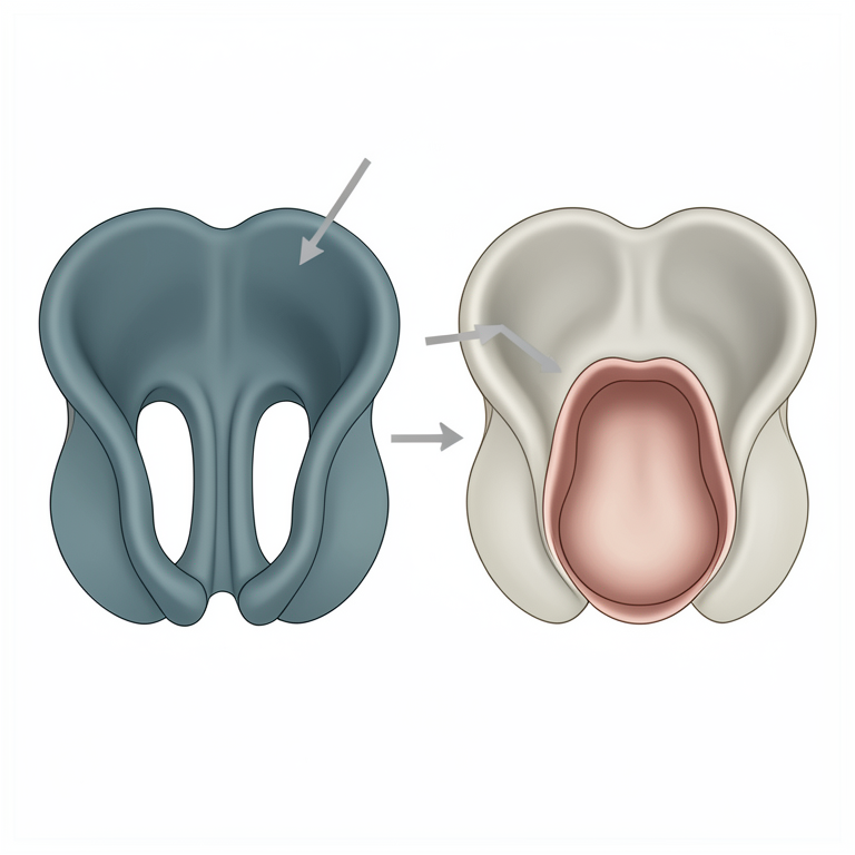
Mechanism of Perineal Hernia and Rectal Diverticulum
AI-generated conceptual illustration (canine pelvic diaphragm and rectum). It explains the idea and does not show study data.
View source evidence ↗Figures & tables
From the original paper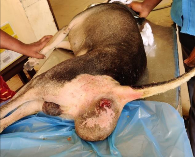 Figure 1
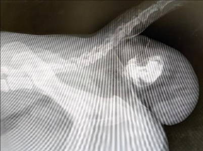 Figure 2
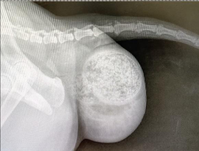 Figure 3
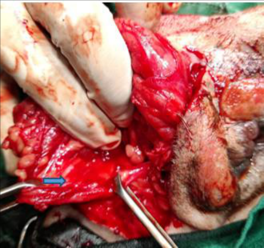 Figure 4
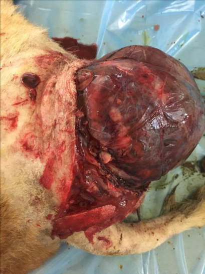 Figure 5
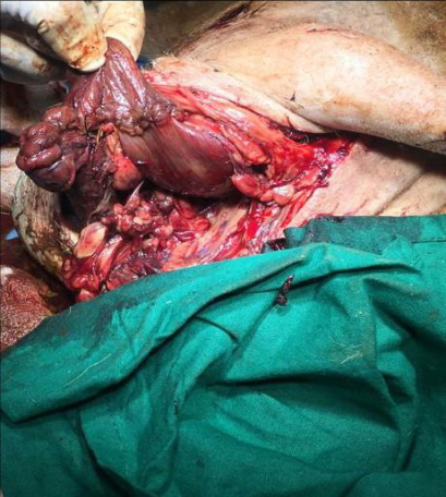 Figure 6
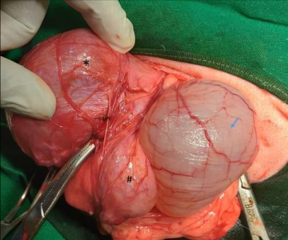 Figure 7
Introduction
Perineal hernia is common in olduncastrated male dogs. The weakness of the levatorani and lateral coccygeus muscles contribute to the formation of perineal hernia. The weakness of these muscles arises from increased tenesmus associated with gonadal hormonal imbalances, rectal abnormalities, prolonged constipation, chronic prostatic disease or myopathy. The weakness of the pelvic diaphragm in turn may cause loss of support to the rectal wall causing a rectal diverticulum. In male dogs, perineal herniorrhaphy and castration are suggested for preventing recurrence as hormonal imbalance is found to have a role (Ragni and Moore, 2011) [4] . The common hernial contents in perineal hernia in male dogs are urinary bladder, prostate, perineal fat and rectum (Gill and Barstad, 2018) [1] . The treatment may vary depending on the organ involved and the strength of the pelvic diaphragm. The present report discusses four cases of perineal hernia with rectal diverticulum alone and with concurrent conditions like cystocele, prostatic abscess and perianal fistula.
Materials and Methods
All the cases were prepared pre-anaesthetically, by food and water withdrawal a day before the surgery. Oral laxatives were given two days before the surgery. Before surgery, manual emptying of the rectum was performed. Case 1: Presented with swelling in the right side of the anal sphincter. There was history of obstipation and dyschezia. Rectal examination revealed that there was weakening and a pronounced pouching of the rectal mucosa on the right side. (Fig. 1). Laxatives diet and drugs didn't give any improvement in the condition. The dog was aggressive and needed weekly emptying of rectum under sedation. The faces was dry and hard. Radiography revealed rectal diverticulum and faecal accumulation. Positive contrast radiography using barium sulphate enaema revealed a solitary left rectal wall diverticulum (Fig. 2). Case 2: Presented with rectal diverticulum and perianal fistula.
The anal sphincter was malpositioned and the faeces passed through the perianal fistula. Per-rectal examination revealed weakening and moderate pouching of the rectal mucosa on the right side. Laxative diet and drugs were advised and the faces became semisolid. Plain radiography confirmed accumulation of faecal material in the swelling (Fig. 3
.).
Case 3: There was history of obstipation and anuria. The dog became recumbent a day before surgery. Per-rectal examination revealed that there was hard faeces in the rectum which was pressed from behind by a fluid filled organ ie., bladder which was confirmed by an ultrasonography. Ultrasonography revealed a hyperechoic mass and an anechoic bladder. Radiography showed fluid opacity cranial to the faecal accumulation. Case 4: The case was brought with a history of obstipation and anuria. Pre-rectal examination revealed hard faeces in the rectum. Ultrasonography revealed hyperechoic mass in the rectum, anechoic bladder and hypoechoic enlarged prostate gland.
Surgical Treatment
Case 1: Perineal incision followed by plication of the rectal diverticulum and suturing of the pelvic diaphragm using polyamide no: 1. Castration was performed. Case 2: Perineal incision followed by debriding and suturing the fistulous tract on the rectal wall followed by plication of the rectal diverticulum. The pelvic diaphragm was re-enforced by rotating a flap of the obturator internus muscle (Fig. 4.) which was sutured to the levatorani and coccygeus muscles. Polyamide no: 1 was used for the herniorrhaphy. Castration was performed. Case 3: Catheterization of the bladder could not be performed.
The perineal incision revealed a large thin walled rectal diverticulum (Fig. 5.). An incision was made on the distended rectum to removed impacted hard faecal ball. The rectal diverticulum was emptied. The distended bladder was seen cranial to the rectal diverticulum. The bladder was emptied by cystocentesis and replaced into the abdomen. The weak portion of the rectum was excised and sutured to the healthy portion using 4-0 PGA sutures in continuous pattern followed by a layer of interrupted catgut 3-0. was done. The pelvic muscles were atrophied and hence polyamide sutures were applied to the pelvic area in a mesh like manner so as to close the perineal ring. Castration was performed. Case 4: The perineal incision revealed rectal diverticulum, bladder and prostatic abscess (Fig. 7.). An incision was made on the distended rectum to removed impacted hard faecal ball. The rectal diverticulum was emptied. The incision was sutured using polyglycolic acid no: 0. Plication of the rectal diverticulum was performed. The distended bladder cranial to the rectal diverticulum was emptied by cystocentesis. The prostatic abscess was also aspirated and evacuated. The pelvic muscles were atrophied and hence polyamide sutures were applied to the pelvic area in a mesh like manner so as to close the perineal ring. Castration was performed. All cases were given antibiotics and analgesics postoperatively for five days and three days respectively. Laxatives (if needed) and dietary management were also advised for a month post-operatively to prevent the recurrence.
Results and Discussions
Skin suture dehiscence due to self-mutilation was the main complication noticed. All the animals were maintained on laxative diet. No recurrence was reported in three cases. In case 3, recurrence was reported and re-operated. Rectal diverticulum is often a chronic condition accompanied by perineal hernia and the clinical signs may include constipation, obstipation, dyschezia, tenesmus, rectal prolapse, stranguria, or anuria. The definitive diagnosis of perineal hernia is based on clinical signs and findings of weak pelvic diaphragm musculature during a per-rectal examination. In dogs, perineal hernias are mostly treated by surgical intervention. Appositional herniorrhaphy is sometimes difficult to perform as the levator ani and coccygeus muscles are atrophied and unsuitable for use. [3,2] .
Conclusion
It could be concluded that the management of perineal hernia and rectal diverticulum with concurrent conditions like perianal fistula, cystocele, and prostatic abscess in four dogs managed by plication of rectum/enterectomy and enteroanastamosis, perineal herniorrhaphy without or with transposition of obturator internus muscle is discussed. Castration was performed in all cases to prevent the recurrence. The temperament of the dogs, use of E-collar, dietary management, duration and extent of involvement of the rectum and the strength of the pelvic diaphragm or the repair technique and the appropriate management of the concurrent conditions were important factors that determined the outcome of the surgery.
Acknowledgement
AcknowledgementThe authors are thankful for the help and support extended by the Dean, VCRI, Tirunelveli, Director of Clinics, Chennai and Associate Professor and Head, VCC, Tirunelveli.
Figures and tables from the paper
References
6 in the original paper- Gill SS, Barstad RD. A review of the surgical management of perineal hernias in dogs. J Am. Anim. Hosp. Assoc. 2018;54:179-187.
- Guerios S, Orms K, Serrano MA. Autologous tunica vaginalis graft to repair perineal hernia in shelter dogs. Vet. Anim. Sci. 2020;9:100122.
- Lee AJ, Chung WH, Kim DH, Lee KP, Suh HJ, Do SH, et al. Use of canine small intestinal submucosa allograft for treating perineal hernias in two dogs. J Vet. Sci. 2012;13(3):327-330.
- Mehrjerdi HK, Mirshahi A, Afkhami A. Rectal diverticulum in a terrier dog: A case report. Vet. Res. Forum. 2013;4(1):63-67.
- Ragni RA, Moore AH. Perineal hernia. Comp. Animal. 2011;16:1-8.
- Sprada AG, Huppes, RR, Feranti JPS, De Souza SW, Coelho LD, Moraes P. Perineal hernia in dogs: which technique should we use?. Acta Sci. Vet. 2017;45:7.