
Research article · 2024 · Full text
International Journal of Advanced Biochemistry ResearchOccurrence of classical swine fever in an organized farm of Madurai, Tamil Nadu
Volume 8(2S) · Pages 344–347 · DOI: 10.33545/26174693.2024.v8.i2se.584 ↗
Research areasAnimal Disease Management and EpidemiologyAnimal Virus Infections StudiesViral Infections and Immunology Research
Abstract
Author-writtenDue to high mortality and reproductive issues, classical swine fever (CSF) is a highly transmissible viral illness that affects pigs and causes significant economic losses. In the current investigation, cross-breed pigs raised on managed farms may have experienced a swine flu outbreak. All of 220 pigs of various ages, thirty-three perished in a span of two to three days. It was then discovered that these animals had not had a CSF vaccination. Clinical manifestations of the illness were fever, huddling, conjunctivitis, acute respiratory distress, and purpura of the chest and abdomen. Reverse transcriptase polymerization chain reaction (RT-PCR) was used to employ the presence of the classical swine fever viral genes 5'UTR and E2 in blood samples taken from the suspicious animals. Blood samples from sick animals tested positive for both genes.
Summary
Source-checkedPlain-language reading of the full paper, prepared with AI and checked against the text. Each point links to the authors' own words.
Why it matters
- Classical swine fever (CSF) is a highly contagious viral disease in pigs that leads to significant economic losses due to reproductive problems and high mortality rates. View evidence ↗
- The disease is caused by the classical swine fever virus (CSFV), which belongs to the Pestivirus genus of the Flaviviridae family. View evidence ↗
- CSF is considered the primary endemic viral disease in most Indian states that practice pig husbandry. View evidence ↗
The question
- The study aims to document and detail the first reported case of a CSF epidemic among cross-breed pigs in the Madurai district of Tamil Nadu. View evidence ↗
What they did
- The investigation took place in June and July 2017 at an organized farm in Pottalputhur village, Madurai District, Tamil Nadu. View evidence ↗
- Whole blood samples were collected from suspected live pigs and stored at 4 °C in EDTA vacutainers. View evidence ↗
- RT-PCR was used to detect the presence of the 5'UTR and E2 viral genes of CSFV in the blood samples. View evidence ↗
What they found
- Affected pigs exhibited clinical signs including fever, huddling, conjunctivitis, acute respiratory distress, and purpura on the chest and abdomen. View evidence ↗
- The investigation revealed that the affected animals had not received a CSF vaccination. View evidence ↗
- RT-PCR analysis of blood samples from sick pigs yielded the expected 271 bp products for both the 5'UTR and E2 regions of the CSFV. View evidence ↗
- Every blood sample tested in the study tested positive for the classical swine fever virus. View evidence ↗
What it means
- The authors conclude that this is the first documented instance of a CSF outbreak in the Madurai district of Tamil Nadu, confirmed using RT-PCR in cross-breed pigs. View evidence ↗
Visual summary
1 visualsIllustrations explain the paper; they do not show study data.
Molecular Detection Process
AI-generated conceptual illustration (domestic pigs and blood samples). It explains the idea and does not show study data.
View source evidence ↗Figures & tables
From the original paper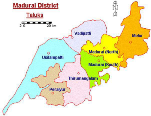 Figure 1
 Table 1
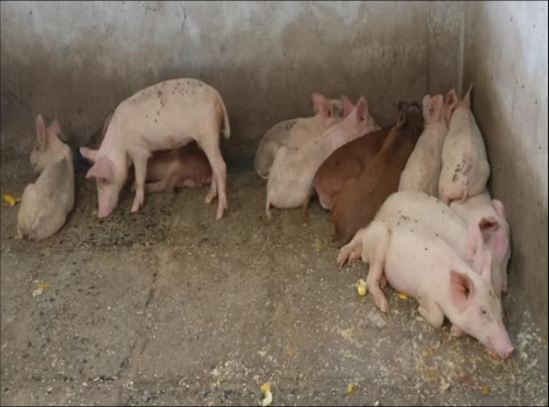 Figure 2
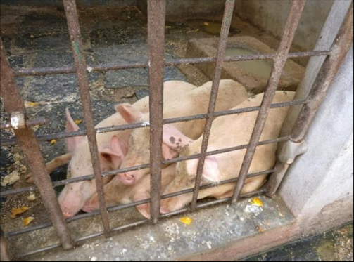 Figure 3
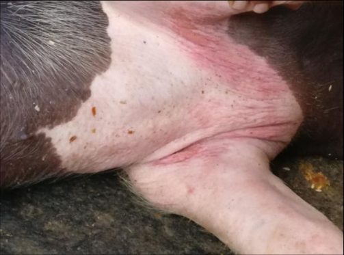 Figure 4
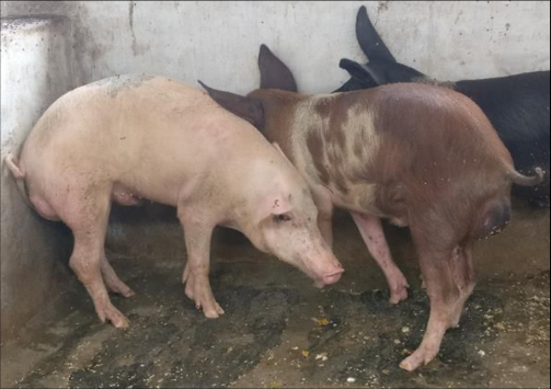 Figure 5
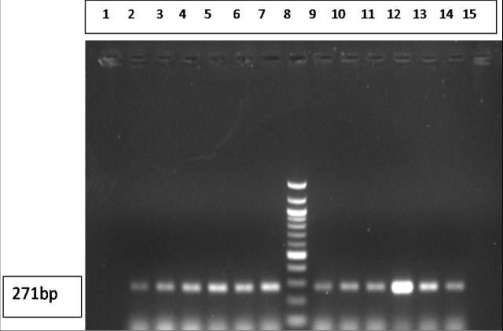 Figure 6
Introduction
Both domestic as well as wild pig populations are susceptible to the extremely contagious and frequently lethal disease known as classical swine fever (CSF). The causative agent responsible for CSF, the classical swine fever virus (CSFV), is a member of the Pestivirus genus within the Flaviviridae family (Wengler et al., 1995) [25] . Pigs can naturally contract both the bovine viral diarrhea virus (BVDV) of cattle and the border disease virus (BDV) of sheep, which are significant animal diseases belonging to the genus Pestivirus (Pringle, 1999) [14] . The World Organization for Animal Health (WOAH, formerly OIE) has listed CSF as a List A disease. The CSF virus is an enclosed virus with a single-stranded RNA genome that is positive-sense and roughly 12,300 nucleotides long. Two untranslated regions (UTRs) are located at the 5′ and 3′ ends of the single open reading frame that the genome encodes. CSF is a serious swine disease that has a major financial impact on the global swine industry. It was discovered that the vast majority of Indian states engaged in pig husbandry have the disease as their primary endemic viral disease (Thakur et al., 1998) [22] . The states of Uttar Pradesh, Maharashtra, Tamil Nadu, Punjab, Kerala, along with the North East Indian states of Arunachal Pradesh, Manipur, Mizoram, Nagaland, as well as West Bengal have all reported illness outbreaks (Ravisankar et al., 2007) [19] . This article discusses the initial case of CSF in the Tamil Nadu district of Madurai and details the CSF epidemic in cross-breed pigs.
Materials Methods Place of the study
The current work is reported from the Madurai district of Tamil Nadu which lies between 9° 56' 20.7348'' N and 78° 7' 18.1884'' E, Place where farm located Pottalpudhur which is very close to Madurai around 15-20 kms.
b). Molecular detection of CSFV by RT-PCR using 5'UTR and E2 gene specific primer
Following the manufacturer's instructions, total RNA was extracted from the blood samples using the GeneipureTM Total RNA isolation kit (BangaloreGenei, India). Complementary DNA (cDNA) was created using the Thermo Scientific Revert Aid H Minus First Strand cDNA Synthesis Kit (Thermo Scientific, USA) with random hexamer primers and M-MuLV reverse transcriptase. As indicated in Table .1, the extracted cDNA was next subjected to PCRs that targeted the CSFV E2 and 5′ UTR genes. 25 μl of reaction volume was used for PCR reactions, and it contained 1.5 units of Taq polymerase (Thermo Scientific), 200 μM of each dNTP, 10 pmol of each primer, 50 ng of each DNA sample, and 2.5 μl of 10x buffer. Amplification was done during the first denaturation stage, which took place at 95 °C for 2 minutes (1 cycle). This was followed by 34 cycles of denaturation, which included 30 seconds at 95 °C, 45 seconds at annealing temperatures, 1 minute of extension at 72 °C, and 1 minute of final extension at 72 °C.
For the 5′ UTR and E2 sections, the annealing temperature (Ta) had been set at 56 °C and 58 °C, respectively. 1%
agarose gel electrophoresis was used to analyze the PCR results.
Results and Discussion
CSF might manifest as an unusual, inapparent, chronic, subacute, acute, or peracute course. Van Oirschot (1999) [23] describes the peracute type as having a quick course without the usual clinical symptoms that point to CSF followed by an abrupt death. The disease known as acute and subacute CSF is associated with a high death rate and pathologic alterations that include many hemorrhages of different diameters (resulting from endothelial cell necrosis) combined with abnormalities in the blood's coagulation mechanism. According to Terpstra (1991) [21] , chronic CSF is a fatal illness that lasts for at least 30 days. The affected animals showed signs of depression during the experiment, including slumped posture, drooping heads (Fig. 2), and cuddling (Fig. 34) in both piglets and adult pigs. On the ventral side of the neck, erythematous lesions were seen (Fig. 5) [5,13] . Since RT-PCR detects the CSFV nucleic acid, it offers great sensitivity and increases the likelihood of getting positive results in circumstances when other assays yield negative results (OIE, 2014) [10] . According to reports, the test is also employed to identify the infectious agent preclinically (Depner et al., 2006; Prathviraj et al., 2022) [3,13] .
Relatively conserved genes are the target of RT-PCR, which is typically used to confirm the diagnosis of CSF. All species of the genus Pestivirus share a highly conserved 5´UTR nucleotide sequence. As a result, it is highly helpful for genotype or species characterization. Three glycoprotein Erns, E1 and E2, are present in the CSFV envelope (Patil et al., 2007; Prathviraj et al., 2022) [11,13] . The predominant glycoprotein, E2, is highly immunogenic as well as raises serum antibody titers that neutralize viruses (Postel et al., 2007) [12] . Reverse transcriptase (RT) PCR testing was therefore used in this investigation to diagnose simultaneously infected and preclinical cases of swine.
Expected 271 bp products of target of 5'UTRand E2 regions of the CSFV were obtained from EDTA blood samples of sick pigs undergoing RT-PCT (Greiser et al., 1998; Lowings et al., 1996) [4,8] . Previous reports from Mizoram and Tamil Nadu (Barman et al., 2010; Rathnapraba et al., 2014) [1,17] support these results. It was discovered that every blood sample tested positive for CSFV (Fig. 6). This proved that there was a direct correlation between the presence of the virus and the suspected samples. Moreover, recent findings of a similar nature from the Bidar district of Karnataka in south India have also been recorded in free-ranging pigs, indicating its virulence as well as incidence in the region (Prathviraj et al., 2022) [13] .
Acknowledgement
AcknowledgementAuthors are thankful to Vice chancellor, Registrar and Director of Centre of Animal Health Studies, TANUVAS, for providing necessary facilities to accomplish the research work
Figures and tables from the paper
References
24 in the original paper- Barman NN, Gupt RS, Bora DP, Kataria RS, Tiwari AK, Roychoudhury P. Molecular Characterization of Classical swine fever virus Involved in the Outbreak in Mizoram. Indian J Virol. 2010;21(1):76-81.
- Cho HS, Lee HI, Park NY. Method of discrimination between vaccine type and wild type viruses of classical swine fever. Indian Vet J. 2006;83:847-849.
- Depner K, Bunzenthal C, Heun Munch B, Streblow G, Hoffmann B, Beer M. Diagnostic evaluation of realtime RT-PCR assay for routine diagnosis of classical swine fever in wild boar. J Vet MedB Infect Dis Vet Public Health. 2006;53:317-20.
- Greiser-Wilke I, Depner K, Fritzemeier J, Haas L, Moennig V. Application of a computer program for genetic typing of classical swine fever virus isolates from Germany. J Virol Methods. 1998;75:141-150. doi: 10.1016/S0166-0934(98)00109-8.
- Handel K, Kehler H, Hills K, Pasick J. Comparison of reverse transcriptase-polymerase chain reaction, virus isolation, and immunoperoxidase assays for detecting pigs infected with low, moderate, and high virulent strains of classical swine fever virus. J Vet Diagn Invest. 2004;16:132-8.
- Horzinek MC. Pestiviruses-taxonomic perspectives. Arch Virol. 1991;3:1-5.
- Katz JB, Ridpath JF, Bolin SR. Presumptive diagnostic differentiation of hog cholera virus from bovine viral diarrhea and border disease viruses by using a cDNA nested-amplification approach. J Clin Microbiol. 1993;31:565-568.
- Lowings P, Ibata G, Needham J, Paton D. Classical swine fever virus diversity and evolution. J Gen Virol. 1996;77(6):1311-1321. doi: 10.1099/0022-1317-77-61311
- McGoldrick A, Lowings JP, Ibata G, Sands JJ, Belak S, Paton DJ. A novel approach to the detection of classical swine fever virus by RT-PCR with a fluorogenic probe (TaqMan). J Virol Methods. 1998;72:125-135.
- Office International des Epizooties. Manual of diagnostic tests and vaccines for terrestrial animals (mammals, birds and bees)., Classical swine fever (Hog Cholera). Paris: Office Internationaldes Epizootie; 2014. p. 244-52
- Patil SS, Hemadri D, Veeresh H, Sreekala K, Gajendragad MR, Prabhudas K. Phylogenetic analysis of NS5B gene of classical swine fever virus isolates indicates plausible Chinese origin of Indian subgroup 2.2 viruses. Virus Genes. 2012;44(1):104-108.
- Postel A, Schmeiser S, Bernau J, Boehmer AM, Pridotkas G, Dirbakova Z, et al. Improved strategy for phylogenetic analysis of classical swine fever virus based on full length E2 encoding sequences. Vet Res. 2012;43:50.
- Prathviraj, Kumar VSS, Ranganath GJ, Chandrashekara N, Raghunath BV, Ramesh D. Incidence of classical swine fever in local domestic pigs of Bidar district of Northern Karnataka. The Pharma Innovation Journal. 2022;SP-11(10):2154-2156.
- Pringle CR. The universal system of virus taxonomy, updated to include the new proposal ratified by the International Committee on Taxonomy of Virus during 1998. Arch Virol. 1999;144:421-429.
- Quinn PJ, Markey BK, Carter ME, Donnelly WJ, Leonard FC. Veterinary Microbiology and Microbial Diseases. Oxford, United Kingdom: Blackwell Science Limited; 2002:431.
- Rajkhowa TK, Hauhnar L, Jamlianthang. Studies on clinico-pathology and diagnosis of CSF in Zovawk pigs: an indigenous pig of Mizoram, India. Indian J Anim Sci. 2013;83(6):620-624.
- Rathnapraba S, Kumanan K, Vijayarani K, Palanisammi A, Gunaseelan L. Molecular typing of swine fever viruses from field outbreaks in Southern India. Indian J Anim Sci. 2014;84:723-727.
- Rathnapraba S, Kumanan K, Vijayarani K, Gunaseelan L, Saravanabava K, Raj DG. Molecular Diagnosis and Typing of Swine Fever Virus by NS5B Gene based RTPCR. Indian Vet J. 2013;90:19-21.
- Ravishankar C, Priya PM, Mini M, et al. First confirmed occurrence of classical swine fever in Kerala state, India. J Swine Health Prod. 2007;15(3):156-159.
- Singh VK, Sai Kumar G, Paliwal OP. Detection of classical swine fever virus in archival formalin-fixed tissues by reverse transcription polymerase chain reaction. Res Vet Sci. 2005;79:81-84.
- Terpstra C. Hog cholera: an update of present knowledge. Br Vet J. 1991;147:397-406.
- Thakur DK, Singh SK, Kumar V. Effect of an outbreak of swine fever on different economic traits in pig. Indian Vet J. 1998;75:185-6.
- Vandeputte J, Chappuis G. Classical swine fever: the European experience and a guide for infected areas. Rev Sci Tech. 1999;18:638-647.
- Wengler G, Bradley DW, Colett MS, Heinz FX, Schlesinger RW, Strauss JH. Flaviviridae. Arch Virol Suppl. 1995;10:415-427.
Cited by 1
From OpenAlex, OpenCitations and reference lists read by Scinovex
- Global prevalence of classical swine fever in domestic pigs: an integrative systematic review and meta-analysis ↗
Nagarajan Siddharthan, Sharanagouda S. Patil, Mouna Venkataramappa, Vijay Muruganantham, Mahadevaswamy Roopa, Kuralayanapalya Puttahonnappa Suresh et al. · Tropical Animal Health and Production · 2026
❝ 0