
Research article · 2024 · Full text
International Journal of Biology SciencesSerum kreb von den lungen-6 (KL-6) as a useful biomarker for evaluation of Covid-19 disease severity
Volume 6(1) · Pages 120–123 · DOI: 10.33545/26649926.2024.v6.i1b.197 ↗
Research areasInterstitial Lung Diseases and Idiopathic Pulmonary FibrosisPleural and Pulmonary DiseasesHematological disorders and diagnostics
Abstract
Author-writtenAim: The study aimed to investigate the role of KL-6 in severe and non severe Covid-19 patients. Method: In our study, 160 patients with Covid-19 were enrolled after being confirmed by RT-PCR. Estimation of KL-6, C-reactive protein (CRP) and lactate dehydrogenase (LDH) were performed, with comparing their levels in severe and non-severe cases. Study of the relationship between KL-6 and inflammatory marker was carried out by using the Spearman correlations. Results: The results showed an increase in CRP, LDH & Kl-6 levels in the sera of Covid-19 patients, and highly significant increases were observed in their levels in severe patients than in non-severe cases (p= 0.001, p= 0.000 & p= 0.000), respectively. Spearman’s correlation showed significant correlation between KL-6 & inflammation. Conclusion: It can be concluded that KL-6 seems to be a useful biomarker in the prognosis of Covid-19.
Summary
Source-checkedPlain-language reading of the full paper, prepared with AI and checked against the text. Each point links to the authors' own words.
Why it matters
- COVID-19 is caused by an enveloped RNA beta-coronavirus (SARS-CoV-2) that leads to a rapidly progressive disease. View evidence ↗
- The primary cause of death in COVID-19 is acute respiratory distress syndrome (ARDS), resulting from widespread damage to alveolar cells. View evidence ↗
- KL-6 (also known as MUC-1) is a high-molecular-weight mucin glycoprotein strongly expressed on the surfaces of respiratory epithelial cells, especially type-2 pneumocytes. View evidence ↗
- When the alveolar epithelium and alveolar-capillary membrane are damaged, KL-6 is released into the bloodstream, making it a potential indicator of lung injury. View evidence ↗
The question
- The study aimed to investigate the role of KL-6 in patients with severe versus non-severe COVID-19. View evidence ↗
What they did
- The study enrolled 160 COVID-19 patients between March and July 2022. View evidence ↗
- Patients were divided equally into severe and non-severe groups based on chest CT scans, respiratory rate, and peripheral oxygen saturation. View evidence ↗
- Serum levels of KL-6 were measured using ELISA kits, while C-Reactive Protein (CRP) and Lactate Dehydrogenase (LDH) were measured using Abbott Laboratories commercial kits. View evidence ↗
- Statistical analysis was performed using SPSS 18, employing independent t-tests for group comparisons and Spearman correlation to analyze relationships between markers. View evidence ↗
What they found
- Serum KL-6 levels were significantly higher in severe patients (1245.38±2706.098 U/ml) compared to non-severe patients (255.826±179.253 U/ml), with a p-value of 0.001. View evidence ↗
- CRP levels were significantly elevated in severe patients (11.111±4.987 mg/dl) compared to non-severe patients (2.94±2.497 mg/dl), with a p-value of 0.000. View evidence ↗
- LDH levels were significantly higher in severe patients (547.435±290.099 U/L) than in non-severe patients (246.13±63.385 U/L), with a p-value of 0.000. View evidence ↗
- While severe patients were older on average (70.84±8.159 years) than non-severe patients (50.28±13.094 years), this variation was reported as not significant (NS). View evidence ↗
- In the severe group, there was a strong significant correlation (0.76, p=0.000) between KL-6 concentration and CRP. View evidence ↗
- Spearman's correlation showed a strong significant relationship between KL-6 levels and inflammatory markers in both the severe and non-severe groups. View evidence ↗
What it means
- The authors conclude that KL-6 could be a helpful biomarker for determining COVID-19 severity and forecasting outcomes related to lung injuries. View evidence ↗
- The study concludes that KL-6 is a useful biomarker for the prognosis of COVID-19. View evidence ↗
Visual summary
3 visualsIllustrations explain the paper; charts are replotted from the paper's own table values.
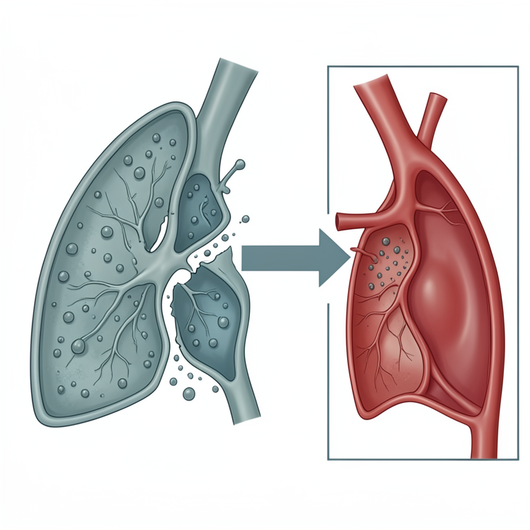
KL-6 Release Mechanism
AI-generated conceptual illustration (human lung alveolus and capillary blood vessel). It explains the idea and does not show study data.
View source evidence ↗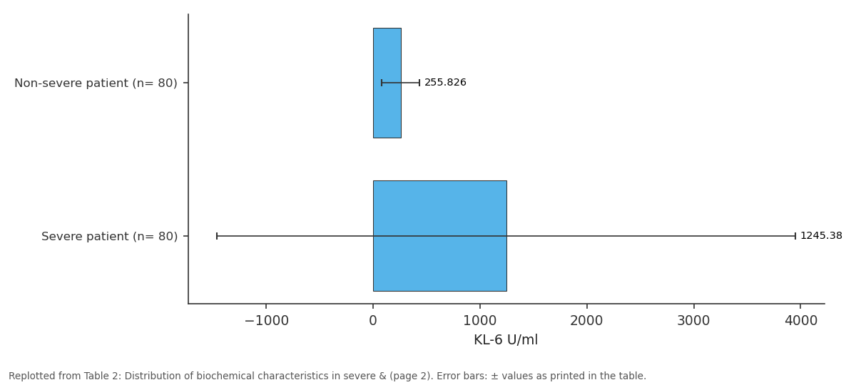
KL-6 U/ml: Non-severe patient (n=... vs Severe patient (n= 80)
Replotted from Table 2 (page 2) of the original paper. Every bar is the paper's own table value.
View source evidence ↗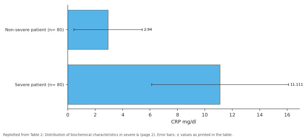
CRP mg/dl: Non-severe patient (n=... vs Severe patient (n= 80)
Replotted from Table 2 (page 2) of the original paper. Every bar is the paper's own table value.
View source evidence ↗Figures & tables
From the original paper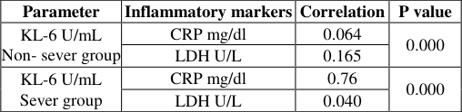 Table 3
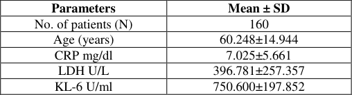 Table 1
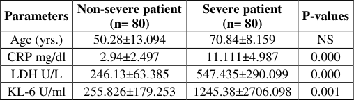 Table 2
Introduction
COVID-19 is an enveloped RNA beta-coronavirus that causes a rapidly progressive disease, and is also termed as (Severe Acute Respiratory Syndrome Coronavirus 2 or SARS CoV2), a member of Corona viride family, and the first outbreak of the infection occurred in the city of Wuhan in China, 2019 [1,2] . The World Health Organization (WHO) classified the disease to mild, moderate, severe & critical in accordance with the clinical symptom severity including; oxygen saturation levels (SpO2) and radiological findings [3,4,5] . The frequently observed COVID-19 symptoms include sore throats, fevers, dry coughs, malaise as well as shortness of breaths, and they range from mild to severe manifestation, e.g. pneumonia to the acute respiratory distresses syndromes (ARDSs), which is the major death cause in COVID-19 due to diffuse damage to the alveolar cells [6,7,8] . One specific indicator of lung alveolar cell damage currently used in studies and researches is Von Den Lungen Kreb or (KL-6) [9,10] . KL-6 is mucin glycoproteins with a highly molecular weight also referred to as Humans Trans membrane Mucin-1(MUC-1) which is expressed strongly on diverse epithelial cell type surfaces, particularly type-2 pneumocyte, respiratory bronchiolar epithelial cell and bronchial gland serus cell [11,12] . KL-6/MUC1 is released into the surrounding tissues and subsequently enters the bloodstream when the alveolar epithelium and alveolar-capillary membrane are destroyed, leading to increased levels of KL-6/MUC1 [13,14] . Many recent studies indicated that the serum concentrations of (KL-6) is raised in severe pulmonary involvement patient and was shown to be correlated with Covid-19 severity in various respiratory diseases especially in acute respiratory distress syndrome, Interstitial Lung Disease (ILD), hypersensitivity pneumonitis and Idiopathic Pulmonary Fibroses (IPF) [15,16] , which could be applied as new biomarkers for assessment of the severity of COVID-19 and prediction of lung injury prognosis [17,18] . Hence, our present study aims at determining KL-6 roles as a marker for pulmonary disease progressions and detecting COVID-19 prognostic values and severity.
Materials and Methods
In the present study, 160 COVID-19 patients were enrolled from March to July 2022. Positive results from the RT PCR (Reverse Transcriptase Polymerase Chain Reactions) testing supported COVID-19 diagnosis. In accordance with the results of a chest CT scanning, peripheral oxygen saturation, and respiratory rate, the disease was equally divided into severe and non-severe group). In severe cases, there were clinical signs of pneumonia with one of the following criteria established by WHO interim guidance for defining severe disease [19] : 1) Respiratory rates >30 breath/minute 2) Oxygen saturations <90% in room air and 3) Partial oxygen pressures in arterial blood ≤300 Patients with non-severe illness showed oxygen saturation levels below 94% with radiographic and clinical signs of lower respiratory tract infection [20] . Blood samples were drawn to measure levels of S. KL-6 using ELISA Kits (Eagle Biosciences, USA), while the inflammatory markers: C-Reactive Proteins (CRP), Lactate Dehydrogenases (LDH) have been estimated using commercial kits produced by Abbott Laboratories (Abbott, Architect, USA). SPSS 18 was used for statistical analysis, and the results were expressed as (Mean± SD). For comparing between severe and non-severe groups, the independent t-test was used. The Spearman correlation was applied to find out the relation between the markers in the study.
Statistical analysis
Statistical analysis
Results
In our current study, 160 COVID-19 patients were involved, and equally categorized into 2 group (severe & non-severe). The average age of COVID-19 patient was 65 years regarding age, a no significant variation was demonstrated between the study group. Table (1) showed the laboratory results for COVID-19 patient.
Discussion
The current study revealed that severely infected Covid19 cases were older than non-severely infected COVID-19 cases. In accordance with our results, [21][22][23][24] reported that age has fundamental roles in COVID-19 mortality and severity due to deterioration of the immune system, which is considered as an immense producer of pro-inflammatory cytokine (Cytokine storms) that leads to cell damage. Additionally, elderly people with heart disease, hypertension and lung illness have higher rates of COVID-19 mortality because these conditions are linked to highly Angiotensin Converting Enzyme2 or (ACE2) production, these enzymes utilized by COVID-19 viruses to adhere to the cell and produce inflammation [25] . Recent investigations revealed that individuals with severe COVID-19 have elevated inflammatory marker level related to endothelial dysfunctions, Cytokine Storm Syndrome (CSS) & coagulopathies [26][27][28][29] . The C-reactive proteins are highly sensitive biomarkers of tissue destruction, inflammations and disease course [30] . Recent studies have shown that high CRP production plays as an indicator of lung lesion and coronavirus severity through excessive cytokine and tissue destruction [31,32] . Nearly all types of human cells contain the intracellular enzyme Lactate Dehydrogenase (LDH), which is also found in large quantities in kidneys, lungs, muscles, cardiac and hepatic cells. Because large amounts of LDH are released into the circulation, LDH is considered an inflammatory indicator of tissue damage. Several evidence-based studies indicate highly significant increased LDH levels in severely infected patient with COVID-19 than in non-severely infected. Furthermore, high blood LDH has a relationship with COVID-19 severity & predicts higher death [33][34][35][36] . Statistical analysis found a significant elevation in CRP & LDH concentrations (p<0.05) in severely infected patients with COVID-19 than in non-severely infected patients. These findings agreed with [37][38][39][40][41] , who revealed a connection between poor COVID-19 outcomes and laboratory biomarkers of disease activity. Serum indicators serve as clinical evaluation tools to predict the level of inflammation and disease progress [42 ]. More recently, antigen of KL-6 was recognized as specific indicator in lung injuries and alveolar epithelial proliferations [43] . It is the mucin like glycoprotein expressed in membranes surfaces of alveolar type-II pneumocyte (AEC-II) with bronchiolar epithelium cell [44] . Increased levels of KL-6 reflect more extensive injury to the alveolar epithelium and type II pneumocyte regeneration [45] .
According to our study, patients with severe infection showed a highly significant increased S. KL-6 levels than those in non-severe patients. These findings were in line with previous studies which reported that there was a highly significant S. KL-6 levels in severe COVID-19 than those with moderate infections or healthy groups [9,[46][47][48][49][50][51] . We also found positive correlations between KL-6 & inflammatory markers (CRP and LDH) in agreement with [48,52,53] . As a result, to determine COVID-19 severity & forecast outcome in lung injuries in patients with COVID-19, the KL-6 could be a helpful biomarker. In conclusion, this study confirmed an increased levels of S. KL-6 in severely infected COVID-19 patient. It may contribute to diagnosing the severity for lung injury.
Conclusion
It can be concluded that KL-6 seems to be a useful biomarker in the prognosis of COVID-19.
Figures and tables from the paper
| Parameter | Inflammatory markers | Correlation | P value |
|---|---|---|---|
| KL-6 U/mL Non- sever group | CRP mg/dl | 0.064 | 0.000 |
| LDH U/L | 0.165 | ||
| KL-6 U/mL Sever group | CRP mg/dl | 0.76 | 0.000 |
| LDH U/L | 0.040 |
Table text extracted from the original PDF; superscript letters are shown inline. The original table image is below.
View original table image
| Parameters | Mean ± SD |
|---|---|
| No. of patients (N) | 160 |
| Age (years) | 60.248±14.944 |
| CRP mg/dl | 7.025±5.661 |
| LDH U/L | 396.781±257.357 |
| KL-6 U/ml | 750.600±197.852 |
Table text extracted from the original PDF; superscript letters are shown inline. The original table image is below.
View original table image
| Parameters | Non-severe patient (n= 80) | Severe patient (n= 80) | P-values |
|---|---|---|---|
| Age (yrs.) | 50.28±13.094 | 70.84±8.159 | NS |
| CRP mg/dl | 2.94±2.497 | 11.111±4.987 | 0.000 |
| LDH U/L | 246.13±63.385 | 547.435±290.099 | 0.000 |
| KL-6 U/ml | 255.826±179.253 | 1245.38±2706.098 | 0.001 |
Table text extracted from the original PDF; superscript letters are shown inline. The original table image is below.
View original table image
References
50 in the original paper- Zhou F, Yu T, Du R. Clinical course and risk factors for mortality of adult inpatients with COVID-19 in Wuhan, China: A retrospective cohort study. Lancet. 2020;395(10229):1054-1062.
- Zhu N, Zhang D, Wang W. A Novel Coronavirus from Patients with Pneumonia in China, 2019. The New England Journal of Medicine. 2020;382(8):727-733.
- World Health Organization. COVID-19 clinical management. Available: https://WHO/2019nCoV/clinical/2021.1 Accessed 30 January 2021.
- Schoeni RF, Wiemers EE, Seltzer JA, Langa KM. Association between risk factors for complications from COVID-19, perceived chances of infection and complications, and protective behavior in the US. JAMA Netw Open. 2021;4(3):e213984.
- Imran MM, Ahmed U, Usman U. Neutrophil/lymphocyte ratio-A marker of COVID-19 pneumonia severity. International Journal of Clinical Practice. 2021;75:e13698.
- D'Angelo A, Giacomin L, Murabito M. Caesarean Section in Preventing Stillbirths in Pregnancy Complicated with COVID-19: A Narrative Review. Clinical Therapeutics. 2021;172(6):570-576.
- Tabassum T, Rahman A, Araf Y, Ullah A, Hosen M. Prospective selected biomarkers in COVID-19 diagnosis and treatment. Biomarkers in Medicine. 2021;15(15):1435-1449.
- Lax SF, Skok K, Zechner P. Pulmonary arterial thrombosis in COVID-19 with fatal outcome: results from a prospective, single-center, clinicopathologic case series. Annals of Internal Medicine. 2020;173(5):350361.
- Michal M, Lukasz S, Zubaid R. Prediction Value of KREBS Von Den Lungen-6 (KL-6) Biomarker in COVID-19 Patients: A Systematic Review and MetaAnalysis. Journal of Clinical Medicine. 2022;11(21):6600.
- Frix AN, Schoneveld L, Ladang A. Could KL-6 levels in COVID-19 help to predict lung disease? Respiratory Research. 2020;21:309.
- Napolitano F, Di Spigna G, Vargas M. Soluble Urokinase Receptor as a Promising Marker for Early Prediction of Outcome in COVID-19 Hospitalized Patients. Journal of Clinical Medicine. 2021;10(21):4914.
- Milara J, Ballester B, Montero P. MUC1 intracellular bioactivation mediates lung fibrosis. Thorax. 2020;75(2):132-142.
- Ballester B, Milara J, Cortijo J. The role of mucin 1 in respiratory diseases. The European Respiratory Review. 2021;30(159):200149.
- D′Alessandro M, Bergantini L, Cameli P. Krebs von den Lungen-6 as a biomarker for disease severity assessment in interstitial lung disease: A comprehensive review. Biomarkers in Medicine. 2020;14(8):665-674.
- Xue M, Zhang T, Chen H. Krebs Von den Lungen-6 as a predictive indicator for the risk of secondary pulmonary fibrosis and its reversibility in COVID19 patients. International Journal of Biological Sciences. 2021;17(6):1565-1572.
- Arnold D, Donald C, Lyon M, Hamilton F, Morley A, Attwood M, et al. Krebs vonden Lungen 6 (KL-6) as a marker for disease severity and persistent radiological abnormalities following COVID-19 infection at 12 weeks. PLoS ONE. 2021;16(4):e0249607.
- Ghati A, Dam P, Tasdemir D. Exogenous pulmonary surfactant: A review focused on adjunctive therapy for severe acute respiratory syndrome coronavirus 2 including SP-A and SP-D as added clinical markers. Current Opinion in Colloid & Interface Science. 2021;51:101413.
- World Health Organization. WHO COVID-19 preparedness and response progress report February to June 30, 2020. Geneva, WHO; c2020, p. 5-6.
- Wu Z, McGoogan JM. Characteristics of and Important Lessons from the Coronavirus Disease 2019 (COVID19) Outbreak in China: Summary of a Report of 72314 Cases from the Chinese Center for Disease Control and Prevention. JAMA. 2020;323(13):1239-1242.
- Farghaly S, Makboul M. Correlation between age, sex, and severity of Coronavirus disease-19 based on chest computed tomography severity scoring system. Egyptian Journal of Radiology and Nuclear Medicine. 2021;52(1):23.
- Liao D, Zhou F, Luo L. Haematological characteristics and risk factors in the classification and prognosis evaluation of COVID-19: A retrospective cohort study. The Lancet Haematology. 2020;7(9):e671-e678.
- Ragaey A, Abdelrahman M. Attia B, Mahmoud Hassan C, Marwa Abdallah Shaker A, Manar Ahmed Kamal D et al. Demographic, clinical, and laboratory characteristics of patients with COVID-19 during the second and third waves of the pandemic in Egypt. Journal of Infection and Public Health. 2021;14(10):1358-1366.
- Crimmins EM. Age-Related Vulnerability to Coronavirus Disease 2019 (COVID-19): Biological, Contextual, and Policy-Related Factors. Public Policy & Aging Report. 2020;30(4):142-146.
- Kermali M, Khalsa RK, Pillai K, Ismail Z, Harky AM. The role of biomarkers in the diagnosis of COVID-19-A systematic review. Life Sciences. 2020;254:117788.
- Fernando C, Javier M, José GC, Jorge B, Ricardo F, Yaimara H-S, et al., Management of COVID-19 based on Risk Features. European Journal of Respiratory Medicine. 2022;4(1):276-284.
- Roshan K, Suchismita P, Vedika R, Sharmistha S, Lalendra Y, Sumesh P et al. The dynamics of inflammatory markers in coronavirus disease-2019 (COVID-19) patients: A systematic review and metaanalysis. Clinical Epidemiology. 2021;11:100727.
- Dogan A, Mustafa G, Dervis T, Orhan A. Diagnostic and early prognostic value of serum CRP and LDH levels in patients with possible COVID-19 at the first admission. The Journal of Infection in Developing Countries. 2021;15(6):766-772.
- Chaudhary R, Garg J, Houghton DE, Murad MH, Kondur A, Chaudhary R, et al. Thromboinflammatory biomarkers in COVID-19: systematic review and metaanalysis of 17,052 patients. Mayo Clinic Proceedings: Innovations, Quality & Outcomes. 2021 Apr;5(2):388402.
- Azar MM, Shin JJ, Kang I, Landry M. Diagnosis of SARS-CoV-2 Infection in the Setting of the Cytokine Release Syndrome. Expert Review of Molecular Diagnostics. 2020;20(11):1087-1097.
- Priyanka A1, Sen D, Wilma Delphine Silvia CR. Inflammatory Profile in Novel Coronavirus Infection: Biochemical Perspective. RJMS. 2021;11(2):75-84.
- Januar W, Arief W, Raymond P. Prognostic value of elevated lactate dehydrogenase in patients with COVID19: A systematic review and meta-analysis. Postgraduate Medical Journal. 2022;98(1160):422-427.
- Bartosz F, Michal P, Jacek S, Rafal J, Mansur RH, Andrea D, et al. Diagnostic value of lactate dehydrogenase in COVID-19: A systematic review and meta-analysis. Cardiology Journal. 2022;29(5):751-758.
- Powanda MC, Moyer ED. A brief, highly selective history of acute phase proteins as indicators of infection, inflammation, and injury. Inflammopharmacology. 2021;29(3):897-901.
- Mei-Ying W, Lin Y, Yi W. Clinical evaluation of potential usefulness of serum Lactate Dehydrogenase (LDH) in 2019 novel coronavirus (COVID-19) pneumonia. Wu. Respiratory Research. 2020;21:171.
- Nayana D, Souparnika S, Mamatha B. Assessment of inflammatory markers and their association with disease mortality in severe COVID-19 patients of a tertiary care hospital in South India. The Egyptian Journal of Bronchology. 2022;16(1):55.
- Rasha M, Hanan Y, Ad'hiah AH. Severity of Coronavirus Disease 19: A Profile of Inflammatory Markers in Iraqi Patients. The Malaysian Journal of Medicine and Health Sciences. 2021;18(1):91-98.
- Sayit AT, Elmali M, Deveci A, Gedikli O. Relationship between acute phase reactants and prognosis in patients with or without COVID-19 pneumonia. Revista do Instituto de Medicina Tropical de São Paulo. 2021;63:e51.
- Semiha O, Bilge B, Tasdemir M, Elif DK, Sinan K, Petek ŞK, et al. The Effect of Acute Phase Reactants on the Survival of COVID-19 Patients in Intensive Care. Progress in Nutrition. 2021;23(S-2):e2021268.
- Ponti G, Maccaferri M, Ruini C. Biomarkers associated with COVID-19 disease progression. Critical Reviews in Clinical Laboratory Sciences. 2020;57(6):389-399.
- Park M, Hur M, Kim H, Lee CH, Lee JH, Nam M et al. Usefulness of KL-6 for Predicting Clinical Outcomes in Hospitalized COVID-19 Patients. Medicina. 2022;58(10):1317.
- Vianello A, Guarnieri G, Braccioni F, Lococo S, Molena B, Cecchetto A, et al. The Pathogenesis, Epidemiology and Biomarkers of Susceptibility of Pulmonary Fibrosis in COVID-19 Survivors. Clinical Chemistry and Laboratory Medicine. 2022;60(3):307-316.
- Asma M, Irina B, Shikha Y, Wanessa F, Amrin K, Mrunanjali G, et al. Post-COVID-19 Pulmonary Fibrosis. Cureus. 2022;14(3):e22770.
- Emanuela A, Lucia M, Elisa, Simone C, Miriam D, Alessandra S, et al. Association of serum Krebs von den Lungen 6 and chest CT as potential prognostic factors in severe acute respiratory syndrome SARS Co V2: A preliminary experience. La Radiologic Medical. 2022;127(7):725-732.
- Nadereh N, Mahsa R. Krebs Von Den Lungen-6 (KL-6) as a clinical marker for severe COVID-19: A systematic review and meta-analyses. Virology. 2022;566:106-113.
- Hazan K, Aslıhan A, Pınar HS, Gonca E, Gülendam B, et al. The Prognostic Value of Lung Injury and Fibrosis Markers, KL-6, TGF-β1, FGF-2 in COVID-19 Patients. Biomarker Insights. 2022;17:1-7.
- Yamaya T, Hagiwara E, Baba T. Serum Krebs von den Lungen-6 levels are associated with mortality and severity in patients with coronavirus disease 2019. Respiratory Investigation. 2021;59(5):596-601.
- Tao Z, Ping Sh, Chunyan D, Lingyun G. KL-6 as an Immunological Biomarker Predicts the Severity, Progression, Acute Exacerbation, and Poor Outcomes of Interstitial Lung Disease: A Systematic Review and Meta-Analysis. Frontiers in Immunology. 2021;12:745233.
- Ding-Hui P, Luo Y, Li-Jun Y, Fan-Lu L, Yan-Yuan L, Peng T, et al. Correlation of Krebs von den Lungen-6 and fibronectin with pulmonary fibrosis in coronavirus disease 2019. Clinica Chimica Acta. 2021;517:48-53.
- Sei W, Heayon L, Sang H, Sung Jin J, Jehoon L, Jihyang L et al. Usefulness of monocyte distribution width and presepsin for early assessment of disease severity in COVID-19 patients. Medicine. 2022;101(27):e29592.
- Henry BM, Aggarwal G, Wong J, Benoit S, Vikse J, Plebani M, et al. Lactate dehydrogenase levels predict coronavirus disease 2019 (COVID-19) severity and mortality: A pooled analysis. The American Journal of Emergency Medicine. 2020;38(9):1722-1726.