
Research article · 2024 · Full text
International Journal of Agriculture and Food SciencePreliminary major health problems and associated risk factors of calves’ mortality in selected districts of Ilubabor and Jimma Zones, Ethiopia
Volume 6(1) · Pages 31–39 · DOI: 10.33545/2664844x.2024.v6.i1a.163 ↗
Research areasRangeland Management and Livestock Ecology
Abstract
Author-writtenIntroduction: Even though good beginnings of initiatives on calves to improve blood level of local breeds in Ethiopia through artificial insemination, health problems and calf mortality are becoming a bottle neck on livestock production and livelihood of poor farmers. The aim of the study was to know preliminary major health problems and associated risk factors of calves’ mortality. Cross-sectional study was conducted on calves from February to June 2023. Binary outcome Logistic Regression data analysis was used to summarize using Stata software version 13. Cluster random sampling technique was used to accomplish the study. Result: A total of 218 farmers and farm owners were interviewed and feacal samples, ectoparasites and skin lesions were collected from 389 calves. The gastrointestinal parasite infection was significantly related with body condition, age and breeds of calves (p
Summary
Source-checkedPlain-language reading of the full paper, prepared with AI and checked against the text. Each point links to the authors' own words.
Why it matters
- Calf mortality and health issues are significant obstacles to livestock production and the livelihoods of poor farmers in Ethiopia. View evidence ↗
- Gastrointestinal parasites are identified as the primary cause of disease in calves. View evidence ↗
- Ectoparasites cause substantial economic losses in Ethiopia by increasing animal mortality, reducing production, and damaging skins and hides. View evidence ↗
The question
- The study aimed to identify the primary health problems and the risk factors associated with calf mortality. View evidence ↗
What they did
- A cross-sectional study was performed between February and June 2023 to evaluate calf morbidity, mortality, and associated risk factors. View evidence ↗
- Data were analyzed using Binary outcome Logistic Regression via Stata software version 13. View evidence ↗
What they found
- The overall prevalence of gastrointestinal parasites was 38% (148 out of 389 calves). View evidence ↗
- Ectoparasite infestation was found in 71.98% (280 out of 389) of the observed calves. View evidence ↗
- Clinical ringworm lesions were present in 4.10% (16 out of 389) of the calves. View evidence ↗
- The overall morbidity rate was 34.90% (196/561) and the crude mortality rate was 12.30% (69/561). View evidence ↗
- Calves weaned before six months were 2.67 times more likely to experience morbidity and mortality. View evidence ↗
- Hand-feeding colostrum increased the odds of calf morbidity and mortality by 2.04 times. View evidence ↗
- Cross-breed calves had a 2.02 times higher risk of morbidity and mortality compared to local breeds. View evidence ↗
What it means
- The authors conclude that gastrointestinal parasites, ectoparasites, and ringworms are the primary health problems driving high morbidity and mortality in the study areas. View evidence ↗
- The authors recommend increasing awareness regarding disease prevention and improved husbandry practices. View evidence ↗
- The authors state that further detailed studies are needed to understand the seasonal occurrence, specific species, and economic burden of ectoparasites in the region. View evidence ↗
Visual summary
3 visualsIllustrations explain the paper; charts are replotted from the paper's own table values.
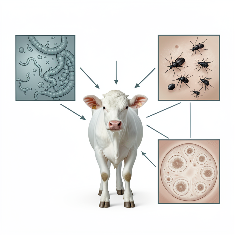
Major Health Threats to Calves
AI-generated conceptual illustration (calf (Bos taurus)). It explains the idea and does not show study data.
View source evidence ↗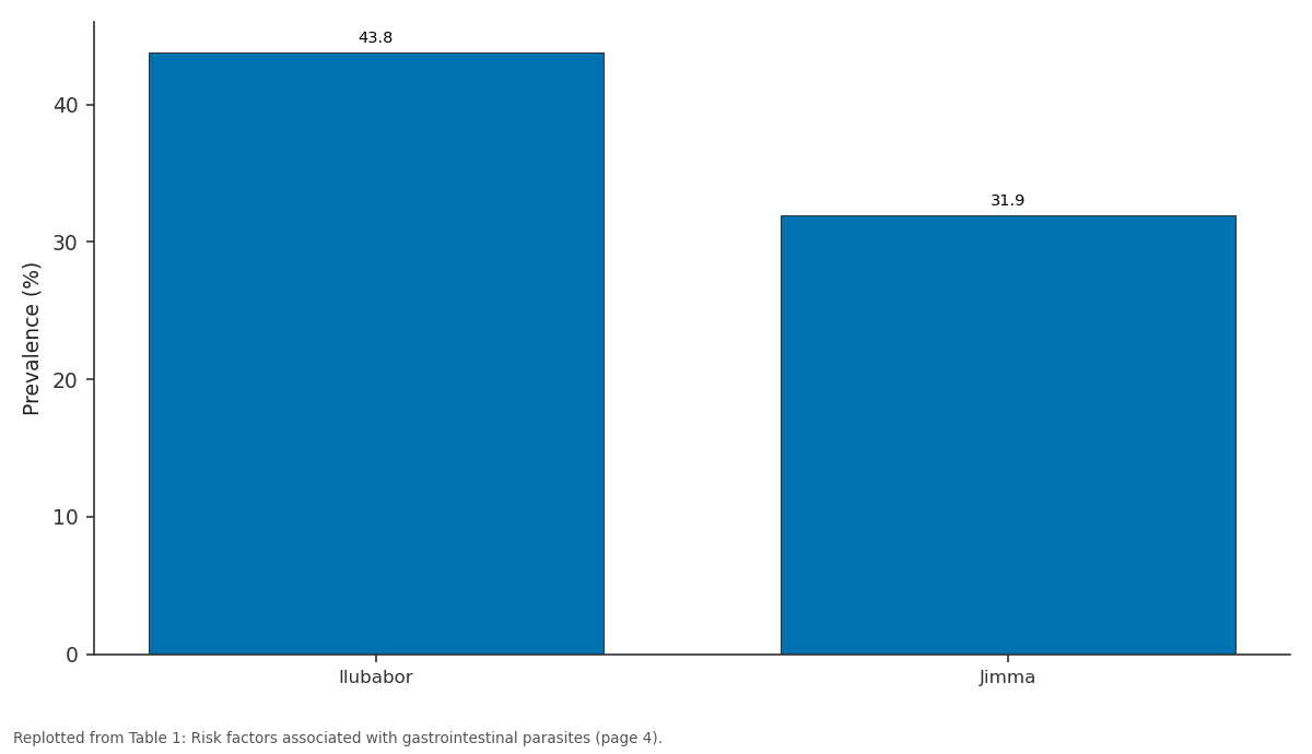
Prevalence (%): Ilubabor vs Jimma
Replotted from Table 1 (page 4) of the original paper. Every bar is the paper's own table value.
View source evidence ↗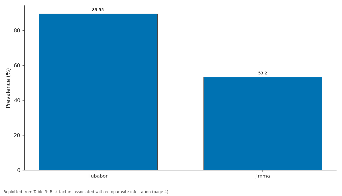
Prevalence: Ilubabor vs Jimma
Replotted from Table 3 (page 4) of the original paper. Every bar is the paper's own table value.
View source evidence ↗Figures & tables
From the original paper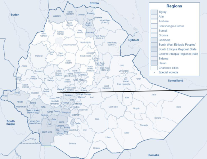 Figure 1
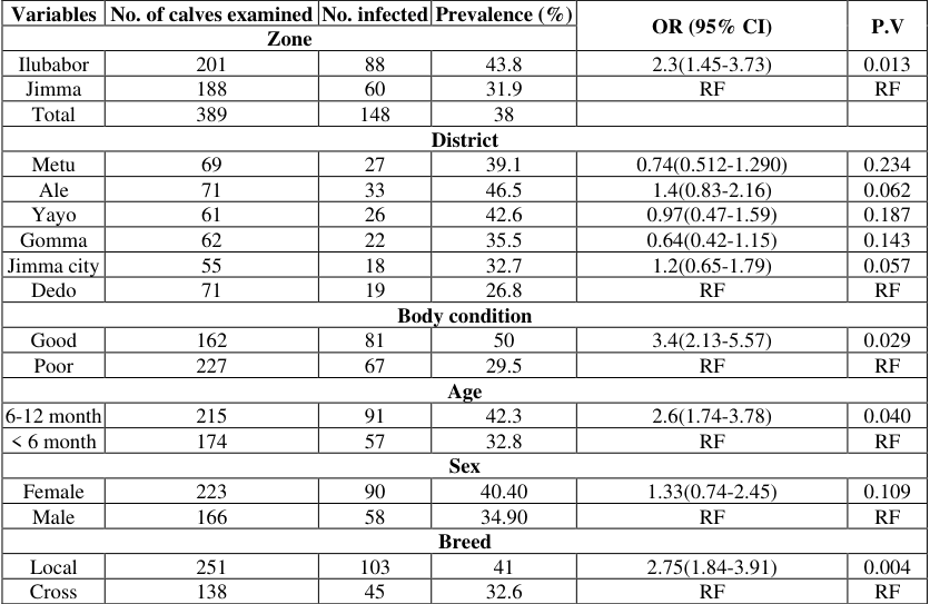 Table 1
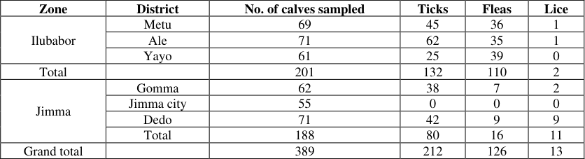 Table 2
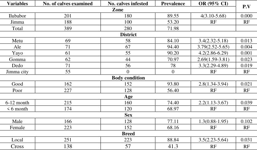 Table 3
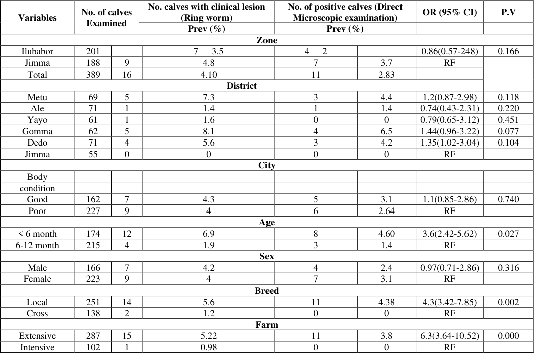 Table 4
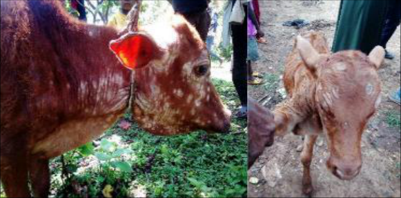 Figure 2
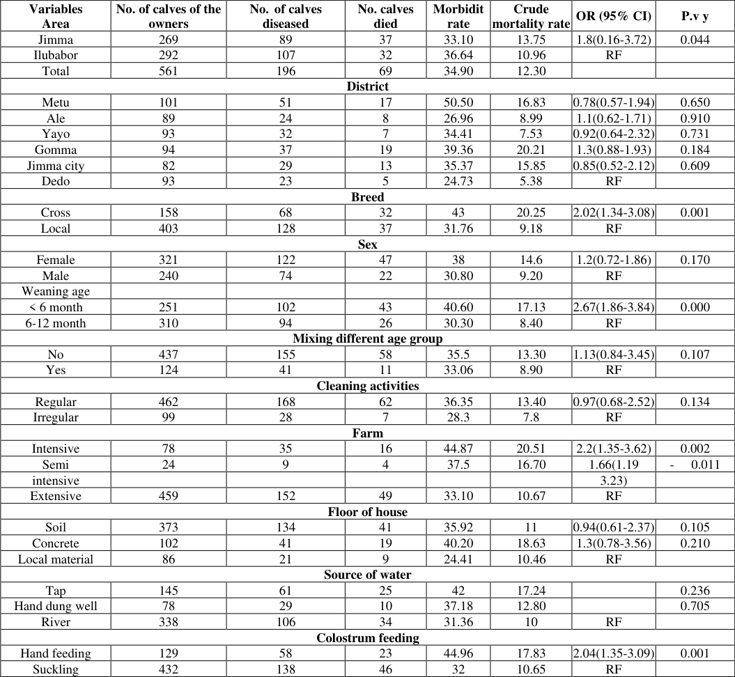 Table 5
Introduction
Ethiopia is first by livestock population from Africa, with an estimated 70.3 million cattle, 9.12% of which are calves under the age of six months [1] . Dairy and beef farming in urban and peri-urban regions is growing swiftly across the country as a result of rising urbanization and increased demand for meat, milk and milk products, even though the extensive production system is still the country's leading method of livestock husbandry [2,3] . Urban and peri-urban dairies are semi-intensive to intensive production systems that maintain exotic and cross-bred cows with comparatively better management practices [4] . Although the dairy industry has grown significantly in recent years, it is reportedly suffer from inefficient reproduction, a low rate of calf survival, high calf morbidity and mortality, and a high incidence of diseases [5] . Inadequate quality feed, poor husbandry practices, and reduced attention to young stock management as a result of diverse responsibilities in small-scale mixed farming systems increase calf morbidity and mortality in tropical regions [6] . Parasitic infection of cattle is major factors responsible for economic losses through reduction in productivity and increased mortality in heavily parasitized animals [7] . Gastrointestinal parasites are the most important agents causing disease in calves [8] . Calves are most vulnerable to gastrointestinal parasites in their first grazing season, although yearlings and, less often, adults are sometimes affected [9] . Gastrointestinal parasitic infections, nematode and Eimeria species, play a key role in the economic losses in that they cause low productivity, delayed growth, declined weight gain and death of the animal, and significant expenses of treatment [8,24] .
Ectoparasites; commonly ticks, mites, flee and lice affect the host by inflammation and through the damage they inflict on the skin and on the host physiology (Thaware et al, 2023) [37] . They are also very important in different disease transmissions such as parasitic, bacterial, rickettsial, and viral diseases to man and animals [10] . The damages inflicted by ectoparasites are annoyance, stress, or blood loss. Lice and tick worry are recognized conditions that reduce feed efficiency and weight gains in livestock [10] . In Ethiopia, ectoparasites in ruminant causes a serious economic loss to small holder farmers, the tanning industry and the country as a whole through mortality of the animals, decreased production, down grading and rejection of skin and hide [11] . Dermatophytosis in domestic animals is an infection of keratinized tissues by one of the two genera fungi, Microsporum and Trichophyton [12] . Although information is lacking in Ethiopia, it is believed to pose the greatest economic and human health consequence in most developed countries [13] . Tartor [14] described that 84.91% calves showed ringworm lesions were positive for fungal elements in direct-microscopy and 79.72% were positive in culture. The detection rates were 84.91% by direct microscopy and 79.72% by fungal culture. As researchers described terbinafine and miconazole were effective antifungal drugs for dermatophytes followed by itraconazole and griseofulvin [15] .
Calf morbidity and mortality are the two most important constraints for improving peri-urban and urban dairy production in Ethiopia. Annual calf mortality in urban and peri-urban dairy production systems is reported to be in the range of 15.3% -25% [16] . Similarly, 62% morbidity and 22% mortality are reported in market-oriented smallholder dairy farms of central Ethiopia [17] . However, in the current study areas there has been no any information on calves' health problems. Therefore the aim of this study was to provide the following objectives. 1. To identify preliminary major health problems of calves 2. To know the magnitude of calf morbidity and mortality 3. To identify associated risk factors of calves' morbidity and mortality
Materials and Methods Description of study area
This study was conducted in purposively selected districts of Ilubabor and Jimma zones (Figure 1). Ilubabor Zone is located in south-western part of Oromia, Ethiopia. In Ilubabor zone the study was done in three purposively selected districts of Metu, Ale and Yayo with cattle population of 146635, 70553 and 63769 respectively. In Jimma zone the study was done in two purposively selected Jimma city, Dedo and Gomma districts with cattle population of 67808, 435281 and 271332 respectively.
Study population
The study population consisted of local and cross breeds of calves. The target population was calves up to 12 months of age in purposively selected districts and Jimma city. The study population was all calves of 12 months in randomly selected kebele and households. Calves were categorized by: weaning age (<6 months, 6-12 months), sex (Male, Female), body condition (good, poor), breed (cross, local), mixing different age group (yes, no), cleaning activity (regular, irregular), farm type (extensive, intensive, semi intensive), floor of house (soil, concrete, made from local tree), source of drinking water (tap, hand dung well, river) and colostrum feeding (suckling, hand feeding). Semi-structured questionnaire survey of selected household owner was used to assess factors associated with calf morbidity and mortality.
Study design and sampling method
A cross-sectional study was conducted from February, 2023 to June, 2023 to know the status of calf morbidity and mortality and also to assess risk factors as well. All calves of farmers or in farm of one year age was observed for any health problem and feacal samples and skin scraping for skin lesions were collected and submitted to regional laboratory to know prevalence and major gastrointestinal parasites of calves. Also all calves of owners were assessed for ectoparasites and submitted to regional laboratory to identify the genera of those ectoparasites.
Sample size determination and sampling technique
The sample size was determined according to [18] for cluster random sampling using an expected animal level prevalence of 50% and a desired absolute precision of 5% with 95% CI, since there was no previously expected prevalence in the study area.
Where, N = sample size, T = linked to 95% confidence interval for cluster sampling (2.045), P = expected prevalence (Fraction of 1), Q = 1-p (expected nonprevalence), D = relative desired precision, DEFF = Design Effect =1. Accordingly, 389 local and cross breeds of calves were sampled and assessed for ecto parasites and scraped if any skin lesion found. First districts were purposively selected based on calves morbidity and mortality problem reports. In Metu, Ale, Yayo, Gomma, Jimma city and Dedo districts kebele and households or farms were randomly selected. At the 2 nd stage household of each kebele was randomly selected. Finally, all caves of one year age were examined for ectoparasite and fecal sample was collected except those did not start feeding grass.
Sample collection and transportation Sedimentation method
A total of 389 fresh fecal samples were collected from the rectum of the animals by wearing plastic hand gloves. The fecal samples were placed in a screw capped universal bottles, preserved with 10% of formalin and transported to Bedelle Regional Veterinary Laboratory. The fecal samples were examined by using standard sedimentation techniques.
The presence of at least one parasite egg in either of the tests indicates that the result is positive. The egg morphology, appearance, size, color and presence of blastomeres were used to identify the parasites.
Tick collection and preservation
Firstly, the selected calves were properly restrained and checked for any tick infestation. Ticks were removed from different body regions of the host skin for identification using hand manually [19] . The collected ticks were preserved in separate pre-filled universal bottles with 70% alcohol before transportation to Bedelle regional veterinary laboratory for identification their genera. The collected ticks were identified in to different genera level by using stereomicroscope, according to standard identification keys given by Walker [38] . Accordingly the most prevalent tick genera was Boophilus, which followed by Amblyomma.
Lice collection and identification
Those detected lice that are unidentified during clinical examination was collected by forceps/hand picking, with hairs from their attachment site, put into a clean separate container (universal bottles), labeled and kept preserved with 70% ethyl alcohol before transportation to Asella regional veterinary laboratory for detailed laboratory examination as described by Urquhart [21] . Then the collected ectoparasites were examined by stereomicroscope and identification was performed according to the identification key given by Wall and Shearer [22] . Linognathus vituli and Damalina bovis were among the major lice species identified in different studies.
Skin scraping lesions
The samples of skin scraping were collected from the periphery of the lesion after cleaning with 70% ethyl alcohol in sterile falcons. Samples were collected from infected animal's suffering from lesions suggesting ring worm infections (Circumscribed areas of hair loss filled with raised white scales on head, neck or all over the body). One or two drops of 20% KOH (potassium hydroxide) were placed on a microscopic slide and a small amount of the specimen was added and then, the slide was gently passed through a low flame and covered by a cover slip. After 2 h, the specimen was examined for the presence of hyphae under a light microscope (40 x objectives) according to [23] .
Data management and analysis
All data obtained from the field was recorded in the record sheet format and later entered into Microsoft Excel worksheet and Binary outcome Logistic Regression data analysis was used to summarize using Stata software version 13. The overall prevalence was calculated by dividing positive samples by the total number of examined samples and multiplied by a hundred. The Odds Ratio was used to assess the association between the dependent and independent variables. P-value of less than 0.05 (p<0.05) was set for the significance of statistical associations [39] .
Results
During the study period 164 small holder farmers, 31 semi intensive dairy farm owners and 23 intensive dairy farm owners with total 561 calves up to 12 months age they owned were interviewed. A total of 389 caves were observed for any ectoparasite infestation, feacal samples and skin lesion samples were collected from up to one year. 1). However, there was no significant association among districts and sex (p>0.05), (Table 1). 3). However, there was no significant association between sex (p>0.05), (Table 3). During surveillance two ticks genera were identified namely Boophilus and Amblyomma. Boophilus was the most tick genera that infested calves. Biting lice (Damalinia bovis) and sucking lice (Linognathus vituli) were identified during surveillance.
| Variables | No. of calves examined | No. calves infested | Prevalence | OR (95% CI) | P.V |
|---|---|---|---|---|---|
| Zone | |||||
| Ilubabor | 201 | 180 | 89.55 | 4(3.10-5.68) | 0.000 |
| Jimma | 188 | 100 | 53.20 | RF | RF |
| Total | 389 | 280 | 71.98 | ||
| District | |||||
| Metu | 69 | 58 | 84.10 | 3.4(2.32-5.18) | 0.013 |
| Ale | 71 | 67 | 94.40 | 3.79(2.52-5.65) | 0.004 |
| Yayo | 61 | 55 | 90.20 | 4.2(2.86-6.29) | 0.001 |
| Gomma | 62 | 44 | 70.97 | 2.69(1.59-3.81) | 0.023 |
| Dedo | 71 | 56 | 78 | 3.3(2.29-4.89) | 0.019 |
| Jimma city | 55 | 0 | 0 | RF | RF |
| Body condition | |||||
| Good | 162 | 152 | 93.80 | 2.8(1.34-3.94) | 0.021 |
| Poor | 227 | 128 | 56.40 | RF | RF |
| Age | |||||
| 6-12 month | 215 | 160 | 74.40 | 2.2(1.13-3.67) | 0.039 |
| < 6 month | 174 | 120 | 68.97 | RF | RF |
| Sex | |||||
| Male | 166 | 128 | 77.11 | 1.3(0.88-1.95) | 0.102 |
| Female | 223 | 152 | 68.16 | RF | RF |
| Breed | |||||
| Local | 251 | 223 | 88.84 | 3.5(2.23-5.64) | 0.031 |
| Cross | 138 | 57 | 41.3 | RF | RF |
Table text extracted from the original PDF; superscript letters are shown inline. The original table image is below.
View original table image
| Variables | No. of calves examined | No. infected | Prevalence (%) | OR (95% CI) | P.V |
|---|---|---|---|---|---|
| Zone | |||||
| Ilubabor | 201 | 88 | 43.8 | 2.3(1.45-3.73) | 0.013 |
| Jimma | 188 | 60 | 31.9 | RF | RF |
| Total | 389 | 148 | 38 | ||
| District | |||||
| Metu | 69 | 27 | 39.1 | 0.74(0.512-1.290) | 0.234 |
| Ale | 71 | 33 | 46.5 | 1.4(0.83-2.16) | 0.062 |
| Yayo | 61 | 26 | 42.6 | 0.97(0.47-1.59) | 0.187 |
| Gomma | 62 | 22 | 35.5 | 0.64(0.42-1.15) | 0.143 |
| Jimma city | 55 | 18 | 32.7 | 1.2(0.65-1.79) | 0.057 |
| Dedo | 71 | 19 | 26.8 | RF | RF |
| Body condition | |||||
| Good | 162 | 81 | 50 | 3.4(2.13-5.57) | 0.029 |
| Poor | 227 | 67 | 29.5 | RF | RF |
| Age | |||||
| 6-12 month | 215 | 91 | 42.3 | 2.6(1.74-3.78) | 0.040 |
| < 6 month | 174 | 57 | 32.8 | RF | RF |
| Sex | |||||
| Female | 223 | 90 | 40.40 | 1.33(0.74-2.45) | 0.109 |
| Male | 166 | 58 | 34.90 | RF | RF |
| Breed | |||||
| Local | 251 | 103 | 41 | 2.75(1.84-3.91) | 0.004 |
| Cross | 138 | 45 | 32.6 | RF | RF |
Table text extracted from the original PDF; superscript letters are shown inline. The original table image is below.
View original table image
Prevalence of gastrointestinal parasites
Prevalence of gastrointestinal parasites
Prevalence of ectoparasites
Prevalence of ectoparasites
Prevalence of ring worm
According to the results of this study among 389 calves, 16 of them (4.10%) were clinically positive for skin ringworm lesions (Figure 2). After microscopic examination, 11 calves (2.83%) were positive for dermatophyte infections. However skin lesions were negative for mangemitis and Dermathophilosis. The variation of ring worm prevalence among risk factors ages, breeds and farms were statistically significant (p<0.05), (Table 4).
| Variables | No. of calves Examined | — | No. calves with clinical lesion (Ring worm) | — | No. of positive calves (Direct Microscopic examination) | — | OR (95% CI) | P.V |
|---|---|---|---|---|---|---|---|---|
| Prev (%) | Prev (%) | |||||||
| Zone | ||||||||
| Ilubabor | 201 | 7 3.5 | 4 2 | 0.86(0.57-248) | 0.166 | |||
| Jimma | 188 | 9 | 4.8 | 7 | 3.7 | RF | ||
| Total | 389 | 16 | 4.10 | 11 | 2.83 | |||
| District | ||||||||
| Metu | 69 | 5 | 7.3 | 3 | 4.4 | 1.2(0.87-2.98) | 0.118 | |
| Ale | 71 | 1 | 1.4 | 1 | 1.4 | 0.74(0.43-2.31) | 0.220 | |
| Yayo | 61 | 1 | 1.6 | 0 | 0 | 0.79(0.65-3.12) | 0.451 | |
| Gomma | 62 | 5 | 8.1 | 4 | 6.5 | 1.44(0.96-3.22) | 0.077 | |
| Dedo | 71 | 4 | 5.6 | 3 | 4.2 | 1.35(1.02-3.04) | 0.104 | |
| Jimma | 55 | 0 | 0 | 0 | 0 | RF | ||
| City | ||||||||
| Body | ||||||||
| condition | ||||||||
| Good | 162 | 7 | 4.3 | 5 | 3.1 | 1.1(0.85-2.86) | 0.740 | |
| Poor | 227 | 9 | 4 | 6 | 2.64 | RF | ||
| Age | ||||||||
| < 6 month | 174 | 12 | 6.9 | 8 | 4.60 | 3.6(2.42-5.62) | 0.027 | |
| 6-12 month | 215 | 4 | 1.9 | 3 | 1.4 | RF | ||
| Sex | ||||||||
| Male | 166 | 7 | 4.2 | 4 | 2.4 | 0.97(0.71-2.86) | 0.316 | |
| Female | 223 | 9 | 4 | 7 | 3.1 | RF | ||
| Breed | ||||||||
| Local | 251 | 14 | 5.6 | 11 | 4.38 | 4.3(3.42-7.85) | 0.002 | |
| Cross | 138 | 2 | 1.2 | 0 | 0 | RF | ||
| Farm | ||||||||
| Extensive | 287 | 15 | 5.22 | 11 | 3.8 | 6.3(3.64-10.52) | 0.000 | |
| Intensive | 102 | 1 | 0.98 | 0 | 0 | RF |
Table text extracted from the original PDF; superscript letters are shown inline. The original table image is below.
View original table image
Morbidity and mortality of calves and semi intensive
Generally the morbidity rate and crude mortality rate were 34.90% (196/561) and 12.30% (69/561) respectively (Table 5). There was a significant difference in crude mortality rate between Jimma zone (13.75%) and Ilubabor zone (10.96%), with an Odds ratio of 1.8 higher mortality risks in Jimma zone.
| Variables Area | No. of calves of the owners | No. of calves diseased | No. calves died | Morbidit rate | Crude mortality rate | OR (95% CI) | P.v y |
|---|---|---|---|---|---|---|---|
| Jimma | 269 | 89 | 37 | 33.10 | 13.75 | 1.8(0.16-3.72) | 0.044 |
| Ilubabor | 292 | 107 | 32 | 36.64 | 10.96 | RF | |
| Total | 561 | 196 | 69 | 34.90 | 12.30 | ||
| District | |||||||
| Metu | 101 | 51 | 17 | 50.50 | 16.83 | 0.78(0.57-1.94) | 0.650 |
| Ale | 89 | 24 | 8 | 26.96 | 8.99 | 1.1(0.62-1.71) | 0.910 |
| Yayo | 93 | 32 | 7 | 34.41 | 7.53 | 0.92(0.64-2.32) | 0.731 |
| Gomma | 94 | 37 | 19 | 39.36 | 20.21 | 1.3(0.88-1.93) | 0.184 |
| Jimma city | 82 | 29 | 13 | 35.37 | 15.85 | 0.85(0.52-2.12) | 0.609 |
| Dedo | 93 | 23 | 5 | 24.73 | 5.38 | RF | |
| Breed | |||||||
| Cross | 158 | 68 | 32 | 43 | 20.25 | 2.02(1.34-3.08) | 0.001 |
| Local | 403 | 128 | 37 | 31.76 | 9.18 | RF | |
| Sex | |||||||
| Female | 321 | 122 | 47 | 38 | 14.6 | 1.2(0.72-1.86) | 0.170 |
| Male | 240 | 74 | 22 | 30.80 | 9.20 | RF | |
| Weaning age | |||||||
| < 6 month | 251 | 102 | 43 | 40.60 | 17.13 | 2.67(1.86-3.84) | 0.000 |
| 6-12 month | 310 | 94 | 26 | 30.30 | 8.40 | RF | |
| Mixing different age group | |||||||
| No | 437 | 155 | 58 | 35.5 | 13.30 | 1.13(0.84-3.45) | 0.107 |
| Yes | 124 | 41 | 11 | 33.06 | 8.90 | RF | |
| Cleaning activities | |||||||
| Regular | 462 | 168 | 62 | 36.35 | 13.40 | 0.97(0.68-2.52) | 0.134 |
| Irregular | 99 | 28 | 7 | 28.3 | 7.8 | RF | |
| Farm | |||||||
| Intensive | 78 | 35 | 16 | 44.87 | 20.51 | 2.2(1.35-3.62) | 0.002 |
| Semi | 24 | 9 | 4 | 37.5 | 16.70 | 1.66(1.19 | - 0.011 |
| intensive | 3.23) | ||||||
| Extensive | 459 | 152 | 49 | 33.10 | 10.67 | RF | |
| Floor of house | |||||||
| Soil | 373 | 134 | 41 | 35.92 | 11 | 0.94(0.61-2.37) | 0.105 |
| Concrete | 102 | 41 | 19 | 40.20 | 18.63 | 1.3(0.78-3.56) | 0.210 |
| Local material | 86 | 21 | 9 | 24.41 | 10.46 | RF | |
| Source of water | |||||||
| Tap | 145 | 61 | 25 | 42 | 17.24 | 0.236 | |
| Hand dung well | 78 | 29 | 10 | 37.18 | 12.80 | 0.705 | |
| River | 338 | 106 | 34 | 31.36 | 10 | RF | |
| Colostrum feeding | |||||||
| Hand feeding | 129 | 58 | 23 | 44.96 | 17.83 | 2.04(1.35-3.09) | 0.001 |
| Suckling | 432 | 138 | 46 | 32 | 10.65 | RF |
Table text extracted from the original PDF; superscript letters are shown inline. The original table image is below.
View original table image
Calves in Jimma zone 1.8 more likely at higher risk of morbidity and mortality. The odds of calf morbidity and mortality were 2.02 times more likely in cross breeds in compare to local breeds which was statistically significant.
The odds of calf morbidity and mortality were 2.67 times more likely in calves weaned in age of less than six months. The odds of calf morbidity and mortality were 2.2 and 1.66 times more likely in calves in intensive production and semi intensive production system respectively. The odds of calf morbidity and mortality were 2.04 times more likely in calves feed colostrum's by hand (Table 5). While risk factors like: districts, body condition, sex, mixing different age groups, cleaning activities, floor of the house and source of water were statistically not significant (p>0.05, Table 5)
Discussion
The current study showed that calves from study area were infected with gastrointestinal parasites, ring worms and infested by ectoparasites. The overall prevalence of gastrointestinal parasites in calves was 38% in dairy farm in towns and rural areas of small holder dairy farms that were agree with Tigist [20,25] who reported to be 41.30% in Amhara regional state. The gastrointestinal garasite infection was significantly related with age of calves (p<0.05) that agree with Cheru [26] who noted that the incidence of gastrointestinal garasites rises with age. In this study, the prevalence of gastrointestinal parasite was found significant (p<0.05) higher in local breed calves (41%) than cross-breed calves (32.6%) which is in agreement with the report of Tigist [25] in and around Gonder town and Gudeta [26] in East Wollega Ethiopia. This may be due to the fact that farmers owned cross-breed or farmers that found in urban area are tend to follow intensive management system where as those farmers that haves our indigenous breed were tend to follow free grazing system mean extensive management system. Thus, the chance of exposure to infective parasitic egg or larvae of local-breed calf is higher than that cross-breed calf. In this study, the prevalence of gastrointestinal parasite was found significant (p<0.05) higher in calves with good body condition (50%) than calves with poor body condition (29.5%) which is in agreement with the report of Hailu [28] .
In the present study, overall ectoparasites prevalence was 71.89% that was agree with Singh and Rath [29] who were reported 72.59% in India. The odds of ectoparasite infestation were 3.5 times more likely in local calf breeds in compare to cross breed calves which was statistically significant (P.V< 0.05) that was in agreement with Assefa [30] who showed significant difference between breeds on prevalence of ectoparasite infestation and recorded higher significant ectoparasite infestation in local calf breed than exotic breeds. In this study, the prevalence of ectoparasite infestation was found significant (p<0.05). In this study, the prevalence of ectoparasite infestation was found significant (p<0.05) higher in calves 6 to 12 months age (74.40%) than calves less six months age (68.97%). The odds of ectoparasite infestation were 2.2 times more likely in calves 6 to 12 months age in compare to calves less six months age. The difference might be due to adult calves can more move from one grazing area to other, from one herd to other herd, from one home to other in compare to younger calves.
In the current study the prevalence of ring worm was 4.10% and 2.83% by clinical lesions and direct microscopic examination respectively. That was in agreement with Zekarias and Berhanu [31] , who described the clinical case prevalence of ring worm as 6.2% in Ethiopia. The ring worm infection was significantly related with age and 3. 6 more likely to occur in calves less than 6 months (6.9%) in compare to calves 6-12 months (1.9%) age that agree with previous Tartor [14] which young animals are particularly susceptible to infection by ringworm fungi specially calves with poor husbandry practice. The odds of ring worm infection were 4.3 times more likely in local calf breeds in comparison to calves in cross breeds that was statistically significant (P.V< 0.05) that was in agreement with Hameed [32] those showed significant difference between breeds on prevalence of ring worm infection. The odds of ring worm infection was 6.3 times more likely occur in calves of extensive production system in compare to calves of intensive and semi intensive calves production system which was statistically significant (P.V< 0.05) and agree with Tartor [14] that showed significant ring worm infection among production system.
In the present study, the estimated morbidity rate and crude mortality rate across all rural areas of districts and towns were 34.94% and 12.30% respectively. That in agreement with Rahmeto [33] with 39.8% and 13.5% morbidity rate and crude mortality rate respectively. There was a significant difference in mortality rate between Jimma zone and Ilubabor zone, with an Odds ratio of 1.8 higher mortality risks in Jimma. The mortality variation can be explained by the difference in environment or production system. The difference may be due to larger number of cross breed calves in Jimma zone those difficult to resist poor husbandry practices and endemic diseases. This finding is consistent with results from previous studies [34] . The odds of calf mortality were 2.2 and 1.66 times more likely in calves in intensive production (20.51%) and semi intensive production systems (16.70%) respectively in compare to calves in extensive production system (10.67%) which was statistically significant. That in agreement with Fentie [34] reported calf crude mortality in the range of 9.4%-14% in mixed crop-livestock production, 15%-25% in urban and peri-urban dairy production. The odds of calf morbidity and mortality were 2.04 times more likely exposed to the risk of death in calves feed colostrums by hand (12,8%) in compare to calves feed colostrums by suckling(10%) which was statistically significant and agreed with Rahmeto [33] and Amold [34] those described importance of colostrum on calf morbidity and mortality. In the current study weaning age of the calves was associated with calf mortality with rates of 17.13% and 8.40% in weaning ages less than six months and six to twelve months respectively. The odds of calf morbidity and mortality were 2.02 times more likely exposed to the risk of death in cross breed calves (20.25%) in compare to local breed calves (9.18%).
Breed was having significant effect on calf mortality in the current study and crossbred were recorded significantly higher mortality than local calve breeds that in agreement with Tadesse [36] . Crossbred calves are not well adapted to the tropical environment and are often subjected to environmental stress which leads them to high risks of health problems.
Conclusion and Recommendations
The overall high prevalence of gastrointestinal parasites in the current study shows that parasites can be considered as one of the production constraints of calves in the study areas. Zones study area, body condition, age and breeds of calves showed a statistically significant variation with the occurrence of gastrointestinal parasites. Ectoparasite infestation in calves has significant economic importance due to direct and indirect loss of production, reduce weight gain, retard growth, skin damage, immunosuppression, nuisance and biological vectors of different bacteria, viruses and protozoa. The present study showed high prevalence of ectoparasites on calves in the study area. The problem of external parasite seems to be crucial as they are widely distributed in relation to breed and management system. Ticks, Fleas and Lice were important ectoparasite investigated study areas. Risk factors: study area, body condition, age and breed were significantly associated with ectoparasite infestation. Even though low ring worm infection in this study, it causes adverse effect on the calf production, tanning industry and health of the public and concomitantly pose huge economic loss. Age, production system and breeds of calves showed a statistically significant variation with ring worm infection.
In the present study, the estimated morbidity rate and crude mortality rate across all rural areas of districts and towns were high. Study area, breeds of calves, weaning age, farm production system and colostrums feeding showed a statistically significant association with Calf morbidity and mortality.
Based above conclusions the following recommendations are forwarded ▪ Awareness should be created for farmers to manage their calves intensively(zero grazing), improve feeding and watering of calves, regular cleaning of calf floor, separate their pen from adults, making appropriate floor, avoiding grazing in marshy areas, improving management of cross breeds. ▪ Anti-parasitic drugs are still an important part of parasite control in the grazing livestock. As study area climatic conditions the grazing animals must be dosed by broad spectrum anthelmintic at least twice in year at the onset rain (March and April) and offset of rain (September and October). ▪ Awareness should be created for owners of intensive and semi intensive production systems to rear in appropriate place with enough land, store feeds in appropriate place, avoid feeding fresh green grass, manage their calves intensively and keep an environmental sanitation in addition to strategic deworming of cattle with effective broad spectrum anthelmintic. ▪ It is better to encourage and adapt deworming supporting by laboratorial investigation ▪ There should be a close attention by all stakeholders; farmers, veterinarians and government on control and prevention measures of losses caused by ectoparasites infestation and transmission of pathogens to domestic animals. ▪ Further detailed study should be conducted to have appropriate information on the seasonal occurrence, species of ectoparasite, burden and the effect of these ectoparasites on calves and economic losses caused by them in the study area. ▪ For effective control of ringworm proper cleaning and disinfection of house and environment is mandatory ▪ Reducing the density of animals and direct contact with infected calves ▪ Isolation and treatment of ring worm infected calves by antifungal drugs like terbinafine and miconazole. ▪ Awareness creation on extension production system to provide supplementary feed, enough water, forage and grass, improve their house, bedding house floor, calves receive colostrum within one hour of life, avoid early winning, separate from adults, early treatment of sick calves, proper disposal of died calves or animals (burring).
Acknowledgement
AcknowledgementThe authors express special thanks to owners calves included in the study for their collaboration during the study period. Members of the veterinary departments in the surveyed districts are also very much appreciated for their cooperation in the field work. The authors are thankful to the editor and an anonymous reviewer for their constructive comments that helped to improve the quality of this paper.
Figures and tables from the paper
| Zone | District | No. of calves sampled | Ticks | Fleas | Lice |
|---|---|---|---|---|---|
| Ilubabor | Metu | 69 | 45 | 36 | 1 |
| Ale | 71 | 62 | 35 | 1 | |
| Yayo | 61 | 25 | 39 | 0 | |
| Total | 201 | 132 | 110 | 2 | |
| Jimma | Gomma | 62 | 38 | 7 | 2 |
| Jimma city | 55 | 0 | 0 | 0 | |
| Dedo | 71 | 42 | 9 | 9 | |
| Total | 188 | 80 | 16 | 11 | |
| Grand total | 389 | 212 | 126 | 13 |
Table text extracted from the original PDF; superscript letters are shown inline. The original table image is below.
View original table image
References
27 in the original paper- CSA (Central Statistical Agency). Federal Democratic Republic of Ethiopia Central Statistical Agency Agricultural Sample survey 2020/21 [2013 E.C.] Volume II Report On livestock and Livestock Characteristics; c2021.
- Tesfaye N. Review on the major causes and associated risk factors of calf morbidity and mortality in dairy farms of Ethiopia. Journal of Dairy, Veterinary & Animal Research. 2019;8(2):83-92.
- Musa SA. Phenotypic characterization and production systems of indigenous chickens in urban, peri urban and rural area of the west Harerghe zone, Ethiopia. Int J Vet Sci Anim Husbandry. 2022;7(5):13-22. DOI:10.22271/veterinary.2022.v7.i5a.437.
- Goshu G, Singh H. Lactation specific and life time demographic parameters in a Holstein Friesian herd in the central highlands of Ethiopia. Livestock Research for Rural Development. 2013;25(0):5.
- Moran JB. Factors affecting high mortality rates of dairy replacement calves and heifers in the tropics and strategies for their reduction. Asian-Australasian Journal of Animal Sciences. 2011;24(9):1318-1328.
- Strydom T, Lavan RP, Torres S, Heaney K. The Economic Impact of Parasitism from Nematodes, Trematodes and Ticks on Beef Cattle Production. Animals. 2023;13(10):1599. DOI:10.3390/ani13101599.
- Hailu M, Asmare K, Gebremedhin EZ, Vincenzo DMLP, Vitale M, Sheferaw D. Calves gastrointestinal nematodes and Eimeria prevalence and associated risk factors in dairy farms, southern Ethiopia. East African Journal of Biophysical and Computational Sciences. 2022;3(1):1-8.
- Constable DP, Hinchcliff WK, Done HS, Grünberg W. Veterinary Medicine: A Textbook of the Diseases of Cattle, Horses, Sheep, Pigs, and Goats. 11 th edition. Elsevier Ltd. Riverport Lane, St. Louis, Missouri. 2017;3251:401-408, 603-617.
- Tiki B, Addis M. Distribution of Ixodid Ticks on Cattle in and Around Holeta Town, Ethiopia. Glob Vet. 2011;7(6):527-531.
- Tartor YH, El-Neshwy WM, Merwad AMA. Ringworm in calves: risk factors, improved molecular diagnosis, and therapeutic efficacy of an Aloe vera gel extract. BMC Veterinary Research. 2020;16:421. DOI: 10.1186/s12917-020-02616-9.
- El Damaty HM, Tartor YH, Mahmmod YS. Species identification, strain differentiation, and antifungal susceptibility of dermatophyte species isolated from clinically infected Arabian horses. Journal of Equine Veterinary Science. 2017;59:26-33. DOI: 10.1016/j.jevs.2017.08.019.
- Fentie T, Guta S, Mekonen G. Assessment of major causes of calf mortality in Urban and Periurban dairy production system of Ethiopia. Veterinary Medicine International. 2020;2020(1):1-7. DOI: 10.1155/2020/3075429.
- SMART. Sampling Methods and Sample Size Calculation for the SMART Methodology. University. 2012;2:20-30.
- Shimelis E. Isolation and antimicrobial sensitivity test of Salmonella isolates from selected intensive dairy farms in Bishoftu town, Central Ethiopia. Int J Biol Sci. 2023;5(1):109-113.
- Urquhart G, Armour J, Duncan J. Veterinary Helminthology. Veterinary Parasitology. 1996;2:35-38.
- Putu, Arjentinia. Macroconidia of Dermatophytes Fungi on Direct Microscopic Examinations. Laboratory of Internal Medicine, Faculty of Veterinary Medicine, Udayana University. Journal of Veterinary Medicine and Animal Sciences. 2017;1(1):41-43.
- Ame MM, Ahmed B, Abdulhamid M, Sayaka FM, Bedruddin M, Jemal M, et al. Condemnation of organ, its impact on public health, and its economic importance in cattle slaughtered at Chiro Municipal Abattoir, Eastern Ethiopia. Zool Entomol Lett. 2023;3(1):52-58.
- Awraris T, Bogale B, Chanie M. Occurrence of gastro intestinal nematodes of cattle in and around Gondar town, Amhara regional state, Ethiopia. Acta Parasitologica Globalis. 2012;3(2):28-33.
- Cheru T, Birhanu A, Diriba L. Prevalence of gastrointestinal parasitism of cattle in East Showa Zone, Oromia regional state, Central Ethiopia. Journal of Veterinary Medicine and Animal Health. 2014;6(2):5462. DOI:10.5897/JVMAH2013.0260.
- Mideksa MG, Duguma HA, Gudeta T. Epidemiological study of gastro intestinal Helminthes parasites of calves in urban, peri urban and rural smallholder dairy of East Wollega Zone, Western Ethiopia. Journal of Biology, Agriculture and Healthcare. 2015;6(17):29-34.
- Hailu D, Cherenet A, Moti Y. Gastrointestinal helminth infections in small-scale dairy cattle farms of Jimma town, Ethiopia. Ethiopian Journal of Science and Technology. 2011;2(1):31-37.
- Singh NK, Rath SS. Epidemiology of ixodid ticks in cattle population of various agro-climatic zones of Punjab, India. Asian Pacific Journal of Tropical Medicine. 2013;6(12):947-951.
- Assefa K, Hika W, Tayu D. Prevalence of Major Ectoparasites of calves and associated risk factors in and around Bishoftu town. African Journal of Agricultural Research. 2014;10(10):1127-1135.
- Fantaye Z, Melake BM. Cross-sectional study on the prevalence and risk factors for major skin diseases of cattle in Hawassa city, Southern Ethiopia. Journal of Veterinary Medicine and Animal Health. 2018;10(3):89-95.
- Hameed K, Ch FR, Nawaz MA, Naqvi SM, Gräser Y, Kupsch C, et al. Trichophyton verrucosum infection in livestock in the Chitral district of Pakistan. The Journal of Infection in Developing Countries. 2017 Apr 30;11(04):326-333. DOI:10.3855/jidc.7925.
- Muluken T, Niguse A, Mu-uz G. Major Causes and Risk Factors Associated with Calf Mortality in Small Scale Dairy Farms in Gondar Town, Ethiopia. Academic Journal of Animal Diseases. 2017;6(3):6774.
- Thaware VH, Mane AM. Integrated pest management: An eco-friendly approach to control and manage ticks in cattle. Acta Entomol Zool 2023;4(1):33-36. DOI: 10.33545/27080013.2023.v4.i1a.93