
Research article · 2024 · Full text
International Journal of Biology SciencesBacterial species prevalence and their antimicrobial susceptibility in diabetic foot infection patients in: Single center study
Volume 6(1) · Pages 100–105 · DOI: 10.33545/26649926.2024.v6.i1b.193 ↗
Research areasDiabetic Foot Ulcer Assessment and ManagementWound Healing and Treatments
Abstract
Author-writtenBackground: One of the most dangerous and costly effects of diabetes is diabetic foot infection (DFI). Many factors, such as peripheral neuropathy and peripheral artery disease, can combine to cause foot infections. Lower limb amputation is frequently necessary for patients with DFI. Objectives: This study aimed to determine which bacteria in Basra City cause DFI and what their antibiotic sensitivity patterns were. Methods: A cohort study was conducted on 40 patients From October 2022 until March 2023 at the AL Mashfa Diabetic Foot Clinic and Laboratory, Basra City. Results: In the current study, there were 40 patients with DFI. Most of the patients (23) were male (57.5%), while females (17) (42.5%). Most of the cases were severe, n = 26 patients (65%), while the others were moderate, n = 14 patients (35%). 24 patients (60%) were gram negative bacteria while others were gram-positive bacteria 16 patients (40%).Most isolated cases were Escherichia coli 14 patients (35%) and the second most cases were Staphylococcus aureus 12 patients (30%). Conclusion: Numerous pathogens can cause infections in diabetic feet. Inadequate glycemic control is the main risk factor for diabetic complications. Therefore, all problems, including DFI, will stop progressing if plasma glucose is kept under ideal control.
Summary
Source-checkedPlain-language reading of the full paper, prepared with AI and checked against the text. Each point links to the authors' own words.
Why it matters
- Diabetic Foot Infection (DFI) is considered one of the most expensive and dangerous complications of diabetes. View evidence ↗
- DFIs are the primary reason for lower extremity amputations in diabetic patients, particularly when the infection reaches the bone. View evidence ↗
- Foot infections in diabetic patients can be caused by a combination of factors, including peripheral artery disease and peripheral neuropathy. View evidence ↗
The question
- The study aimed to identify the bacteria causing DFI in Basra City and determine their patterns of sensitivity to antibiotics. View evidence ↗
What they did
- This cross-sectional descriptive study was conducted at the AL Mashfa Diabetic Foot Clinic and Laboratory in Basra City from October 2022 to March 2023, involving 40 adult patients with DFI. View evidence ↗
- Wound swabs were collected using the Levine technique, which involves cleaning the wound with sterile normal saline and swabbing a 1.2-cm section. View evidence ↗
- Samples were inoculated on MacConkey and blood agar, incubated at 37 °C for 24-48 hours, and then differentiated using Gram staining. View evidence ↗
What they found
- The study population consisted of 57.5% males (23 patients) and 42.5% females (17 patients). View evidence ↗
- The majority of DFI cases were classified as severe (65%, n=26), while 35% (n=14) were moderate. View evidence ↗
- Gram-negative bacteria were found in 60% (24 patients) of the cases, while gram-positive bacteria were found in 40% (16 patients). View evidence ↗
- The most common isolated bacterium was Escherichia coli (35%, 14 patients), followed by Staphylococcus aureus (30%, 12 patients). View evidence ↗
- Imipenem, meropenem, and vancomycin were the antibiotics to which the isolates showed the highest sensitivity. View evidence ↗
- Cefepime and Amikacin were identified as the most common antibiotics to which the bacteria were resistant. View evidence ↗
What it means
- The authors conclude that poor glycemic control is the primary risk factor and that maintaining ideal plasma glucose levels can stop the progression of DFI. View evidence ↗
- The authors recommend performing bacteriological culture and sensitivity tests before initiating empirical antibiotic therapy for DFIs. View evidence ↗
Visual summary
2 visualsIllustrations explain the paper; charts are replotted from the paper's own table values.
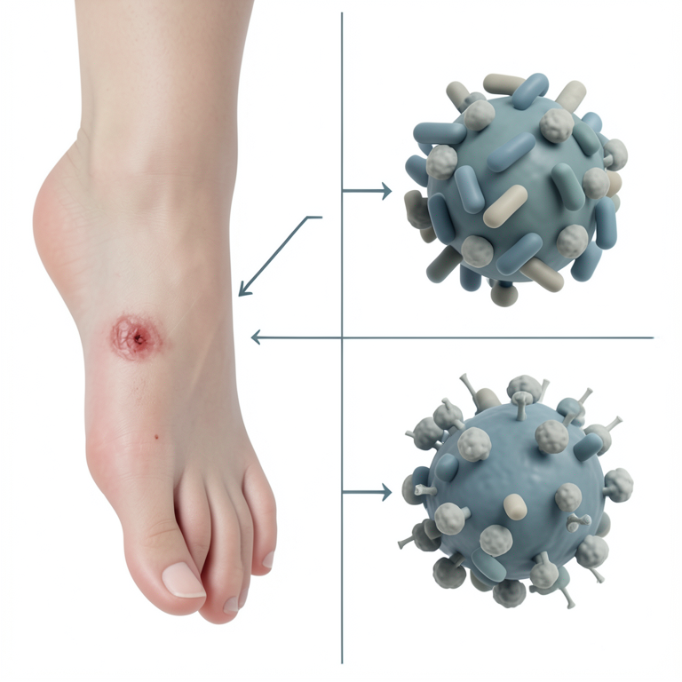
Bacterial Distribution in DFI
AI-generated conceptual illustration (Human foot skin and bacterial cells (Escherichia coli and Staphylococcus aureus)). It explains the idea and does not show study data.
View source evidence ↗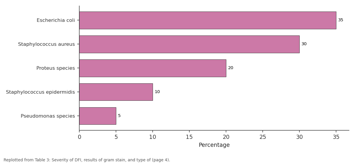
Percentage: Escherichia coli vs Staphylococcus aureus vs Proteus species vs Staphylococcus... vs Pseudomonas species
Replotted from Table 3 (page 4) of the original paper. Every bar is the paper's own table value.
View source evidence ↗Figures & tables
From the original paper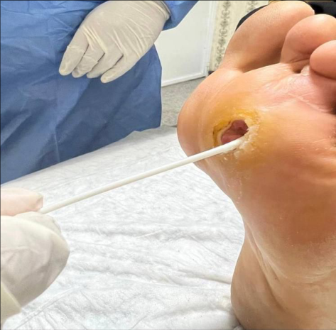 Figure 1
 Table 1
 Table 2
 Table 3
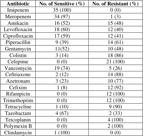 Table 4
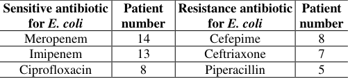 Table 5
Introduction
Diabetes Mellitus (DM) is a metabolic group of disorders characterized by chronic hyperglycemia (high blood sugar levels) resulting from a deficiency of insulin secretion, insulin action, or both. Insulin is a hormone manufactured by the beta cells of the pancreas [1] . One of the biggest new health risks of the twenty-first century is thought to be diabetes mellitus. More than 439 million people are diagnosed with DM, which causes a high rate of mortality and morbidity around the world [2] . The International Diabetes Federation (IDF) 2015 research projects that number to increase to 640 million (1 in 10) by 2040 [3] . In 2019, the straight reason of 1.5 million deaths is due to Diabetes Mellitus (DM). 48% of all deaths happened before age of 70 years [4] . The incidence of DFIs and peripheral arterial disease due to increase higher number of diabetic patients [5] . Most diabetic patients were admitted to the hospital due to DFIs. DFIs cause an increase in the use of antibiotics [6] . DFIs are the main cause of Lower Extremity Amputation (LEA) in individuals with Diabetes Mellitus (DM), especially those that reach the bone. This leads to a higher risk of death, a greater financial burden, and a worse quality of life. To escape these harmful results, it is important to protect against DFIs or, if that is not feasible, to handle wounds that have not been treated [5] . Individuals with diabetes mellitus have an increased risk of developing skin sores due to neuropathy, vascular disease, trauma, and persistent infections. Individuals with diabetes mellitus frequently experience peripheral neuropathy and peripheral artery disease.
People with diabetes in general have immune system deficiencies that are not yet known, which limits their capacity to prevent or cure infections. The most common condition in people with DM is foot infection, because they are probably to develop this condition [7][8] .
DM is classified into two major classes: Type 1 DM (T1DM) and Type 2 DM (T2DM). About 5%-10% of people with diabetes have T1DM and it is mostly diagnosed during childhood. Whereas T2DM accounting for 90%-95% of all diabetes and it is usually developing after the age of 40 years, but it may occur at any age [9][10] . DM has been linked to the traditional symptoms of the illness as well as reduced T-cell responses, disruption of humoral immunity, and neutrophil function [11][12] . Diabetic retinopathy, diabetic nephropathy, diabetic neuropathy, and DFI are among the complications that patients with uncontrolled diabetes mellitus are most likely to experience [13] . DFIs often start when there is a breach in the protective cutaneous envelope, typically in an area where there has been trauma or ulceration. People with peripheral neuropathy and peripheral arterial disorders are the most common cases of this [14] . Clinically speaking, DFI is the existence of signs of an inflammatory process in any tissue in a diabetic patient below the malleoli [15] . There are three types of foot infections in diabetic patients: mild, moderate and severe [16] . Acute infection in a previously untreated patient is usually caused by aerobic gram-positive cocci (Staphylococcus aureus), but deep or chronic wounds often harbor polymicrobial flora, including aerobic gramnegative (Escherichia coli) and obligate anaerobic bacteria (Clostridium perfringens) [17] . The biggest problem after DFI is the heightened susceptibility to many possible infections that can cause dangerous consequences like infection, gangrene, osteomyelitis, amputation, or even death [18] . Studies conducted by the Infectious Diseases Society of America (IDSA) and others indicate that individuals with DFI infection have a 50% higher risk of amputation than those with uninfected foot infections [19][20] . Around 20% of the general public who create a DFI will require LEA, each of two minor (below the ankle), major (above the ankle), or both, and 10% will die within 1 year of their first DFI diagnosis [5] . Conversely, the development of diabetic foot osteomyelitis is observed in around 44-68% of hospitalized patients, and it is the primary cause of amputation in these individuals [21] . This research aims to determine the antibiotic resistance profile and the causative microbe in Basra City DFIs.
Materials and Methods
Study design: The AL Mashfa Diabetic Foot Clinic and Laboratory is the site of this cross-sectional descriptive study in Basra City. From October 2022 until March 2023. It included 40 adult patients suffering from DFI: 23 (57.5%) were male patients and 17 (42.5%) were female patients. A questionnaire form is completed. It includes the patient's name, age, sex, type of DM, severity, type of bacteria, and random blood sugar. The patient as a whole, the affected limb, and a local evaluation of the wound were the three levels at which patients were examined.
Local examination of the wound
Local examination of the wound
Diabetic foot wound classification according to the
Diabetic foot wound classification according to the
University of Texas Classification (4 grades and 4 stages).
University of Texas Classification (4 grades and 4 stages).
The classification was
The classification was
Investigations:
The counts of white blood cells and hemoglobin were performed on each subject.
Samples collection
In this study, the wound swabs were collected according to the Levine technique. The Levine technique involves turning on the wound swab over a 1.2-cm section of the wound after cleaning the wound surface with copious sterile normal saline, then inserting the swab immediately into a sterile container. The swabs that were taken from patients were inoculated on blood agar and MacConkey agar media, and then Petri dishes were incubated for 24-48 hours at 37 °C. After that, Gram stain was used to differentiate the growing bacterial species. Then, the cultured samples were taken to the AL Mashfa laboratory, where they diagnosed the bacteria on the plate and did an antibiotic sensitivity test.
Statistical analysis
We used the SPSS software (version 260, SPPS, Chicago, IL, USA) for statistical analyses. Quantitative variables were expressed as mean ± Standard Deviation (SD) or median (Minimum and maximum), while categorical variables were expressed as percentages. To determine whether the distribution of the quantitative variables was normal, we performed the Kolmogorov-Smirnov test.
Results
The current study showed different species of gram positive and gram negative bacteria as shown in Table 1.
| No. | Sex | Age | Gram stain | Type of bacteria | Severity |
|---|---|---|---|---|---|
| 1 | F | 42 | Positive | Staphylococcus aureus | Severe |
| 2 | M | 52 | Positive | Staphylococcus aureus | Severe |
| 3 | F | 57 | Positive | Staphylococcus epidermidis | Severe |
| 4 | F | 71 | Negative | Escherichia coli | Severe |
| 5 | F | 50 | Positive | Staphylococcus epidermidis | Moderate |
| 6 | F | 51 | Positive | Staphylococcus aureus | Moderate |
| 7 | F | 48 | Negative | Proteus spp. | Moderate |
| 8 | M | 45 | Negative | Escherichia coli | Moderate |
| 9 | M | 56 | Negative | Escherichia coli | Moderate |
| 10 | F | 57 | Negative | Escherichia coli | Moderate |
| 11 | F | 60 | Positive | Staphylococcus aureus | Moderate |
| 12 | F | 50 | Negative | Proteus spp. | Severe |
| 13 | M | 55 | Negative | Escherichia coli | Moderate |
| 14 | M | 65 | Negative | Pseudomonas spp. | Severe |
| 15 | M | 40 | Negative | Proteus spp. | Severe |
| 16 | M | 60 | Negative | Escherichia coli | Severe |
| 17 | F | 55 | Negative | Pseudomonas spp. | Moderate |
| 18 | F | 52 | Positive | Staphylococcus aureus | Severe |
| 19 | F | 56 | Negative | Escherichia coli | Severe |
| 20 | M | 76 | Negative | Proteus spp. | Severe |
| 21 | F | 53 | Positive | Staphylococcus aureus | Severe |
| 22 | M | 56 | Negative | Escherichia coli | Severe |
| 23 | M | 54 | Positive | Staphylococcus aureus | Moderate |
| 24 | M | 62 | Positive | Staphylococcus aureus | Severe |
| 25 | M | 60 | Positive | Staphylococcus aureus | Severe |
| 26 | F | 35 | Negative | Proteus spp. | Severe |
| 27 | M | 63 | Negative | Proteus spp. | Moderate |
| 28 | F | 75 | Positive | Staphylococcus epidermidis | Severe |
| 29 | M | 51 | Negative | Proteus spp. | Moderate |
| 30 | M | 63 | Positive | Staphylococcus aureus | Moderate |
| 31 | M | 70 | Positive | Staphylococcus aureus | Severe |
| 32 | M | 53 | Positive | Staphylococcus aureus | Severe |
| 33 | M | 70 | Negative | Escherichia coli | Moderate |
| 34 | F | 66 | Negative | Escherichia coli | Severe |
| 35 | M | 57 | Negative | Proteus spp. | Severe |
| 36 | M | 53 | Negative | Escherichia coli | Severe |
| 37 | M | 61 | Negative | Escherichia coli | Severe |
| 38 | M | 56 | Negative | Escherichia coli | Severe |
| 39 | M | 62 | Negative | Escherichia coli | Severe |
| 40 | F | 55 | Positive | Staphylococcus epidermidis | Severe |
Table text extracted from the original PDF; superscript letters are shown inline. The original table image is below.
View original table image
The most frequent sensitive antibiotics were imipenem, meropenem, and vancomycin. The most common resistance antibiotics in order were Cefepime and Amikacin.
Discussion
The current study was carried out in Basra City to assess the susceptibility profile and pathogen prevalence of microorganisms isolated from infected wounds in patients with diabetic foot disease. For defense, the immune system is essential. The conditions of hyperglycemia and hyperinsulinemia in diabetes modify the innate immune system's typical functioning, but they have little to no influence on the adaptive immune system.
In DM, there are several changes to immune cell activities [22] . In the current study, most patients were males, as compared with other studies, which reported a higher frequency of DFI in males than in females. In our study, the mean age group was 56.83±8.83, while eight of the 12 published studies documented that the maximum number of DFI mostly occurred within the age group of 40-60 years. Both male gender and age were considered risk factors for DFI. Male predominance was explained in several previous studies due to outdoor activity by males, hard physical activity, being at higher risk for trauma, higher alcohol consumption, higher smoking behavior, and less compliance with foot care practices. The elderly patients who have spent longer with DM will have decreased immunity and nutritional deficiencies and are at risk for the development of certain complications such as peripheral neuropathy and vascular diseases [23][24][25][26] . 60% of the microorganisms in our investigation were gram negative, which is in line with findings from a study done at the Haji Adam Malik General Hospital in Medan by Bulolo et al., which showed that gram-negative bacteria were the most often detected in DFI [27] .
In contrast to a prior research conducted in Ethiopia that found Klebsiella species to be the most common bacterium at 23.9%, followed by Proteus species at 18.47%, the current investigation found that E. coli was the predominant isolate in 35% of cases [28] .
According to another Egyptian research, Proteus mirabilis (16.8%) is the most prevalent isolate [29] .
In Saudi Arabia studies found Pseudomonas species in 15.6% of cases, while the most prevalent bacteria in the South America study was Pseudomonas species 18.8%. In Kenya research 17.5% of cases was Pseudomonas also Pseudomonas found in Nigeria cases 32.9%, India 24.42%, 65.2% in China and 28% in Iran [30][31][32][33][34][35][36] . This demonstrates how various environments may have different prevalent bacteria that cause DFI illnesses.
The results of this study can be taken into account while creating an empirical antibiotic treatment plan for the local management of DFI.
The most common microorganism was E. coli, while the most sensitive antibiotics were Meropenem (14 cases), Imipenem (13 cases), and Ciprofloxacin (8 cases), while the most resistant antibiotics were Cefepime (8 cases), Ceftriaxone (7 cases), and Piperacillin (5 cases). Gram-negative bacteria outer membrane performs the vital function of providing an additional layer of defense without interfering with the flow of materials necessary to maintain life [37] . Bacterial porins are a prominent topic of study in bacterial pathogenesis due to their functional significance in microbehost interactions throughout various bacterial infections [38] . The beta-lactam antibiotic family includes the carbapenem class of antibiotics, which includes the antimicrobials meropenem and imipenem. Because most gram-negative bacteria are resistant to these drugs, they have a demonstrated clinical efficacy against a variety of pathogens, including extended-spectrum beta-lactamaseproducing organisms like Escherichia coli [39] .
Conclusion
1. Various microorganisms can cause diabetic foot infections.
2. The most common isolate was E. coli, which was followed by other gram-positive bacterial species. 3. A current study showed that isolated bacterial species are more sensitive to imipenem, meropenem, amikacin, and levofloxacin. 4. The results indicate the importance of bacteriological culture and sensitivity tests before starting imiprical antibiotic therapy for DFIs. 5. Theemergence of resistance appeared in the use of antibiotics, alerting the careful use of antibiotics.
Recommendation
1. Having poorly controlled blood sugar is the primary cause of problems from diabetes. As a result, all problems, including DFI, will stop progressing if plasma glucose is optimally controlled. 2. Therefore, the patient must follow the doctor's instructions optimum control of blood sugar, follow a healthy diet and drink water. 3. Exercising, protecting the foot from wounds and trauma, wearing appropriate shoes, cutting nails, taking care of wounds immediately upon injury, and seeing a doctor to regulate sugar. 4. Doing analyzes to ensure the patient's health and adherence to medications and all the doctor's instructions. 5. In order to start treatment as soon as possible with the right medicines, it is required to identify the particular bacteria and their susceptibility pattern.
Figures and tables from the paper
| Variable | Frequency | Percentage |
|---|---|---|
| Severity | ||
| Moderate | 14 | 35.0 |
| Severe | 26 | 65.0 |
| Gram stain | ||
| Positive | 16 | 40.0 |
| Negative | 24 | 60.0 |
| Type of bacteria | ||
| Escherichia coli | 14 | 35.0 |
| Staphylococcus aureus | 12 | 30.0 |
| Proteus species | 8 | 20.0 |
| Staphylococcus epidermidis | 4 | 10.0 |
| Pseudomonas species | 2 | 5.0 |
| Total | 40 | 100.0 |
Table text extracted from the original PDF; superscript letters are shown inline. The original table image is below.
View original table image
| Antibiotic | No. of Sensitive (%) | No. of Resistant (%) |
|---|---|---|
| Imipenem | 35 (100) | 0 (0) |
| Meropenem | 34 (97) | 1 (3) |
| Amikacin | 16 (52) | 15 (48) |
| Levofloxacin | 18 (60) | 12 (40) |
| Ciprofloxacin | 17 (59) | 12 (41) |
| Piperacillin | 9 (39) | 14 (61) |
| Gentamycin | 11(52) | 10 (48) |
| Colistin | 3 (14) | 18 (86) |
| Cefepime | 0 (0) | 21 (100) |
| Vancomycin | 19 (74) | 5 (26) |
| Ceftriaxone | 2 (12) | 14 (88) |
| Azetronam | 3 (23) | 10 (77) |
| Cefixim | 1 (8) | 12 (92) |
| Rifampicin | 0 (0) | 12 (100) |
| Trimethoprim | 0 (0) | 12 (100) |
| Tetracycline | 1 (10) | 9 (90) |
| Tazobactam | 4 (67) | 2 (33) |
| Teicoplanin | 0 (0) | 4 (100) |
| Polymexin B | 0 (0) | 2 (100) |
| Clindamycin | 1 (100) | 0 (0) |
Table text extracted from the original PDF; superscript letters are shown inline. The original table image is below.
View original table image
| Sensitive antibiotic for E. coli | Patient number | Resistance antibiotic for E. coli | Patient number |
|---|---|---|---|
| Meropenem | 14 | Cefepime | 8 |
| Imipenem | 13 | Ceftriaxone | 7 |
| Ciprofloxacin | 8 | Piperacillin | 5 |
Table text extracted from the original PDF; superscript letters are shown inline. The original table image is below.
View original table image
References
37 in the original paper- Atkins RC, Zimmet P. Diabetic kidney disease: act now or pay later. Nephrology Dialysis Transplantation. 2010 Feb 1;25(2):331-333.
- Olokoba AB, Obateru OA, Olokoba LB. Type 2 diabetes mellitus: A review of current trends. Oman Medical Journal. 2012;27(4):269-273.
- Jiménez PG, Martín-Carmona J, Hernández EL. Diabetes Mellitus. Medicine. 2020;13:883-890.
- Meloni M, Izzo V, Giurato L, Lazaro-Martínez JL, Uccioli L. Prevalence, clinical aspects and outcomes in a large cohort of persons with diabetic foot disease: comparison between neuropathic and ischemic ulcers. Journal of Clinical Medicine. 2020;9(6):1780.
- American Diabetes Association. Economic Costs of Diabetes in the U.S. in 2017. Diabetes Care. 2018;41(5):917-928.
- Aynalem SB, Zeleke AJ. Prevalence of diabetes mellitus and its risk factors among individuals aged 15 years and above in Mizan-Aman town, Southwest Ethiopia, 2016: A cross sectional study. International Journal of Endocrinology. 2018;2018(1):9317987.
- Ubeid MH. Prevalence of leukocytes in type 2 diabetic patients in Erbil City. Medical Journal of Babylon. 2020;17(1):19.
- Saleh RH, Hadi B. Bacterial profile in patients with diabetic foot infections and its association with TNF-α. Plant Archives. 2019;19(Supplement 1):222-228.
- Rajana VK. Immune Dysfunction in Diabetes Mellitus (DM). International Journal of Health Sciences Research. 2017;7(12):256-275.
- Peleg AY, Weerarathna T, McCarthy JS, Davis TM. Common infections in diabetes: Pathogenesis, management and relationship to glycemic control. Diabetes and Metabolism Research and Reviews. 2007;23(1):3-13.
- Johani K, Fritz BG, Bjarnsholt T, Lipsky BA, Jensen SO, Yang M, et al. Understanding the microbiome of diabetic foot osteomyelitis: Insights from molecular and microscopic approaches. Clinical Microbiology and Infection. 2019;25(3):332-339.
- Anvarinejad M, Pouladfar G, Japoni A, Bolandparvaz S, Satiary Z, Abbasi P, et al. Isolation and antibiotic susceptibility of the microorganisms isolated from diabetic foot infections in Nemazee Hospital, Southern Iran. Journal of Pathogens. 2015;2015(1):328796.
- Peters EJ, Lipsky BA. Diagnosis and management of infection in the diabetic foot. Medical Clinics of North America. 2013;97(5):911-946.
- Lipsky B, Berendt A, Cornia P, Pile J, Peters E, Armstrong D, et al. Infectious Diseases Society of America Clinical Practice Guideline for the Diagnosis and Treatment of Diabetic Foot Infections. Clinical Infectious Diseases. 2012;54(12):e132-e173.
- Katherine EM, Sophie B, Helen J, Stacey K, Joshua DJ. The microbiology of diabetic foot infections: A metaanalysis. Infectious Diseases. 2021;21:1-10.
- Hitam SAS, Hassan SA, Maning N. The significant association between polymicrobial diabetic foot infection and its severity and outcomes. Malaysian Journal of Medical Sciences. 2019;26(1):107-114.
- Benjamin A, Lipsky B, Éric S, Zulfiqarali G, et al. Guidelines on the diagnosis and treatment of foot infection in persons with diabetes (IWGDF 2019 update). Diabetes/Metabolism Research and Reviews. 2020;36(S1):1-24.
- Noor S, Zubair M, Ahmad J. Diabetic foot ulcer-a review on pathophysiology, classification and microbial etiology. Diabetes and Metabolic Syndrome. 2015;9(3):192-199.
- Van Asten SA, La Fontaine J, Peters EJ, Bhavan K, Kim PJ, Lavery LA et al. The microbiome of diabetic foot osteomyelitis. European Journal of Clinical Microbiology & Infectious Diseases. 2016;35(2):293298.
- Marzoq A, Shiaa N, Zaboon R. Assessment of the outcome of diabetic foot ulcers in Basra, Southern Iraq: A cohort study. International Journal of Diabetes and Metabolism. 2019;25(1-2):33-38.
- Amjad SS, Zafar J, Shams N. Bacteriology of diabetic foot in tertiary care hospital; Frequency, antibiotic susceptibility and risk factors. Journal of Ayub Medical College Abbottabad. 2017;29(2):234-240.
- Neama NA, Darweesh MF, Al-Obiadi AB. Prevalence and antibiotic susceptibility pattern in diabetic foot ulcer infection with evaluation the role of biomarker il12 in disease. Biochemistry and Cell Archives. 2018;18(2):2321-2328.
- Qadir A, Nasih M, Bakhtiar M, Mahwi TO, Al-Attar DM, Raoof A, et al. Prevalence of microorganisms and antibiotic sensitivity among patients with diabetic foot ulcer in Sulaimani City, Iraq. Hospital Practice Research. 2020;5(2):56-63.
- Ullah I, Ali SS, Ahmad I, Khan MN, Ur Rehman M, Malik SA et al. Bacteriological profile and antibiotic susceptibility patterns in diabetic foot infections, at lady reading hospital, Peshawar. Journal of Ayub Medical College Abbottabad. 2020;32(3):382-388.
- Bulolo BA, Pase MA, Ginting F. Antibiotic sensitivity pattern of bacteria from diabetic foot infections Haji Adam Malik central general hospital. IOP Conference Series: Earth and Environmental Science. 2018;125(1):012052.
- Amogne W, Reja A, Amare A. Diabetic foot disease in Ethiopian patients: A hospital-based study. Ethiopian Journal of Health Development. 2011;25(1):17-21.
- Dwedar R, Ismail D, Abdulbaky A. Diabetic foot infection: Microbiological causes with special reference to their antibiotic resistance pattern. Egyptian Journal of Medical Microbiology. 2015;24(3):95-102.
- Al Ayed MY, Ababneh M, Alwin RA, Alzaid A, Ahmed RA, Salman A, et al. Common pathogens and antibiotic sensitivity profiles of infected diabetic foot ulcers in Saudi Arabia. International Journal of Lower Extremity Wounds. 2018;17(3):161-168.
- Ponce de Leon A, Merchant S, Raman G, Avendano E, Chan J, Tepichin Hernandez G, et al. Pseudomonas infections among hospitalized adults in Latin America: a systematic review and meta-analysis. BMC Infectious Diseases. 2020;20(1):250.
- Mutonga DM, Mureithi MW, Ngugi NN, Otieno FCF. Bacterial isolation and antibiotic susceptibility from diabetic foot ulcers in Kenya using microbiological tests and comparison with RT-PCR in the detection of S. aureus and MRSA. BMC Research Notes. 2019;12:(244):1-6.
- Ogba OM, Nisan E, Eyam ES. Aerobic bacteria associated with diabetic foot ulcers and their susceptibility pattern. Biomedical Dermatology. 2019;3:1.
- Aleem S, Multani H, Bashir H. Bacteriological profile and antimicrobial sensitivity pattern of isolates from the diabetic foot of patients attending a teaching hospital in northern India. Asian Journal of Medical Sciences. 2021;12(5):83-87.
- Xie X, Bao Y, Ni L, Liu D, Niu S, Lin H, et al. Bacterial profile and antibiotic resistance in patients with diabetic foot ulcer in Guangzhou, Southern China: focus on the differences among different Wagner's grades, IDSA/IWGDF grades, and ulcer types. International journal of endocrinology. 2017;2017(1):8694903.
- Akhi MT, Ghotaslou R, Asgharzadeh M, Varshochi M, Pirzadeh T, Memar MY, et al. Bacterial aetiology and antibiotic susceptibility pattern of diabetic foot infections in Tabriz, Iran. GMS Hygiene and Infection Control. 2015;10:Doc02.
- Armstrong T, Fenn SJ, Hardie KR. JMM Profile: Carbapenems: A broad-spectrum antibiotic. Journal of Medical Microbiology. 2021;70(12):1-5.
- Senchyna F, Gaur RL, Sandlund J, Truong C, Tremintin G, Kültz D, et al. Diversity of resistance mechanisms in carbapenem-resistant Enterobacteriaceae at a health care system in Northern California, from 2013 to 2016. Diagnostic Microbiology and Infectious Disease. 2019;93(3):250-257.
- Harris PNA, Tambyah PA, Lye DC. MERINO Trial Investigators and the Australasian Society for Infectious Disease Clinical Research Network (ASID-CRN). Effect of piperacillin-tazobactam vs meropenem on 30day mortality for patients with E coli or Klebsiella pneumoniae bloodstream infection and ceftriaxone resistance: A randomized clinical trial. JAMA. 2018;320(23):984-994.