
Research article · 2023 · Full text
International Journal of Biology SciencesMechanisms of development of type 2 diabetes
Volume 5(1) · Pages 198–208 · DOI: 10.33545/26649926.2023.v5.i1c.187 ↗
Research areasDiabetes and associated disordersDiet, Metabolism, and Disease
Abstract
Author-writtenType 2 Diabetes Mellitus (T2DM) has been defined as one of the commonly occurring metabolic disorders arising due to the combination of 2 primary factors, namely, inadequate insulin response in the insulin-sensitive tissues and insufficient insulin secretion through pancreatic beta-cells. The precise regulation of insulin release, detection, and synthesis is imperative for glucose homeostasis maintenance, and involves intricate molecular mechanisms. The onset of the disease may be attributed to a metabolic imbalance arising from deficiencies in any of the mechanisms in processes. This research examines the primary characteristics of T2DM, including the molecular mechanisms and pathways contributing to the insulin resistance (IR) and T2DM development. The information is compiled with the aim of examining insulin release, sensing, synthesis, and resulting effects on various insulin-sensitive organs. Special attention is paid to these aspects. This paper examines the various pathogenic factors contributing to T2DM development and progression, including diet, exercise, gut dysbiosis, and metabolic memory. The present discourse delves into the molecular pathways which establish a connection between IR and T2DM. Furthermore, the discussion highlights the noteworthy implications of T2DM, particularly the hastened progression of atherosclerosis, on cardiovascular risk.
Summary
Source-checkedPlain-language reading of the full paper, prepared with AI and checked against the text. Each point links to the authors' own words.
Why it matters
- Type 2 Diabetes Mellitus (T2DM) is a common metabolic disorder caused by a combination of insufficient insulin secretion from pancreatic beta-cells and an inadequate response to insulin in sensitive tissues. View evidence ↗
- T2DM is the most prevalent form of diabetes, accounting for more than 90% of all diabetes cases. View evidence ↗
- T2DM significantly increases health risks, including a 15% higher risk of all-cause mortality compared to non-diabetics. View evidence ↗
The question
- The aim of this research is to examine the primary characteristics of T2DM, specifically the molecular mechanisms and pathways that lead to insulin resistance and the development of the disease. View evidence ↗
What they did
- The authors compiled information to analyze the processes of insulin synthesis, sensing, and release, as well as the subsequent effects on insulin-sensitive organs. View evidence ↗
- The review evaluates several pathogenic factors, including metabolic memory, gut dysbiosis, exercise, and diet. View evidence ↗
What they found
- The prevalence of diabetes in adults aged 20 to 79 was estimated at 463 million in 2019 and is projected to reach 700 million by 2045. View evidence ↗
- Diabetes is correlated with increased risks of coronary heart disease (HR 2), ischemic stroke (HR 2.27), and other vascular disease-associated deaths (HR 1.73). View evidence ↗
- Prevalent genetic variations related to glycemic levels in T2DM patients only account for 10% of the overall trait variability, indicating that rare variants are likely very important. View evidence ↗
- Walking for at least 40 minutes daily or 2-3 hours weekly can reduce T2DM susceptibility by 34% and 56%, respectively. View evidence ↗
- Moderate physical activity is associated with a 40% increase in glucose absorption. View evidence ↗
- Excessive free fatty acids (FFAs) and hyperglycemia activate apoptotic unfolded protein response (UPR) pathways, causing endoplasmic reticulum (ER) stress and beta-cell dysfunction. View evidence ↗
- Metabolic memory, which causes complications to persist even after glycemic control is restored, is driven by chronic inflammation, epigenetics, oxidative stress, and non-enzymatic protein glycation. View evidence ↗
What it means
- The authors conclude that a better understanding of T2DM pathophysiology should lead to the implementation of precision medicine and individualized treatment plans. View evidence ↗
- The authors state that more research is needed to establish the causal link between the development of T2DM and intestinal microbiota, as well as to test potential treatments. View evidence ↗
Visual summary
1 visualsIllustrations explain the paper; they do not show study data.
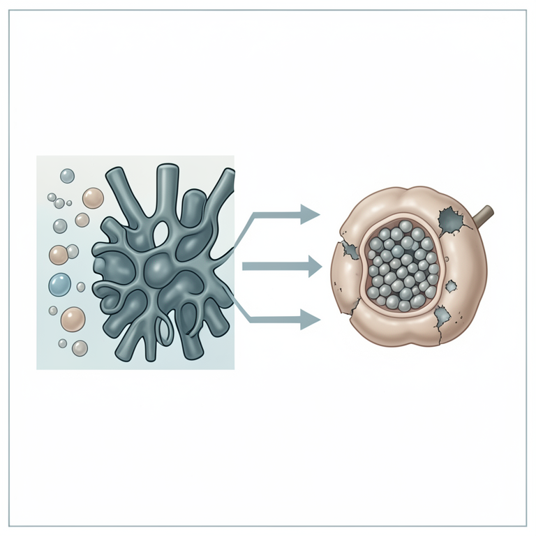
Pathways to Beta-Cell Dysfunction
AI-generated conceptual illustration (pancreatic beta-cells). It explains the idea and does not show study data.
View source evidence ↗Figures & tables
From the original paper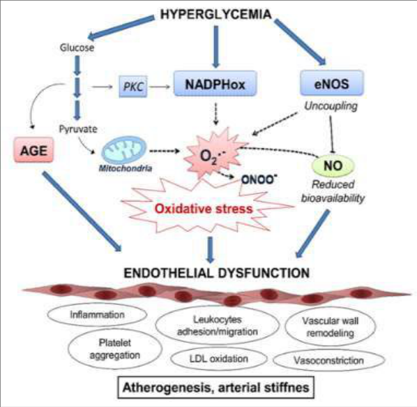 Figure 1
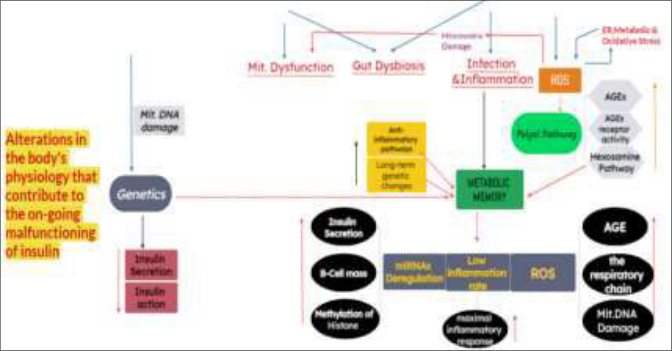 Figure 2
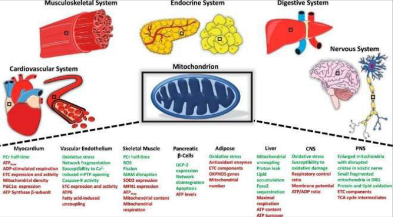 Figure 3
Introduction
The onset of T2DM, a highly prevalent metabolic disorder on a global scale, is primarily attributed to the interplay of two factors: compromised insulin sensitivity in tissues and deficient insulin secretion by the pancreatic beta-cells [1] . The regulation of molecular processes that govern insulin release and synthesis, in addition to insulin response in tissues, is crucial for optimal metabolic function. Consequently, any deficiencies in the aforementioned pathways may result in metabolic disturbance and the onset of T2DM. This study investigates the primary characteristics of T2DM, including the underlying molecular mechanisms as well as pathways associated with insulin metabolism. Additionally, the relation between T2DM and cardiovascular pathology is explored in detail. This work examines the global prevalence of T2DM and the primary risk factors associated with its development. These risk factors include lifestyle choices, gut dysbiosis, inherited susceptibilities, obesity, mitochondrial dysregulation, and epigenetics. Our focus is on elucidating the molecular and physiological mechanisms underlying the development of T2DM and its associated complications.
Development and Prevalence of Type 2 Diabetes
Based on reports by WHO, DM is a persistent metabolic condition which has been characterized through elevated glucose levels in bloodstream, and that can lead to the detrimental effects on heart, kidneys, eyes, nerves and blood vessels during an extended period of time. T2DM is a medical condition that is distinguished by inadequate secretion of the insulin via pancreatic islet β-cells, tissue insulin resistance, and a sufficient compensatory insulin secretory response. This diabetes form accounts for over 90% of all the cases of DM. With the progression of the illness, the insulin secretion becomes insufficient to maintain glucose homeostasis that results in the development of hyperglycemia. The primary characteristics of individuals with T2DM are obesity or a higher percentage of body fat, particularly in abdominal part. Adipose tissue fosters IR in this scenario via diverse inflammatory mechanisms, including but not limited to heightened release of FFA and dysregulation of adipokines. The sedentary lifestyles, global increases in obesity, highcalorie diets, and population aging have led to a fourfold increase in prevalence as well as incidence of T2DM, as it has been reported in sources [4,5] T2DM. development involves various organs, including but not limited to adipose tissue, skeletal muscle, brain, kidneys, liver, and small intestine [6] . Recent research has indicated that significant pathophysiological factors include adipokine dysregulation, alterations in the microbiota of the gut, immunological dysregulations, and inflammation. These findings had been identified as emerging in nature. The epidemiological data presents concerning statistics that suggest a potentially bleak outlook for individuals with T2DM in the future. Based on International Diabetes Federation (IDF), prevalence ofdiabetes amongst the adults who are aged 20 to 79 has been estimated to be 463 million in 2019, with projected increase to 700 million by 2045. According to global statistics from 2019, diabetes was responsible for causing 4.2 million fatalities across the globe. In 2019, medical expenses associated with diabetes amounted to at least 720 billion USD. Moreover, it is worth to note that a significant proportion of diabetics, specifically one in three individuals or approximately 232 million individuals, have received an inaccurate diagnosis of their condition. As a result, the actual prevalence and impact of type 2 diabetes mellitus may be underestimated. Individuals who are afflicted with diabetes are commonly found to be within age range of 40 to 59 years. The prevalence and incidence of T2DM exhibit geographic variability, with a minimum of 80% of patients inhabiting low-to middleincome countries, thereby exacerbating the challenges associated with delivering efficacious healthcare. Cardiovascular diseases (CVD) represent the primary causes of mortalities and morbidities related to T2DM. Individuals with T2DM exhibit a 15% higher risk of all-cause mortality when compared to those who do not have diabetes [8]. According to a meta-analysis [9], there exists a correlation between diabetes and an increase in the likelihood of experiencing ischemic stroke (HR 2.27; 1.95-2.65), coronary heart disease (CHD) (hazard ratio [HR] 2; 95% CI 1. 83-2.19), and other vascular disease-associated fatalities (HR 1.73; 1.51-1.98). The epidemiology of T2DM is impacted by both genetic as well as environmental factors. Genetic factors are influenced by exposure to an environment that is identified by a sedentary behaviour and high intake of calories. The findings of genome-wide association studies had revealed existence of the prevalent genetic variations that are related to glycaemic levels in individuals with T2DM. However, these variations are only accountable for 10% of the overall variability in traits, suggesting that rare variants hold considerable importance. Individuals exhibiting a range of phenotypic variations may possess a greater propensity towards specific groupings of CVD risk factors, which include IR, dyslipidemia and hypertension [11] .
Pathophysiology and Risk Factors
Development of T2DM is influenced by the interaction of metabolic, environmental factors, and genetic factors. Whilst non-modifiable risk factors such as ethnicity, genetic predisposition, and history of family have a significant impact on an individual's susceptibility to T2DM, there exists a strong genetic component.
However, epidemiological studies have demonstrated that a multitude of T2DM cases may be preventable by addressing main modifiable risk factors, which include obesity, inactivity and unhealthy food [12,13] .
Ethnicity and Genetic Predisposition / Family History
The populations of Hispanics, Native Americans, and Japanese have been identified as having the greatest susceptibility to T2DM on a global scale. The prevalence and incidence of this condition, however, exhibit significant variation based on both geographic location and ethnicity, as evidenced by previous research [14][15][16] . Research has shown that Asians have higher incidence rates compared to White Americans [17,18] or White Britons [19] , while Black individuals have the highest risk [20] . Although the etiology of the condition remains uncertain, pertinent factors have been detected, like contemporary lifestyle factors that promote adiposity, socioeconomic factors, geneenvironment interactions, and direct genetic predispositions. The genetic predisposition exerts a significant impact on the probability of developing T2DM. Several genome-wide association researches that have been carried out in the past decade had demonstrated the intricate polygenic nature of T2DM [21,22] . Most of those loci augment the risk of T2DM by primarily affecting insulin secretion. Dimas et al. classified variants into 4 categories that exhibit a distinct IR pattern, 9 categories that decrease insulin secretion while maintaining normal fasting glycemia, 2 categories that decrease insulin secretion with fasting hyperglycemia, and 1 category which alters insulin processing, based upon their possible involvement as intermediate mechanisms in T2DMpathophysiology. The aforementioned findings suggest that genetic architecture regarding T2DM is very polygenic, and further association studies are necessary for the identification of almost all of the T2DM loci [24] . Numerous clinical trials as well as observational studies indicated that the impacts of specific genetic variant could be modified by some environmental aspects, and conversely, environmental factors could be influenced by susceptibility loci. This suggests that the absence of heritability in T2DM might be attributed to interactions between environmental factors and susceptibility loci [25] .
Obesity, inactivity and Unhealthy food
Obesity (BMI=30 kg/m 2 ) represents the most important factor ofrisk for the T2DM and is also related to metabolic anomalies which result in the IR [28] . According to a previous study [29] , there exists a linear inverse relation of the BMI with age at T2DMdiagnosis. Several factors were shown to play a significant role in developing this pathological process that encompasses inter-organ interactions as well as cell-auto nomousmechanisms. The specific pathways through which obesity induces the development of T2DM and IR remain unclear. Kuipio Ischemic Heart Disease Risk Factor Study and Women's Health Study have identified a sedentary lifestyle as a contributing risk factor for the T2DM. The findings of aforementioned studies suggest that individuals who engage in a minimum of 40 mins of walking daily or 2-3 hours weekly may experience a reduction in their susceptibility to T2DM by 34% and 56%, respectively [30,31] . Engaging in physical activity offers 3 primary benefits in terms of postponing T2DM on set. At the onset, during skeletal muscle cell contractions, there is an increase in the blood flow to the muscle, resulting in enhanced glucose uptake from plasma. Physical activity has been shown to reduce intra-abdominal adiposity that is a well-established IR risk factor [32,33] . Finally, it was indicated that engaging in moderate physical activity leads to a 40% increase in the glucose absorption, as reported in reference 34. Physical activity had been shown to enhance glucose uptake and insulin sensitivity, while also potentially mitigating oxidative stress and inflammation, both of which are related to increased T2DM risks [32] .
Pathophysiology
The pathogenesis of the disease involves a disruption in feedback mechanisms between insulin action and secretion, resulting in abnormally elevated level of blood glucose [2] . Beta-cell dysfunction results in the decrease of insulin secretion, which impairs the capacity of the body to regulate physiological glucose levels. Simultaneously, IR mitigates glucose absorption in adipose tissue, liver and muscle, whereas augmenting glucose production in liver. Although various pathophysiological mechanisms play a role in disease manifestation, beta-cell dysfunction is generally more severe when compared to the IR. None-theless, the presence of both IR as well as beta-cell dysfunction exacerbates hyperglycemia and leads to T2DM progression [35, 36] .
Mechanisms Resulting to Pathophysiology and T2DM
Secretion of Insulin: Dysfunctional and Physiological Mechanisms Resulting in T2DM.
β-Cell Physiology
It is imperative to safeguard cellular integrity and exercise stringent control over the mechanisms and pathways that govern beta-cell physiology to ensure optimal beta-cell function [35] . Beta-cells are responsible for the synthesis of insulin that undergoes an initial conversion into preproinsulin. Furthermore, pre-proinsulin experiences a structural modification during its maturation process, facilitated by numerous proteins within ER, resulting in proinsulin production [37] . Subsequently, proinsulin is translocated from ER to Golgi apparatus (GA), where it is incorporated into immature secretory vesicles and subjected to proteolytic processing to yield insulin and C-peptide [38,39] . Upon attaining maturity, insulin is stored in the form of granules until its release is necessitated. The primary trigger for the secretion of insulin is a response to heightened concentrations of glucose. It should be emphasized that the release regarding insulin [40] may be influenced by supplementary variables like amino acids, fatty acids and hormones. The glucose transporter 2 (GLUT2) can be defined as one of the solute carrier proteins that functions as glucose sensor for beta-cells. It represents primary mechanism by which beta-cells uptake glucose as circulating levels of glucose increase. Upon glucose entry, the glucose catabolism process is started. This leads to intracellular ATP/ADP ratioelevation, leading to closure of the ATP-dependent potassium channels that are located in plasma membrane. Consequently, de-polarization of the membrane occurs, opening voltage-dependent Ca 2+ channels, then let Ca 2+ into cell. The process of priming and fusing insulin-containing granules with plasma membrane is triggered by an elevation in intra-cellular concentration of Ca 2+ . This, in turn, leads to insulin exocytosis [38][39][40][41][42] (Fig1-A). This study aims to investigate the signaling pathways involved in secretion of insulin in beta-cells under the normal physiological conditions (A) and the underlying mechanisms that lead to the dysfunction(B). Insulin is predominantly triggered by elevated concentrations of the glucose, whereas glucose uptake is primarily facilitated by the GLUT2 transporter. The catabolism of Glucose enhances the ratio of ATP to ADP through inhibiting ATP-dependent potassium channels, which results in de-polarization of the membrane and consequent activation regarding voltage-dependent Ca 2+ channels. Influx of Ca 2+ resulting from the latter process facilitates the exocytosis of insulin. The facilitation of Ca 2+ mobilization as well as insulin secretion is supported by additional Ca 2+ channels, namely P2Y, SERCA, RYR, and P2X. (B) Elevated levels of blood glucose and cholesterol have been found to induce oxidative stress, leading to the generation of ROS. And that, in turn, leads to the inhibition of Ca 2+ mobilization and activates the proapoptotic signaling. Apoptosis activation and unfolded protein response (UPR) mechanisms leading to the ER stress is caused by an overabundance of hyperglycemia and FFAs. The production of ROS is observed in the case when there is an elevated synthesis of proinsulin and IAAP, which is attributed to the persistent hyperglycemic state. The aforementioned abbreviations correspond to GLUT2, which stands for glucose transporter 2, P2Y and P2X, which denote purinergic receptors Y and X, respectively. Additionally, IP3 and IP2 refer to inositol 1, 4, 5-trisphosphate and inositol 1,3-bisphosphate, while SERCA represents sarco-endoplasmic reticulum Ca 2+ -ATPase. RYR designates ryanodine receptor channel, ROS stands for reactive oxygen species, FFA denotes free fatty acid, and UPR refers to the unfolded protein response.
Mechanisms resulting in dysfunction
RYR receptors, known for their ability to facilitate Ca 2+ signals, are located within the cell and possess the capability for the mediation of Ca 2+ induced Ca 2+ release (CICR). As a result, they may have a noteworthy impact on the coupling between stimulus and insulin secretion. RYR, which is implicated in the augmentation of insulin secretion [43] , enhances Ca 2+ signals in the case where channel is sensitized through messenger molecules that are generated by ligand-binding and nutrient metabolism (as illustrated by Figure 1A). The provision of supplementary cellular signals may potentially enhance the efficacy of beta-cell insulin secretion. Of the various messengers, cAMP appears to hold the greatest significance in its ability to augment insulin release. Emerging evidence suggests that cAMP facilitates the movement of secretory vesicles that transport insulin by decreasing intracellular Ca 2+ reserves and increasing intracellular Ca 2+ levels. Furthermore, there exists compelling evidence that that extra-cellular ATP plays an important role in regulation of the function of beta-cells. The exocytosis of insulin granules is a well-established mechanism by which B-cells release ATP in response to glucose stimulation. Irrespective of glucose, the activation of P2X and P2Y purinergic receptors facilitates Ca 2+ mobilization and regulates the exocytosis of the insulin via purinergic signaling. P2X receptors are a type of ligand-gated ion channel which is activated by the ATP and exhibits non-selectivity towards cations. Conversely, P2Y purinoreceptors were observed to be associated with Gproteins, as reported in previous studies [44,45,46] . The proposition has been put forth that the release of insulin from P2Y receptors could be facilitated by means of intracellular Ca 2+ mobilization, which is triggered by synthesis of inositol-1, 4, 5-trisphosphate (IP3). Which, in turn, leads to Ca 2+ releasefrom ER storage and increase Ca 2+ signal that result in exocytosis [48,49] (Figure1A).
Mechanisms Resulting in β-Cell Dysfunction
Traditionally, there were an association between beta-cell death and beta-cell dysfunction [50] . According to recent study, there appears to be a complex network of the interactions between environment and different molecular path-ways that are related to cell biology that may be responsible for dysfunction of beta-cells in individuals with T2DM [51] . The co-occurrence of hyperlipidemia and hyperglycemia is commonly observed in a state of dietary excess, akin to that observed in obesity, which promotes IR as well as chronic inflammation. Beta-cells are subject to various toxic pressures, which include inflammation, metabolic/oxidative stress, ER stress, amyloid stress and inflammatory stress, which can result in eventual loss of islet integrity. This susceptibility is due to genetic variations. This information is supported by previous research (reference 50). The activation of apoptotic UPR pathways due to an excess of hyperglycemia and FFAs leads to ER stress, ultimately resulting in beta-cell dysfunction [52] . Metabolic and oxidative stress, which are caused by obesity-related lipotoxicity, glucotoxicity, and glucolipotoxicity, have been found to be detrimental to betacells [51] . The pathway of the UPR can be activated by the stress that is induced by elevated levels of the saturated FFAs through various mechanisms like inhibiting sarco/endoplasmic reticulum Ca 2+ ATPase (SERCA) that is responsible for the mobilization of ER Ca 2+ , activating the receptors ofIP3, or the direct ER homeostasis impairment. Furthermore, it has been observed that extended exposure to elevated glucose levels leads to heightened production of IAAP and proinsulin biosynthesis in beta-cells. This results in buildup of the misfolded insulin and IAAP, along with an increase of generation of ROS through oxidative protein folding mechanisms [52] . The aforementioned activities induce alterations in physiological mobilization regarding ER Ca 2+ , facilitate degradation of the proinsulin mRNA, promote proapoptotic signals, as well as triggering beta-release of IL-1 that attracts macrophages and exacerbates local islet inflammation (as it has been illustrated in Fig1-B). As it has been previously established, the regulation of insulin secretion is crucial for meeting metabolic requirements. Preservation of islet integrity is essential for β-cells to effectively respond to metabolic demands. The mechanism that has been described above has the potential to cause disruption in islet integrity and organization under pathogenic conditions. This disruption can lead to suboptimal cell-to-cell communication within pancreatic islets, and that can further result in poor regulations of insulin and glucagon release, exacerbating hyperglycemia ultimately. Insulin secretory dysfunction is the primary beta-cell failure cause and forms the foundation of T2DM. This dysfunction may arise due to errors in synthesis regarding insulin precursors or actual insulin, in addition to the disruptions in secretion. The diminished expression of GLUT2 glucose transporter may have an effect on signaling pathway that follows [53] . Additionally, the failure of proinsulin folding is a commonly observed outcome that is linked to insufficient insulin synthesis and the development of diabetes [51,54] .
Pathological Conditions Perpetuate T2DM Nutrition Factors
The consumption of high-calorie Western diet results in elevated blood glucose levels and increased levels of circulating the VLDLs, chylomicrons, and their remnants (CMRs), which are particles that are rich in TGs and carbohydrates. The elevation of ROS levels induces the aberrant synthesis of pro-inflammatory molecules. The interaction between oxidative stress and inflammation is widely recognized, and after consuming a substantial meal, these two processes work together in a synergistic manner, thereby exacerbating any adverse postprandial effects. The development of T2DM and IR is facilitated by the prolonged and significant increase in the steady-state ROS levels. The generation of superoxide (O2-), activation of NADPH oxidase (NOX), ER stress, and mitochondrial malfunction are consequences of a pro-oxidant environment. An elevation in O2-production triggers the activation of 5 primary pathways implicated in diabetes complications' pathogenesis, which is polyol path-way, the accelerated production of advanced glycation end products (AGE), the increased expression of the AGE receptor and its activating ligands, the PKC isoforms' activation, and the overactivity of hexosamine pathway. Through those mechanisms, heightened intra-cellular ROS induces certain proinflammatory pathways as a reaction to ischemia, hinders angiogenesis, and leads to persistent epigenetic modifications that persistently impact the expression of the pro-inflammatory genes, even after glycemia normalizes. In addition, elevated concentrations of FFAs in the bloodstream can lead to impairment of mitochondrial function through two separate pathways: (a) the byproducts regarding FFA metabolism can disrupt electron flow throughout mitochondrial respiratory chain; (b) FFA integration into mitochondrial membranes may promote electron leakage [55][56][57][58][59] .
Physical Activity
T2DM as well as obesity are correlated due to a reduction in physical activity, a reduction in exercise, increasing sedentary behavior, and an elevation in markers of chronic low-grade systemic inflammation (references 60 and 61). Throughout such disease, proinflammatory molecules like CRP, TNF-alpha, IL-6, and IL-1 are released into bloodstream, leading to a state of metabolic inflammation [37] . The inhibition of the B-cell function and activation of nuclear factor κ-light-chain-enhancer regarding the activated B cells (NF-κB) transcription factor by IL-1 results in inhibition of β-cell function and promotion of apoptosis in auto-immune response to beta-cells in pancreas, as indicated by previous research [32] . The preclinical animal data indicates that the removal of macro-molecular complex NLRP-3 inflammasome, responsible for generating IL-1 beta and IL-18, had led to the improvement of insulin sensitivity. This finding supports previous preclinical data that suggests resolution of inflammation may prevent onset of T2DM in individuals with prediabetes and obesity. (Reference: 62) For individuals who have prediabetes and obesity, deliberate weight reduction remains the fundamental approach for improving insulin sensitivity and, in certain instances, averting the onset of T2DM [63] . Elevated levels of physical activity and consistent exercises were found to stimulate generation of anti-inflammatory cytokines, such as soluble TNF receptor (s-TNF-R) that acts as an antagonist of IL1 and TNF-beta, and IL-1 receptor antagonist (IL-1Ra) that acts as an antagonist of IL-1. Elevated physical activity levels had been observed to be concomitant with decreased leptin levels, a substance related to the CRP, and also with the circulating IL-6, IL-18, and CRP levels, as per a previous study [64] . Exercise has the potential to reduce oxidative stress which leads to T2DM by promoting the synthesis of antioxidants, like GSH, which is a notable non-enzymatic antioxidant, and other antioxidant enzymes. This reduction in free radical levels could be sustained over time. Finally, irisin can be defined as a myokine which is secreted by adipose tissue [67] and skeletal muscle [57] in reaction to physical activity [68] and amplifies glucose tolerance [66] . Compared with control participants, it was observed that individuals with T2DM exhibit reduced circulating irisinlevels. In addition, it was observed that diabetic patients who have CVD had much lower levels of serum irisin in comparison with patients without CVD [69] . Individuals diagnosed with T2DM and exhibiting reduced levels of serum irisin are at a 1.6-fold increased risks of developing CVD [70] .
Metabolic Memory
Metabolic memory is the term used to describe the ongoing issues related to diabetes, even with consistent glycemic management. The aforementioned notion was derived from the outcomes of several extensive clinical trials, which had demonstrated that despite the administration of medication intervention for restoring glycemic control in early diabetes stages, complications persist and deteriorate [71][72][73] . Studies such as UKPDS post-trial research as well as Steno-2 trial have shown that initiating glycemic treatments early could effectively prevent diabetic complications and considerably reduce the occurrence of CVD endpoints in patients receiving either intensive or standard therapy following diagnosis [73] . Studies involving in vitro cell cultures and animal models of diabetes have demonstrated that the initial hyper-glycemic phase results in enduring impairments in target organs/cells, including abnormal gene expression (References) [74][75][76][77] . Metabolic memory is influenced by four distinct pathways, namely oxidative stress, epigenetics, chronic inflammation, and nonenzymatic protein glycation. Epigenetics has the potential to regulate gene expression and determine the proteins that are synthesized by utilizing factors beyond DNA sequences [78] . Diverse epigenetic regulatory mechanisms are available, such as higher-order chromatin architecture, non-coding RNAs, direct cytosine or adenine residue methylation, covalent modifications of the histone proteins, and additional approaches. Epigenetic imbalances or disruptions might lead to the pathogenesis of diabetes, as stated in reference [79] . To summarize, T2DM represents progressive and heterogeneous disease identified by a range of metabolic abnormalities that are related to elevated blood glucose levels, resulting from intricate pathological mechanisms that impair insulin secretion and/or function. There exist multiple interrelated pathways that exhibit mutual reinforcement, thereby augmenting the susceptibility to different ailments such as peripheral arterial disease, cerebro-vascular disease, heart disease, non-alcoholic fatty liver disease, obesity, and several others. The activation of those pathways is affected by a variety of environmental and genetic factors. Figure 2 presents a comprehensive overview of the complicated network of clinical variables contributing to the pathogenesis of T2DM.
Mitochondrial Dysfunction
The body of evidence supporting the association between mitochondrial dysfunction and development of age-related IR, T2DM, and T2DM comorbidities is expanding [80] . The onset of T2DM is associated with mitochondrial dysfunction, which is expedited by oxidative stress, genetic modifications that affect mitochondrial integrity, inadequate mitochondrial biogenesis, and the process of aging (as illustrated in Figure 3) [81,82] . Mitochondria's principal function is to produce ATP through oxidative phosphorylation in response to the metabolic requirements [83] . Furthermore, mitochondria play a crucial role in biosynthesis of numerous metabolites that act as precursors for diverse macromolecules such as lipids, DNA, and proteins. Furthermore, mitochondria play a critical role in the maintenance of ion homeostasis, responding to stress, ROS clearance, and integrating multiple signaling pathways [84, 85] .
Insulin Resistance
IR can be defined as a diminished response or impairment to insulin that circulates through the bloodstream and regulates blood glucose levels. It can also involve the decrease in insulin-responsive cells' metabolic response to the insulin on systemic levels. This is supported by research findings [86] . IR conditions can be broadly classified to three classes, which are: (i) diminished insulin secretion via beta-cells; (ii) plasma insulin antagonists, that may arise as a result of the counter-regulatory hormones or the non-hormonal substances which hinder insulin receptors or signalling; (iii) lowered insulin response in the target tissues [87] . Interaction between growth hormone and IGF1 during fed state has an impact on insulin actions. In order to prevent hypoglycemia induced through insulin during fasting, the suppression of insulin response is achieved through the action of glucagon, glucocorticoids, and catecholamines. Downstream enzymes' regulation in regulatory signaling path--ways is dependent on the relative level of phosphorylation, making the insulin/glucagon ratio a critical factor in this process. Glucocorticoids have been found to promote muscle catabolism, gluconeogenesis, and lipolysis, whereas catecholamines have been observed to enhance lipolysis and glycogenolysis. Consequently, excessive secretion of those hormones could be attributed to IR onset [88,89] . Regarding the last group, adipose tissue, skeletal muscle, and liver represent the three primary extra-pancreatic insulin-sensitive organs that play a vital role in aforementioned physiological processes. The inadequate functioning of insulin in these specific tissues often precedes the manifestation of widespread IR, which leads to the gradual development of T2DM.
Outcomes/Complications of T2DM: Cardio-vascular Risks T2DM is a complex medical condition that has been found to have a significant correlation with the development of CVD, as elaborated in the preceding sections [90] . T2DM is related to macro-vascular as well as micro-vascular complications. The former encompasses accelerated atherosclerosis, which leads to premature CAD, severe peripheral vascular disease, and an elevated susceptibility to cerebrovascular diseases [91][92][93] . Additionally, it results in a two to four-fold rise in the fatality rate among adult individuals with stroke and heart disease. The aforementioned elements give rise to the potentiality that several molecular mechanisms as well as pathogenic pathways play a role in T2DMonset, which is a significant factor of risk for CVD [94] . Several factors have been identified in relation to vascular function, hypertension, oxidative stress, atherosclerosis, macrophage accumulation, and inflammations, as it has been reported in references 95-98. The subsequent sections provide an in-depth coverage of the primary variables linked to cardio-vascular risk outcomes stemming from the T2DM and their corresponding interactions, as illustrated in Figure 6. This study examines the risk factors associated with T2DM and underlying pathological changes that contribute to impaired insulin function. The risk factors for a particular condition could be categorized into 2 groups: modifiable and non-modifiable. Modifiable risk factors include obesity, low physical activity, and unhealthy diet, whereas the non-modifiable risk factors include ethnicity and genetic predisposition/family history. These risk factors result from complicated interactions between genetic, metabolic, and environmental factors. The aforementioned conditions exert an influence on cellular activity, which can result in a complicated set of the pathological modifications that interconnect and sustain impaired insulin functionality. The following abbreviations are commonly used in academic literature: ER, which stands for endoplasmic reticulum; ROS, which refers to reactive oxygen species; PKC, which stands for protein kinase C; AGEs, which are advanced glycation end products; LPS, which refers to lipopolysaccharide; and miRNA, which stands for microRNA.
Fig 3:
The correlation between mitochondrial dysfunction and the onset of T2DM. Mitochondrial dysfunction is caused by a combination of factors including inadequate mitochondrial biogenesis, oxidative stress, and impaired mitophagy. The generation of ROS is associated with the impairment of both the mitochondrial function and the insulin signaling pathway. The mitochondrial ETC experiences an upsurge in electron supply as a result of excessive food intake. The excess electrons are subsequently conveyed to produce oxygen and generate O2and H2O2. ROS have the capability to oxidize various biomolecules such as DNA, proteins, and membrane lipids. The observed phenomenon entails a decrease in the expression of PGC-1 and mitofusin-2, leading to a reduction in the process of mitochondrial biogenesis. Cellular stress and ROS generation have been found to induce an elevation in mitochondrial fission and a decrease in mitophagy. PCG 1α refers to the coactivator protein that has been referred to as the peroxisome proliferator-activated receptor-gamma coactivator-1.
Conclusion
The significance of research in insulin, diabetes, and glucose homeostasis fields remains substantial. There is a pressing need for further research on this topic due to the increasing prevalence of sedentary lifestyles, the accelerated pace of globalization, the surge in diabetes and obesity rates, and the associated co-morbidities. In order to effectively address the pathophysiology and associated complications of T2DM, it is essential to possess a comprehensive understanding of the underlying mechanisms at play throughout the various stages of its development. While it is widely acknowledged that early T2DM detection through screening as well as intensive patient-centered management can enhance the quality outcomes regarding patients, further research is necessary to determine the causal factors underlying the correlations among stvarious demographic sub-sets, along with the associated variable risks for the T2DM and the factors that elevate the risk for individuals with low socioeconomic status. With an improved comprehension of the fundamental mechanisms and pathophysiology related to T2DM, it is imperative to implement precision medicine and individualize treatment approaches to cater to the distinctive requirements of each patient. Molecular genetic tools could facilitate the identification of specific variants that contribute to development of a disease as well as the search for bio-markers which could monitor disease progression and response to the therapeutic interventions. Further investigation is necessary to determine the causal correlation between the intestinal microbiota and development of T2DM, as well as the efficacy of potential treatments. Based on the aforementioned overview, it is apparent that there remains a significant amount of knowledge to be gained regarding the various entities implicated in glucose homeostasis regulation.
References
97 in the original paper- Roden M, Shulman GI. The integrative biology of type 2 diabetes. Nature. 2019;576(7785):51-60.
- Stumvoll M, Goldstein BJ, Van Haeften TW. Type 2 diabetes: Principles of pathogenesis and therapy. The Lancet. 2005;365(9467):1333-1346.
- Weyer C, Bogardus C, Mott DM, Pratley RE. The natural history of insulin secretory dysfunction and insulin resistance in the pathogenesis of type 2 diabetes mellitus. The Journal of clinical investigation. 1999;104(4):787-794.
- Chatterjee S, Khunti K, Davies MJ. Type 2 diabetes. Lancet. 2017;389(10085):2239-2251.
- NCD Risk Factor Collaboration. Worldwide trends in diabetes since 1980: A pooled analysis of 751 population-based studies with 4.4 million participants. Lancet. 2016;387:1513-1530.
- Defronzo RA. From the triumvirate to the ominous octet: A new paradigm for the treatment of type 2 diabetes mellitus. Diabetes. 2009;58(4):773-795.
- Schwartz SS, Epstein S, Corkey BE, Grant SF, Gavin JR 3 rd , Aguilar RB. The Time Is Right for a New Classification System for Diabetes: Rationale and Implications of the beta-Cell-Centric Classification Schema. Diabetes Care. 2016;39(2):179-186.
- Gaede P, Vedel P, Larsen N, Jensen GV, Parving HH, Pedersen O. Multifactorial intervention and cardiovascular disease in patients with type 2 diabetes. New England Journal of Medicine. 2003;348(5):383393.
- Sarwar N, Gao P, Seshasai SR, Gobin R, Kaptoge S, Di Angelantonio E, et al. Diabetes mellitus, fasting blood glucose concentration, and risk of vascular disease: A collaborative meta-analysis of 102 prospective studies. Lancet. 2010;375(9733):22152222.
- Grarup N, Sandholt CH, Hansen T, Pedersen O. Genetic susceptibility to type 2 diabetes and obesity: From genome-wide association studies to rare variants and beyond. Diabetologia. 2014;57(7):1528-1541.
- Wong ND, Zhao Y, Patel R, Patao C, Malik S, Bertoni AG, et al. Cardiovascular Risk Factor Targets and Cardiovascular Disease Event Risk in Diabetes: A Pooling Project of the Atherosclerosis Risk in Communities Study, Multi-Ethnic Study of Atherosclerosis, and Jackson Heart Study. Diabetes Care. 2016;39(5):668-676.
- Hu FB, Manson JE, Stampfer MJ, Colditz G, Liu S, Solomon CG. Diet, lifestyle, and the risk of type 2 diabetes mellitus in women. New England Journal of Medicine. 2001;345(11):790-797.
- Schellenberg ES, Dryden DM, Vandermeer B, Ha C, Korownyk C. Lifestyle interventions for patients with and at risk for type 2 diabetes: A systematic review and meta-analysis. Annals of Internal Medicine. 2013;159(8):543-551.
- Chan JC, Cheung CK, Swaminathan R, Nicholls MG, Cockram CS. Obesity, albuminuria and hypertension among Hong Kong Chinese with non-insulindependent diabetes mellitus (NIDDM). Postgraduate Medical Journal. 1993;69(809):204-210.
- Dabelea D, DeGroat J, Sorrelman C, Glass M, Percy CA, Avery C, et al. Search for Diabetes in Navajo youth: Prevalence, incidence, and clinical characteristics: The Search for Diabetes in Youth Study. Diabetes Care. 2009;32(Suppl.2):S141-S147.
- Liu LL, Yi JP, Beyer J, Mayer-Davis EJ, Dolan LM, Dabelea DM, et al. Type 1 and Type 2 diabetes in Asian and Pacific Islander U.S. youth: The SEARCH for Diabetes in Youth Study. Diabetes Care. 2009;32(Suppl.2):S133-S140.
- Karter AJ, Schillinger D, Adams AS, Moffet HH, Liu J, Adler NE. Elevated rates of diabetes in Pacific Islanders and Asian subgroups: The Diabetes Study of Northern California (DISTANCE). Diabetes Care. 2013;36(3):574-579.
- Sattar N, Gill JM. Type 2 diabetes in migrant south Asians: Mechanisms, mitigation, and management. Lancet Diabetes Endocrinol. 2015;3(12):1004-1016.
- McKeigue PM, Shah B, Marmot MG. Relation of central obesity and insulin resistance with high diabetes prevalence and cardiovascular risk in South Asians. The Lancet. 1991;337(8738):382-386.
- Haines L, Wan KC, Lynn R, Barrett TG, Shield JPR. Rising incidence of type 2 diabetes in children in the U.K. Diabetes Care. 2007;30(5):1097-1101.
- Fuchsberger C, Flannick J, Teslovich TM, Mahajan A, Agarwala V, Gaulton KJ, et al. The genetic architecture of type 2 diabetes. Nature. 2016;536(7614):41-47.
- McCarthy MI. Genomics, type 2 diabetes, and obesity. New England Journal of Medicine. 2010;363(24):2339-2350.
- Dimas AS, Lagou V, Barker A, Knowles JW, Magi R, Hivert MF, et al. Impact of type 2 diabetes susceptibility variants on quantitative glycemic traits reveals mechanistic heterogeneity. Diabetes. 2014;63(6):2158-2171.
- Flannick J, Florez JC. Type 2 diabetes: Genetic data sharing to advance complex disease research. Nature Reviews Genetics. 2016;17(9):535-549.
- Franks PW, Pearson E, Florez JC. Gene-environment and gene-treatment interactions in type 2 diabetes: Progress, pitfalls, and prospects. Diabetes Care. 2013;36(5):1413-1421.
- Bellou V, Belbasis L, Tzoulaki I, Evangelou E. Risk factors for type 2 diabetes mellitus: An exposure-wide umbrella review of meta-analyses. PLoS ONE. 2018;13(3):e0194127.
- Carey VJ, Walters EE, Colditz GA, Solomon CG, Willett WC, Rosner BA. Body fat distribution and risk of non-insulin-dependent diabetes mellitus in women. The Nurses' Health Study. American journal of Epidemiology. 1997;145(7):614-619.
- Sinha R, Dufour S, Petersen KF, LeBon V, Enoksson S, Ma YZ. Assessment of skeletal muscle triglyceride content by ^1H nuclear magnetic resonance spectroscopy in lean and obese adolescents: Relationships to insulin sensitivity, total body fat, and central adiposity. Diabetes. 2002;51(4):1022-1027.
- Hillier TA, Pedula KL. Complications in young adults with early-onset type 2 diabetes: Losing the relative protection of youth. Diabetes Care. 2003;26(11):29993005.
- Weinstein AR, Sesso HD, Lee IM, Cook NR, Manson JE, Buring JE. Relationship of physical activity vs body mass index with type 2 diabetes in women. JAMA. 2004;292(10):1188-1194.
- Lynch J, Helmrich SP, Lakka TA, Kaplan GA, Cohen RD, Salonen R. Moderately intense physical activities and high levels of cardiorespiratory fitness reduce the risk of non-insulin-dependent diabetes mellitus in middle-aged men. Archives of Internal Medicine. 1996;156(12):1307-1314.
- Venkatasamy VV, Pericherla S, Manthuruthil S, Mishra S, Hanno R. Effect of Physical activity on Insulin Resistance, Inflammation and Oxidative Stress in Diabetes Mellitus. Journal of Clinical and Diagnostic Research. 2013;7(8):1764-1766.
- Strasser B. Physical activity in obesity and metabolic syndrome. Annals of the New York Academy of Sciences. 2013;1281(1):141-159.
- Ross R. Does exercise without weight loss improve insulin sensitivity? Diabetes Care. 2003;26(3):944945.
- Cerf ME. Beta cell dysfunction and insulin resistance. Front Endocrinol (Lausanne). 2013;4:37.
- Zheng Y, Ley SH, Hu FB. Global aetiology and epidemiology of type 2 diabetes mellitus and its complications. Nature Reviews Endocrinology. 2018;14(2):88-98.
- Bunney PE, Zink AN, Holm AA, Billington CJ, Kotz CM. Orexin activation counteracts decreases in nonexercise activity thermogenesis (NEAT) caused by high-fat diet. Physiology & Behavior. 2017;176(1):139-148.
- Fu Z, Gilbert ER, Liu D. Regulation of insulin synthesis and secretion and pancreatic Beta-cell dysfunction in diabetes. Current Diabetes Reviews. 2013;9(1):25-53.
- Halban PA. Proinsulin processing in the regulated and the constitutive secretory pathway. Diabetologia. 1994;37(Suppl. 2):S65-S72.
- Boland BB, Rhodes CJ, Grimsby JS. The dynamic plasticity of insulin production in beta-cells. Molecular Metabolism. 2017;6(9):958-973.
- Rorsman P, Ashcroft FM. Pancreatic beta-Cell Electrical Activity and Insulin Secretion: Of Mice and Men. Physiological Reviews. 2018;98(1):117-214.
- Seino S, Shibasaki T, Minami K. Dynamics of insulin secretion and the clinical implications for obesity and diabetes. Journal of Clinical Investigation. 2011;121(6):2118-2125.
- Islam MS. The ryanodine receptor calcium channel of beta-cells: Molecular regulation and physiological significance. Diabetes. 2002;51(5):1299-1309.
- Cuinas A, Garcia-Morales V, Vina D, Gil-Longo J, Campos-Toimil M. Activation of PKA and Epac proteins by cyclic AMP depletes intracellular calcium stores and reduces calcium availability for vasoconstriction. Life Science. 2016;155:102-109.
- Lustig KD, Shiau AK, Brake AJ, Julius D. Expression cloning of an ATP receptor from mouse neuroblastoma cells. Proceedings of the National Academy of Sciences. 1993;90(11):5113-5117.
- Simon J, Webb TE, King BF, Burnstock G, Barnard EA. Characterisation of a recombinant P2Y purinoceptor. European Journal of Pharmacology. 1995;291(3):281-289.
- Valera S, Hussy N, Evans RJ, Adami N, North RA, Surprenant A, Buell G. A new class of ligand-gated ion channel defined by P2x receptor for extracellular ATP. Nature. 1994;371(6497):516-519.
- Blachier F, Malaisse WJ. Effect of exogenous ATP upon inositol phosphate production, cationic fluxes and insulin release in pancreatic islet cells. Biochimica et Biophysica Acta (BBA)-Molecular Cell Research. 1988;970(2):222-229.
- Li GD, Milani D, Dunne MJ, Pralong WF, Theler JM, Petersen OH. Extracellular ATP causes Ca 2+ - dependent and -independent insulin secretion in RINm5F cells. Phospholipase C mediates Ca 2+ mobilization but not Ca 2+ influx and membrane depolarization. Journal of Biological Chemistry. 1991;266:3449-3457.
- Christensen AA, Gannon M. The Beta Cell in Type 2 Diabetes. Current Diabetes Reports. 2019;19:81.
- Halban PA, Polonsky KS, Bowden DW, Hawkins MA, Ling C, Mather KJ, Powers AC, Rhodes CJ, Sussel L, Weir GC. β-cell failure in type 2 diabetes: postulated mechanisms and prospects for prevention and treatment. The Journal of Clinical Endocrinology & Metabolism. 2014;99(6):1983-1992.
- Yamamoto WR, Bone RN, Sohn P, Syed F, Reissaus CA, Mosley AL, et al. Endoplasmic reticulum stress alters ryanodine receptor function in the murine pancreatic beta cell. Journal of Biological Chemistry. 2019;294(1):168-181.
- Hoang Do O, Thorn P. Insulin secretion from beta cells within intact islets: Location matters. Clinical and Experimental Pharmacology and Physiology. 2015;42(4):406-14.
- Liu M, Weiss MA, Arunagiri A, Yong J, Rege N, Sun J. Biosynthesis, structure, and folding of the insulin precursor protein. Diabetes, Obesity and Metabolism. 2018;20(Suppl. 2):28-50.
- Dali-Youcef N, Mecili M, Ricci R, Andres E. Metabolic inflammation: Connecting obesity and insulin resistance. Annals of Medicine. 2013;45(3):242-253.
- Hummasti S, Hotamisligil GS. Endoplasmic reticulum stress and inflammation in obesity and diabetes. Circulation Research. 2010;107(5):579-591.
- Roca-Rivada A, Castelao C, Senin LL, Landrove MO, Baltar J, Belen Crujeiras A. FNDC5/irisin is not only a myokine but also an adipokine. PLoS ONE. 2013;8(4):e60563.
- Giacco F, Brownlee M. Oxidative stress and diabetic complications. Circulation Research. 2010;107(9):1058-1070.
- Graciano MF, Valle MM, Kowluru A, Curi R, Carpinelli AR. Regulation of insulin secretion and reactive oxygen species production by free fatty acids in pancreatic islets. Islets. 2011;3(5):213-223.
- Esser N, Legrand-Poels S, Piette J, Scheen AJ, Paquot N. Inflammation as a link between obesity, metabolic syndrome and type 2 diabetes. Diabetes Diabetes Research and Clinical Practice. 2014;105(2):141-150.
- Pradhan AD, Manson JE, Rifai N, Buring JE, Ridker PM. C-reactive protein, interleukin 6, and risk of developing type 2 diabetes mellitus. JAMA. 2001;286(3):327-334.
- Vandanmagsar B, Youm YH, Ravussin A, Galgani JE, Stadler K, Mynatt RL. The NLRP3 inflammasome instigates obesity-induced inflammation and insulin resistance. Nature Medicine. 2011;17(2):179-188.
- Association AD. Prevention or Delay of Type 2 Diabetes: Standards of Medical Care in Diabetes-2019. Diabetes Care. 2019;42(Supplement_1):S29-S33.
- Shamsuzzaman AS, Winnicki M, Wolk R, Svatikova A, Phillips BG, Davison DE. Independent association between plasma leptin and C-reactive protein in healthy humans. Circulation. 2004;109(18):2181-2185.
- Leeuwenburgh C, Fiebig R, Chandwaney R, Ji LL. Aging and exercise training in skeletal muscle: Responses of glutathione and antioxidant enzyme systems. Integrative and Comparative Physiology. 1994;267(2):R439-R445.
- Polak K, Czyzyk A, Simoncini T, Meczekalski B. New markers of insulin resistance in polycystic ovary syndrome. Journal of Endocrinological Investigation. 2017;40:1-8.
- Bostrom P, Wu J, Jedrychowski MP, Korde A, Ye L, Lo JC, et al. A PGC1-alpha-dependent myokine that drives brown-fat-like development of white fat and thermogenesis. Nature. 2012;481(7382):463-468.
- Handschin C, Spiegelman BM. The role of exercise and PGC1alpha in inflammation and chronic disease. Nature. 2008;454 (7203):463-469.
- Park KH, Zaichenko L, Brinkoetter M, Thakkar B, Sahin-Efe A, Joung KE, et al. Circulating irisin in relation to insulin resistance and the metabolic syndrome. The Journal of Clinical Endocrinology and Metabolism. 2013;98(12):4899-4907.
- El-Lebedy DH, Ibrahim AA, Ashmawy IO. Novel Adipokines vaspin and irisin as risk biomarkers for cardiovascular diseases in type 2 diabetes mellitus. Diabetology & Metabolic Syndrome. 2018;12(5):643648.
- Turner RC, Cull CA, Frighi V, Holman RR. Glycemic control with diet, sulfonylurea, metformin, or insulin in patients with type 2 diabetes mellitus: Progressive requirement for multiple therapies (UKPDS 49). UK Prospective Diabetes Study (UKPDS) Group. JAMA. 1999;281(21):2005-2012.
- Gaede PH, Jepsen PV, Larsen JN, Jensen GV, Parving HH, Pedersen OB. The Steno-2 study. Intensive multifactorial intervention reduces the occurrence of cardiovascular disease in patients with type 2 diabetes. Ugeskrift for Laeger. 2003;165(26):2658-61..
- Holman RR, Paul SK, Bethel MA, Matthews DR, Neil HA. 10-year follow-up of intensive glucose control in type 2 diabetes. New England Journal of Medicine. 2008;359(15):1577-1589.
- Ihnat MA, Thorpe JE, Kamat CD, Szabo C, Green DE, Warnke LA, et al. Reactive oxygen species mediate a cellular 'memory' of high glucose stress signalling. Diabetologia. 2007;50:1523-1531.
- Ceriello A, Ihnat MA, Thorpe JE. The metabolic memory: Is more than just tight glucose control necessary to prevent diabetic complications? The Journal of Clinical Endocrinology & Metabolism. 2009;94(2):410-415.
- Engerman RL. Pathogenesis of diabetic retinopathy. Diabetes. 1989;38(10):1203-1206.
- Olsen AS, Sarras MP Jr, Leontovich A, Intine RV. Heritable transmission of diabetic metabolic memory in zebrafish correlates with DNA hypomethylation and aberrant gene expression. Diabetes. 2012;61(2):485491.
- Simmons D. Epigenetic Influences and Disease. Nat Education. 2008;1:6.
- Rosen ED, Kaestner KH, Natarajan R, Patti ME, Sallari R, Sander M. Epigenetics and Epigenomics: Implications for Diabetes and Obesity. Diabetes. 2018;67(10):1923-1931.
- Kim JA, Wei Y, Sowers JR. Role of mitochondrial dysfunction in insulin resistance. Circulation Research. 2008;102(4):401-414.
- Stump CS, Short KR, Bigelow ML, Schimke JM, Nair KS. Effect of insulin on human skeletal muscle mitochondrial ATP production, protein synthesis, and mRNA transcripts. Proceedings of the National Academy of Sciences USA. 2003;100(13):7996-8001.
- Petersen KF, Befroy D, Dufour S, Dziura J, Ariyan C, Rothman DL. Mitochondrial dysfunction in the elderly: Possible role in insulin resistance. Science. 2003;300(5622):1140-1142.
- Sazanov LA. A giant molecular proton pump: Structure and mechanism of respiratory complex I. Nature Reviews Molecular Cell Biology. 2015;16(6):375-388.
- Anonymous. Focusing on mitochondrial form and function. Nature Cell Biology. 2018;20:735.
- Spinelli JB, Haigis MC. The multifaceted contributions of mitochondria to cellular metabolism. Nature Cell Biology. 2018;20(7):745-754.
- Czech MP. Insulin action and resistance in obesity and type 2 diabetes. Nature Medicine. 2017;23(7):804-814.
- Pearson T, Wattis JA, King JR, MacDonald IA, Mazzatti DJ. The Effects of Insulin Resistance on Individual Tissues: An Application of a Mathematical Model of Metabolism in Humans. Bulletin of Mathematical Biology. 2016;78:1189-1217.
- Wilcox G. Insulin and insulin resistance. Clinical Biochemist Reviews. 2005;26(2):19-39. [PubMed]
- Gast KB, Tjeerdema N, Stijnen T, Smit JW, Dekkers OM. Insulin resistance and risk of incident cardiovascular events in adults without diabetes: MetaAnalysis. PLoS ONE. 2012;7(12):e52036.
- Haffner SM, Lehto S, Ronnemaa T, Pyorala K, Laakso M. Mortality from coronary heart disease in subjects with type 2 diabetes and in nondiabetic subjects with and without prior myocardial infarction. The New England Journal of Medicine. 1998;339(4):229-234.
- Beckman JA, Creager MA, Libby P. Diabetes and atherosclerosis: Epidemiology, pathophysiology, and management. JAMA. 2002;287(19):2570-2581.
- Nesto RW. Correlation between cardiovascular disease and diabetes mellitus: Current concepts. The American Journal of Medicine. 2004;116(Suppl.5):11S-22S.
- National Cholesterol Education Program (NCEP) Expert Panel on Detection, Evaluation, and Treatment of High Blood Cholesterol in Adults (Adult Treatment Panel III). Third Report of the National Cholesterol Education Program (NCEP) Expert Panel on Detection, Evaluation, and Treatment of High Blood Cholesterol in Adults (Adult Treatment Panel III) final report. Circulation. 2002;106:3143-3421.
- Reaven G. Insulin resistance and coronary heart disease in nondiabetic individuals. Arteriosclerosis, Thrombosis, and Vascular Biology. 2012;32(8):17541759.
- Bornfeldt KE, Tabas I. Insulin resistance, hyperglycemia, and atherosclerosis. Cell Metabolism. 2011;14(5):575-585.
- Davidson JA, Parkin CG. Is hyperglycemia a causal factor in cardiovascular disease? Does proving this relationship really matter? Yes. Diabetes Care. 2009;32(Suppl.2):S331-S333.
- Laakso M, Kuusisto J. Insulin resistance and hyperglycaemia in cardiovascular disease development. Nature Reviews Endocrinology. 2014;10(5):293-302.
Cited by 11
From OpenAlex, OpenCitations and reference lists read by Scinovex
- A subjective study of diabetes mellitus, its complications and homeopathic management ↗
Vaishali, Nutan Sharma, Aditi Chauhan · International Journal of Homoeopathic Sciences · 2024
Diabetes mellitus is one of the metabolic disorder which is characterized by high level of glucose in blood caused by insufficiency of insulin secretion and defective insulin action. This comprehensive abstract purpose to explain a concise summary of diabetes mellitus, including its epidemiology, etiology, classification, clinical manifestations, and management strategies. Epidemiologically, diabetes mellitus has reached epidemic proportions globally, with an
- Association of Type 2 Diabetes Mellitus With Dietary Vitamin B12 Intake in Individuals With Different Fat Mass and Obesity‐Associated ( FTO ) Genotypes ↗
Elham Nikoei Foshtomi, Atefeh Tahavorgar, Ali Shamsi‐Goushki, Mahsa Vahdat, Pegah Samani, Maryam Shojaei et al. · Food Science & Nutrition · 2026
ABSTRACT Type 2 diabetes mellitus (T2DM) is a multifactorial disease influenced by genetic and environmental factors. The fat mass and obesity‐associated (FTO) gene has been implicated in obesity and metabolic disorders, while vitamin B12 plays a role in glucose metabolism and insulin sensitivity. This study aimed to assess the interaction between dietary vitamin B12 intake and FTO genotypes in relation…
❝ 0PDF - Maturity-onset diabetes of the young (MODY): Pathophysiology and dietary management ↗
Satadal Das, Gargi Ckakrabarty, Souvik Chakraborty, D. Chatterjee · Journal of Current Research in Food Science · 2025
Maturity-onset diabetes of the young (MODY) is a rare, monogenic form of diabetes characterized by autosomal dominant inheritance, early onset, and primary defects in pancreatic β-cell function. Unlike type 1 and type 2 diabetes, MODY arises from mutations in genes such as HNF1A, HNF4A, and GCK, which impair insulin secretion without causing insulin resistance or autoimmune β-cell destruction. Understanding the
- The relationship between insulin and obesity molecular and physiological insights ↗
Priyanka Saini · Journal of Entomology and Zoology Studies · 2025
Obesity is a global health crisis intricately linked with insulin dysfunction, which plays a pivotal role in metabolic regulation. This paper explores the molecular and physiological mechanisms underpinning the relationship between insulin and obesity, focusing on how insulin resistance develops in key tissues such as muscle, liver, and adipose tissue. Chronic low-grade inflammation, adipokine imbalance, and elevated free fatty acids
- Overview on type 5 diabetes mellitus: Causes, symptoms and dietary management ↗
Nasir Nabi Batt, Sabyasachi Sen, Mohima Das, D. Chatterjee, Swati Nakhale · International Journal of Pharmaceutical and Clinical Research · 2025
Type 5 Diabetes Mellitus is an emerging subtype of diabetes, classified under "secondary diabetes," primarily associated with pancreatic diseases such as pancreatitis, pancreatic cancer, and cystic fibrosis. Unlike the classical types Type 1 and Type 2, Type 5 diabetes arises from direct damage to the exocrine and endocrine functions of the pancreas, affecting insulin production and glucose metabolism. This review
- Exploring diabetes mellitus: Anatomy, genetics, and cuting-edge treatments ↗
Kothwala, Deveshkumar Mahendralal, Shaikh Amirhamzah Mahmadiqbal, Khalifa Haroonmohammad Rasidmohammad, Pruthavi Bhima Biradar · International Journal of Diabetes Research · 2025
Diabetes Mellitus is a chronic metabolic disorder characterized by elevated blood glucose levels due to defects in insulin production, insulin action, or both. It has become a major global health issue with rising prevalence across all age groups. This article explores the anatomical basis of diabetes involving pancreatic dysfunction, the genetic factors contributing to both Type 1 and Type 2
❝ 0Found by Scinovex - Review on diabetes mellitus type-2 in adults: Pathophysiology, prevention and medications ↗
S Yameen, Nikam Saurabh Bibhishan, Amol Dongre, Vaishali S Jagtap · International Journal of Pharmacognosy and Life Science · 2025
Type 2 diabetes is a chronic metabolic disorder affecting millions worldwide, and recent medical advancements have led to improved therapeutic options. This review provides an overview of current management strategies, focusing on modern treatments and emerging trends. It covers the biological basis, symptoms, and complications of the disease, along with new drug classes such as GLP-1 receptor agonists, DPP-4 inhibitors,
- Exploring natural interventions for mitigating diabetes complications: An updated review of therapeutic potential in natural substances ↗
Arshu P Patel, Sayee Promod Dhavan, Pradnya A Sukhadhane, Nikita R Kature · Journal of Pharmacognosy and Phytochemistry · 2024
The growing prevalence of type 2 diabetes presents substantial threat to human health on a global scale. Diabetes mellitus (DM) is a chronic metabolic disorder that arises from a lack or ineffective response in insulin secretion, leading to increased blood glucose levels, known as hyperglycaemia which has the potential to give rise to various illnesses and serious complications. Compared to
- The role of chromium in gestational diabetes Mellitus: Mechanisms and impacts ↗
Dalal Alkadhily, Omer M Ameen · International Journal of Advanced Community Medicine · 2024
Gestational Diabetes Mellitus (GDM) is a complex condition characterized by glucose intolerance and insulin resistance that develops during pregnancy. Chromium, an essential trace mineral, plays a critical role in modulating insulin sensitivity and glucose metabolism. This review examines the potential of chromium supplementation as a therapeutic intervention in GDM. By analyzing its biochemical mechanisms, the study highlights chromium's ability to
- Understanding the relationship between diabetes mellitus & cardiovascular disease: Pathophysiology and prevention strategies ↗
Srikant Ojha, R. Bera · International Journal of Applied Research · 2024
One of the most prevalent chronic illnesses in the world is diabetes mellitus. It is occurring concurrently with the aging of the population and the rising prevalence of overweight and obesity worldwide. Diabetes and cardiovascular illnesses are strongly correlated. Individuals with diabetes, particularly those with type 2 diabetes mellitus, are at a higher risk of developing cardiovascular illnesses, which are
- Exploring natural interventions for mitigating diabetes complications: An updated review of therapeutic potential in natural substances ↗
Arshu P Patel, Sayee Promod Dhavan, Pradnya A Sukhadhane, Nikita R Kature · Journal of Pharmacognosy and Phytochemistry · 2024
The growing prevalence of type 2 diabetes presents substantial threat to human health on a global scale. Diabetes mellitus (DM) is a chronic metabolic disorder that arises from a lack or ineffective response in insulin secretion, leading to increased blood glucose levels, known as hyperglycaemia which has the potential to give rise to various illnesses and serious complications. Compared to