
Research article · 2024 · Full text
International Journal of Advanced Chemistry ResearchStudy the effectiveness of paraoxonase-1 and creatine kinase enzymes in acute myocardial infarction patients
Volume 6(1) · Pages 15–22 · DOI: 10.33545/26646781.2024.v6.i1a.163 ↗
Research areasParaoxonase enzyme and polymorphisms
Abstract
Author-writtenMyocardial Infarction (MI) is a medical emergency, life-threatening, time-sensitive and non-infectious that occurs due to ischemia or stoppage of perfusion caused by pathological changes or abnormalities in the walls of coronary arteries which the coronary arteries cannot supply to part to the heart for long periods, causing damage to the heart tissue and its possible death. The aim of the present study was to the measure concentration of Paraoxonase-1 (PON1) enzyme and Creatine Kinase (CKMB) enzyme in patients with Acute Myocardial Infarction (AMI), Acute Myocardial infarction with Diabetes Mellitus (AMI+DM)) and controls, as well as identify the association between the parameters that were measured and the increased risk of Cardiovascular (CV) risk of MI patients. This study that was done during two months, in Iraq which enrolled 100 sample for persons who met the participation criteria from patients and controls. The statistical evaluation was achieved with the aid of a Statistical Package for Social Sciences (SPSS). Where this study found PON1 level raised in the total of patients significantly at (P = 0.001), CKMB concentration increased highly significant in the serum total of patients (P = 0.0003), when compared between patients and healthy and according to T-Test analysis. The conclusion of this study is increased concentration of PON1 and CKMB with statistical differences significant appeared in AMI and (AMI + DM) patients compared to the control group. Correlation analysis revealed no statistically significant correlation between concentrations of the PON1 and CKMB (r = -0.140, P = 0.258).
Summary
Source-checkedPlain-language reading of the full paper, prepared with AI and checked against the text. Each point links to the authors' own words.
Why it matters
- Myocardial Infarction (MI) is a life-threatening medical emergency caused by ischemia or the stoppage of blood flow due to abnormalities in the coronary artery walls. View evidence ↗
- Diabetes Mellitus (DM) can lead to complications in large blood vessels, which increases the risk of Cardiovascular Disease (CVD) by 2 to 4 times. View evidence ↗
- Paraoxonase-1 (PON1) is an enzyme involved in vasodilation and the regulation of reverse cholesterol transport, providing protection against oxidation, inflammation, and atherosclerosis. View evidence ↗
The question
- The study aimed to measure the concentrations of PON1 and Creatine Kinase (CK-MB) enzymes in healthy controls and patients with Acute Myocardial Infarction (AMI), both with and without Diabetes Mellitus (DM). View evidence ↗
What they did
- The study enrolled 100 males aged 40-80 years, consisting of 34 AMI patients, 33 AMI+DM patients, and 33 healthy controls. View evidence ↗
- Samples were collected over two months from patients in the Internal Resuscitation Division of hospitals in Kirkuk Governorate, Iraq, who had been diagnosed within one day. View evidence ↗
- PON1 levels were measured using an ELISA kit from LABISKOMA, and CK-MB activity was measured using a diagnostic kit from Shijiazhuang Hipro Biotechnology Co., Ltd. View evidence ↗
What they found
- PON1 levels were significantly higher in AMI patients (10.22±1.50 ng/mL, P=0.0002) and AMI+DM patients (9.45±1.70 ng/mL, P=0.038) compared to the control group (8.53±1.82 ng/mL). View evidence ↗
- CK-MB levels were significantly increased in AMI patients (70.5±14.4 U/L, P=0.0003) and AMI+DM patients (50.6±13.1 U/L, P=0.0003) compared to the control group (20.5±1.22 U/L). View evidence ↗
- There was no statistically significant correlation between the concentrations of PON1 and CK-MB (r = -0.140, P = 0.258). View evidence ↗
What it means
- The authors conclude that elevated PON1 concentrations in AMI and AMI+DM patients may suggest a role in mitigating oxidative stress. View evidence ↗
- The authors state that the increased CK-MB levels found in AMI patients support the enzyme's clinical utility for diagnosing AMI. View evidence ↗
- The authors indicate a need for further exploration of the mechanisms underlying the roles of PON1 and CK-MB in cardiovascular pathology. View evidence ↗
Visual summary
1 visualsIllustrations explain the paper; they do not show study data.
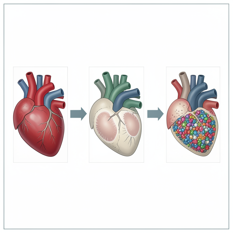
Comparison of Enzyme Levels
AI-generated conceptual illustration (human heart tissue and blood serum). It explains the idea and does not show study data.
View source evidence ↗Figures & tables
From the original paper Table 1
 Table 2
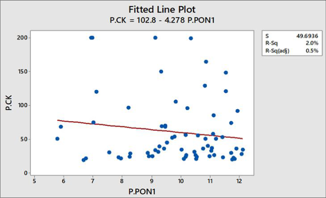 Figure 1
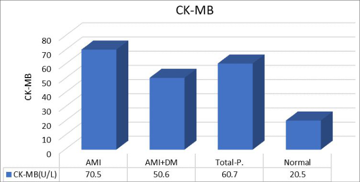 Figure 2
Introduction
Heart Diseases (HDs) are difficult to recognize due to risk factors such as DM, Hypertension (HTN), total Cholesterol (TC), abnormal pulse rate and others. Therefore, HDs must be dealt with carefully, because their nature is complex and it is predicted based on symptoms [1] . Heart consists of three coronary arteries [2] , the coronary arteries main (left and right) that branch into smaller arteries [3] . The left coronary artery divides into the left anterior descending artery, which is the largest coronary artery [4] . Coronary Heart Disease (CHD), It is one of the most common heart diseases it is the result of Atherosclerosis (AS) changes in the vessels supplying the heart and describes a range of clinical disorders from asymptomatic AS and stable angina pectoris to Acute Coronary Syndrome (ACS) that is classified as elevated MI (STEMI) and non-elevated MI (NSTEMI) and unstable angina pectoris [5] and Sudden Coronary Death (SCD) [6] , It is one of the most dangerous results of coronary Artery [5] , arrhythmia and Heart Failure (HF) [7] . CHD is characterized by a complete violation of the heart's blood supply, because of a damage [8] or narrowing coronary arteries [7] and it's the largest cause of death, disease and disability. Rarely a single etiology is found in the majority of elderly affected [9] . The AS or thromboembolism for left anterior descending artery cause MI involving large areas of the anterior, septal, and apical portions of the heart [4] . Studies have reported that arterial blockage is accompanied by blood vessels inflammation. Once the blood vessel wall is damaged, immune cells collect at the site of infection and produce proinflammatory cytokines that activate circulating white blood cells that will engage more immune cells that cause fat accumulation and gradually blockage of the artery, because these areas are smooth for the development of clots, which It clogs the important arteries of the heart, causing MI or stroke [10] . It has been proven that many chemotherapy agents cause CHD, and CV complications during chemotherapy, and radiotherapy became an increasing problem [11] also, it was found that kounis syndrome is an allergic reaction or hypersensitivity leads to coronary spasm and ACS [12] .
Acute myocardial infarction
The MI is a medical emergency [13] life-threatening, timesensitive [14] , and non-infectious that occurs due to ischemia or stoppage of perfusion caused by pathological changes or abnormalities in the walls of coronary arteries [15] which the coronary arteries cannot supply to part of the heart for long periods, causing damage to the heart tissue and its possible death [13] . Sudden occlusion occurs due to AS plaque [16] causing reduced force of heart contraction. If the thrombus ruptures before complete necrosis for the distal tissue will occur MI [15] . Approximately 90% of the consequences of MI are caused by an acute clot that prevents atherosclerosis of the coronary arteries [17] the heart muscle becomes inflamed and necrotic at the point of blockage, and the damaged area rapidly loses its ability to contract and conduct electrical impulses and depletes the supply of O 2 this damage is irreversible and the area of necrosis is eventually replaced by fibrous scar tissue [16] . MI is clinically defined as myocardial injury detected by cardiac biomarkers with evidence of acute cardiac ischemia as changes in the Electrocardiogram (ECG) such as the appearance of pathological Q waves [18] . Complete obstruction of the coronary artery is the main mechanism of MI [8] . MI causes cardiac remodelling, which are changes in heart size, shape, and function, it is a group of pathophysiological changes including electrophysiological changes, ventricular dilatation, myocyte hypertrophy, and interstitial fibrosis [19] . Most information on prevention, diagnosis, and treatment is still based on studies conducted predominantly in males, although predictive risk factors differ between the sexes [20] . The five common symptoms of MI, are chest pain or discomfort, shortness of breath, pain or discomfort in the arms or shoulders, feeling weak, dizzy or faint, and jaw, neck, or back pain [21] . MI can occur in menopause, family history of (HTN and DM) has a significant contribution, where smoking, obesity and dyslipidemia are modifiable factors, non-modifiable factors are age, sex, genetics [15] , age for males over 40, females over 50 years of age, excessive alcohol consumption, high TC, chronic high stress, previous MI and arrhythmia [22] . Diagnosis and treatment of patients depends on the precise classification of the infarction [14] and AMI is divided into modes with respect to period, treatment, and prognosis [20] . ACS is characterized by a sudden decrease in cardiac blood flow and can be diagnosed and categorized into STEMI and NSTEMI based on ECG [23] and generally infarcts are classified on the basis of microscopic size or location (anterior, lateral, inferior, etc.), in the pathological context, an acute, curable or healed infarction should be used [14] .
The fourth global definition of the disease of MI emphasized the difference between AMI and cardiac injury, which divided MI into five types [23] . The incidence of AMI in the age group (40-60) year is (8) times higher than in persons of younger age [24] . Percutaneous Coronary Intervention (PCI) is a strategy preferred for reperfusion for STEMI patients. The 2013 STEMI guidelines recommend that hospitals capable of performing PCI treat patients within (90 minutes) of contact with the medical system [25] .
Diabetes Mellitus
The diabetes mellitus (DM) is classified into three types according to etiology and clinical presentation Diabetes Mellitus Type (1 and 2) (DMT1), (DMT2), gestational diabetes, and subtypes are monogenetic and secondary DM [26] , because of the ambiguities that arose after the discovery of subtypes, for that the classification included new names, which are DM type 1, 2 and 3 [27] . DM is a chronic metabolic disorder [26] involves either a disturbance in insulin secretion, action, or both [27] , It is a multifactorial disease that includes hyperglycemia and insulin resistance [28] . which can lead to direct and indirect effects on large and minute blood vessels [29] , complications of large blood vessels lead to (2-4) times the risk of Cardiovascular Disease (CVD) [26] . The risk factors are advanced age, duration of DM, insulin use, CHD, elevated serum creatinine [30] , poor glucose control, smoking, HTN, dyslipidemia and CVD [29] . The risk of CVD in patients with DM is increased due to the association between insufficient insulin action and the various mechanisms of AS that occur in DM. The mechanism that increases CV damage in DM is oxidative stress. Chronic hyperglycemia leads to endothelial or mitochondrial dysfunction, metabolic disturbances, and damage to the extracellular matrix causing a functional deterioration of the heart called cardiopathy [28] . In DM a whole cascade of pathological interactions appears in the endothelium of blood vessels caused by glucose toxicity, excessive action to stimulate HTN, thrombotic activators and the intensification of oxidative stress leading to Endothelial Dysfunction (ED), The damaged endothelium itself causes DM and inflammatory factors [31] . The anatomical substrate of inflammation in DM is the endothelium of blood vessels. The deficiency of the enzyme lipoprotein lipase enhances the production of Very Low-Density Lipoprotein cholesterol (VLDLs) enriched with cholesterol esters, LDL enriched in Triglycerides (TGs) and increases small dense Low-Density Lipoprotein cholesterol) (sdLDL-c) and Reactive Oxygen Species (ROS). The overproduction of ROS eventually leads to a severe decrease in the bioavailability of Nitric Oxide (NO), especially in endothelial cells. Oxidation of high Density Lipoproteins (HDLs) by ROS reduces their anti-inflammatory capacity and Reverse Cholesterol Transporter (RCT). Insulin causes increased blood flow and glucose availability in skeletal muscle and additional vasodilation, depending on NO. Hyperinsulinemia can promote cardiac hypertrophy and a significant increase in left ventricular mass with an antiproteolytic effect on the heart [32] . It was observed that the presence of DM doubles the risk of CVD in men and triples it (3) times in women [33] . The risk of developing MI among DM patients is equivalent to that of normal people with a previous history of MI [34] . In 2017, DM was estimated to affect 452 million patients worldwide, a number that is expected to rise to 693 million by 2045 [35] .
Paraoxonase-1
Paraoxonases are a family of three specific enzymes: PON1, PON2 and PON3. PON1 is the most studied among them and is not necessarily the most important, that PON2 is the oldest of them, from which it arose PON3 and then PON1 [36] . PON a polygenic family of lipolactonases, their genes are located next to each other on human chromosome 7 [37] and their protein structure is similar, the name paraoxonase is due to the ability to hydrolyze the compound (diethyl pnitrophenyl phosphate), which is a metabolite from oxon, parathion organophosphate. PON1 is a Ca-dependent glycoprotein with a molecular weight of (43KDa) and composed of (355 amino acid) and has a structure consisting of six helical sheets with a central tunnel containing two Ca 2+ to (7.4Å) apart. The ion in the bottom of the tunnel is structural and gives conformational stability. The ion present in the cavity of the active site has a catalytic function by locating the substrate and activating the ester bonds for the substrate the helixes 1(H1), 2(H2) participate in the PON1-HDL reaction, loop 1(L1) is the cap of the active site and the extended helix 3(H3) called loop 2(L2) participates in substrate recognition [38] . The PON1 genes contain many polymorphisms in the gene coding region [39] . Its enzymatic activity depends on the substrate and varies with different backgrounds [40] . CHD has been shown to be closely related to a wide range of genetic variants. One of the major biomarkers of AS is the PON1 gene [41] . PON1 is synthesized in the liver and binds to HDL-c before it is released into the blood, and smaller amounts are made by the kidneys and colon, where a small amount binds to VLDL-c and postprandial chylomicrons [38] and it is found to a lesser extent in LDLs. PON1 possesses three enzymatic activities: Lactonase, arylsterase, and paraoxonase. PON1 polymorphisms play an important role in drug metabolism and prevention of CVD and neuro-degeneration [36] . PON1 plays a role in the regulation of RCT anti (Oxidation, inflammation, urination, coagulation, DM, microbes and AS) and vasodilation [42] . PON1 hydrolytic enzyme [39] because PON1 and (PONAryl) are believed to have the ability to degrade organophosphates and aromatic carboxylic esters and protects HDL-c and LDL-c from H 2 O 2 and its antioxidant effects as it slows down the oxidation of LDL-c [43] through the hydrolysis for fatty acids oxidized, phospholipids, TC and hydroperoxides triglycerides [38] . The arylsterase activity of PON1 is involved in the detoxification of lipid peroxides that is associated with endothelial dysfunction of blood vessels and CHD [40] . In DM, complications in CV due to inhibition and disturbance of stability of PON1, PON1 may be affected by binding to HDL-c resulting in decreased efficiency in its antioxidant and H 2 O 2 properties [38] .
Creatine Kinase isoenzymes MB
The Creatine Kinase (CK) a two-dimensional enzyme [44] has a critical effect on the transfer of energy from mitochondria to sites of use Adenosine Triphosphate (ATP) in the brain, skeletal muscles, and heart. In cardiac cells, it is expressed as three identical enzymes: creatine kinases type M and B, and sarcomeric mitochondrial creatine kinase.
Creatine + ATP ↔ Phosphocreatine + ADP + H +
In patients with MI, ATP and phosphocreatine levels are rapidly depleted resulting in tissue damage and increased CK-MB levels [45] . In stimulated tissues, CK conjugates to ATP-consuming enzymes and performs reverse transmission of the phosphoryl group (P) between phosphocreatine and Adenosine Diphosphate (ADP) in the presence of Mg the CK is also able to act as a cellular metabolic balancer by reducing the rise in intracellular ADP and H + which prevents ADP-induced ATPase inactivation and acidification of cells [46] . Myocardial death due to MI leads to the release of many molecules such as CK into the blood. Since CK-MB is the isoform found in cardiac, it is the most specific and accurate for detecting MI among the three isoforms [47] , its elevation is relatively specific to myocardial injury especially in patients with ischemic symptoms when skeletal muscle damage is not present, elevated CK-MB can be due to non-cardiogenic causes such as hypothermia [48] . CK-MB is the first visceral biomarker of heart damage identified in 1979 [49] when released into the blood, CK-MB can be divided into two groups, MB1 and MB2, and when AMI occurs, MB2 passes into the blood with a significant change in the (MB2:MB1) ratio. Considered as the (MB2:MB1) ratio ≥1.5 is an indicator of AMI [44] . The sensitivity level of CK-MB in the diagnosis of MI is about 90% [50] . The relative index (CK-MB/total CK × 100) can be used to diagnose MI ࣣ if this indicator is 2.5% or higher, then CK-MB has a great potential to launch from the heart. Total levels of CK and CK-MB correlate with infarct size, and CK-MB cannot detect minor damage to the heart [44] . Some studies have shown that asphyxiated newborns have significantly higher CK-MB values than healthy infants in the first 24 hours of life [46] . CK-MB is found in small amounts in skeletal muscle and may be elevated during physical exercise or in certain diseases [51] . CK-MB is not relatively sensitive to detecting small MI and cannot be used for late diagnosis of AMI but can be used to suggest infarct extension if levels rise again after declining [52] .
Materials and Methods Collection and preservation of research samples
Individual information was collected on a questionnaire after obtaining official approval, venous blood samples were drawn blood samples (100 samples) were collected, that included of male sex only from the ages of (40-80) years of smokers and nonsmokers, which included (34) samples for AMI patients, (33) samples for (AMI + DM) patients and (33) samples for healthy people. Patient samples were collected in the period two months in the Internal Resuscitation Division after they were diagnosed by specialized doctors, of those who did not exceed their stagnation a full day of Kirkuk Governorate hospitals/Iraq. AMI patients who underwent drug therapy or coronary artery bypass grafting were excluded. After separating the blood sample and obtaining the serum, it was frozen at a temperature of (-20 ℃) until the variables were measured.
Determination level of paraoxonase-1 in blood serum
Using Human PON1 (serum paraoxonase/arylesterase 1) enzyme-linked immunosorbent assay (ELISA) kit supplied by the korean company (LABISKOMA). The device has been used for absorbance microplate reader-ELX800TM -BioTekTM and the automated microplate strip washer-ELX50-BioTek. Absorbance was read at 450nm, and then, the concentration of PON1 was calculated.
2.2.b. Determination level of CK-MB in blood serum
The diagnostic kit was used to measure the CKMB enzyme prepared by the Chinese company (Shijiazhuang Hipro Biotechnology Co., Ltd).This product is used to determine the activity of creatine kinase (isoenzyme MB) in human serum, it is mainly used for the adjuvant diagnosis of MI, muscular dystrophy and other diseases.
Statistical analysis
A statistical analysis program known as the SPSS was used (Tow-Sample T-test or independent samples T-test) by comparing the group patients of AMI and (AMI+DM), both of them separately, with a healthy group, for the extraction the mean, Standard Deviation (SD), probability level Pvalue, and correlation analysis. 2), it was found that the level of concentration of PON1 by unit (ng/mL) using the mean standard deviation rises in AMI patients with higher statistical differences appear at the level of probability (P=0.000), also rises in (AMI + DM) patients with normal statistical differences appear at (P = 0.038), and it rises in the total of patients significantly higher at (P = 0.001) when compared to the healthy control. CK-MB concentration in units (U/L), using the mean standard deviation. It rises in the sera of both AMI, (AMI + DM) patients and total patients and shows high statistical differences at the level of probability (P = 0.0003) compared to the control group. A negative correlation between the concentration of each (PON1 and CKMB) has no statistical significance (r = -0.140, P = 0.258).
Results
Results
Discussions
In this study the level of PON1 concentration was rises in AMI, (AMI + DM) and total patients when compared them with control group they are not compatible with both Sowmya Varadhan et al., who found a lower level of PON1 in ACS patients as compared to the control group [53] , conflict with the study by Lorena Ciumărnean et al., which showed decreased activity of PON1 arylsterase in patients with consider as an independent predictor of 1-year survival after AMI [54] and the study by Anna Wysocka et al., which found that DM patients with Coronary Artery Disease (CAD) had lower PON1 activity than in DM patients, although similar lipid properties [55] , and conflict with the study by Mike Mackness and Eser Sozmen that confirmed that low PON is associated with AS, by comment by them on some of the data's which they called inaccurate from realistic side [56] , including that high PON levels are associated with AS and that are supporting the our results and which are showed up in (citation 19) from the study of Yaser Said Cetin et al., [57] . PON1 gene contains approximately 200 different single nucleotide polymorphisms [40] that alter the oxidation state of lipoproteins [38] , the polymorphism independently affects the enzyme's ability to protect LDL oxidation [40] and affects its hydrolytic activity with natural and synthetic substrates [39] . Its activities show differences between individuals and multiracial due to genetic polymorphism ࣣ even among individuals with the same genotype, its activity may show differences 13-40 times due to external factors, lifestyle, age, different physiology, or diseases that can affect the levels of PON1 [54] . The elucidation of the physiological roles of PON, the active center, and all applications in medical fields depend on its substrates [58] and homocysteine thiolactone is one of its basic internal substrates that can negatively affect protein structure/function through posttranslational modification and has recently been shown to be predictive of MI in CAD patients [39] . PON is also known as (aromatic esterase 1) encoded by the PON1 gene. Polymorphisms in the PON1 gene, (L55M and Q192R) on exon (3 and 6) respectively are known to pose a risk for CVD [59] . Oxidative stress is a defect resulting from deficits in defense systems or excessive production of ROS associated with decreased antioxidant activity. This deficiency could be the result of genetic factors, environmental, metabolic imbalance, toxicity or direct attacks by accumulation of free radicals, cause nonspecific and irreversible oxidation of biological molecules thus loss of function and can cause metabolic imbalance who will affect biological molecules in their structures or activities. By physiology, the neutralization of free radicals is ensured by defence systems enzymatic and non-enzymatic antioxidants. PON plays an important role in equilibrium redox similarly, individuals do not have the same antioxidant potential. Is prepared a repair process for each pathological condition as a result, the endogenous defence induces many signalling mechanisms to adapt to the new physiological situation [60] . Our study showed that the level of cardiac creatine kinase was elevated in all AMI and (AMI + DM), and total patients when compared with the control group. Our study found a significant increase in CK-MB levels in patients with AMI, and this is consistent with the study of Haider Raad Hashim [17] , and the study of Salih Hamzah [61] and a study (Mehmet Oguzhan Ay et al.) whereas, CK-MB levels were found in the STEMI (p< 0.001) and NSTEMI (p< 0.001) group of AMI patients significantly higher than the control group [62] . And the results of our study are identical with the study (Dr. Kiran Kumar Akka and Dr. Pampa Reddy), which found a significant increase in the level of CK-MB in patients with DMT2 and MI [63] . While it did not agree with the study (Mingdan Zhu et al.), which did not show any significant change in the level of CK-MB in the group MI [64] . when the myocardial membrane loses its integrity due to muscle cell death, the first injuries caused by AMI lead to an increase in the concentration of CK-MB in the surrounding blood to measurable values after approximately 4 or 6 hours. Concentrations can reach peaks of (39-185) ng/mL in the period 18-24 hours [65] since it is cleared from the circulation faster than cTn, this makes it the best biomarker for detecting re-infarction in patients [66] . CK-MB is the most cardiac-specific enzyme [67] . Disease states usually result in moderate or extensive tissue damage (depending on the time and severity of disease onset) which eventually leads to the release of enzymes (non-functional enzymes specific to the diseased organ or tissue) into the circulation resulting in an increase in the activity of these enzymes in fluids body [47] .
Conclusion
On Paraoxonase-1 (PON1) and Creatine Kinase-MB (CK-MB) levels in patients with Acute Myocardial Infarction (AMI), with or without Diabetes Mellitus (DM), reveals significant findings amidst a complex landscape of prior research. Contrary to some studies, we observed elevated PON1 concentrations in AMI and (AMI+DM) patients compared to controls, suggesting a potential role in oxidative stress mitigation. This contrasts with reports of decreased PON1 activity in coronary artery disease contexts. Meanwhile, our findings of increased CK-MB levels in AMI patients align with established biomarker trends, supporting its clinical utility in AMI diagnosis. These insights underscore the multifaceted roles of PON1 and CK-MB in cardiovascular pathology, highlighting both their diagnostic potential and the need for further mechanistic exploration.
Figures and tables from the paper
| Patients | — | AMI | — | AMI+DM | — | Control | — |
|---|---|---|---|---|---|---|---|
| No. | % | No. | % | No. | % | No. | % |
| 67 | 67 | 34 | 50.746 | 33 | 49.2537 | 33 | 33 |
Table text extracted from the original PDF; superscript letters are shown inline. The original table image is below.
View original table image
| Parameters | P-value | — | Mean±SD | — |
|---|---|---|---|---|
| AMI n=34 | AMI+DM n=33 | Total Patients n=67 | Control n=33 | |
| PON1 | (10.22±1.50)** 0.0002 | (9.45±1.70)* 0.038 | (9.84±1.63)** 0.001 | 8.53±1.82 |
| CK-MB | (70.5±14.4)** 0.0003 | (50.6±13.1)** 0.0003 | (60.7±9.8)** 0.0003 | 20.5±1.22 |
Table text extracted from the original PDF; superscript letters are shown inline. The original table image is below.
View original table image
References
48 in the original paper- Mohan S, Senthilkumar, Thirumalai C, Srivastava G. Effective Heart Disease Prediction Using Hybrid Machine Learning Techniques. IEEE Access. 2019;7:81542-81554.
- Bharath Kumar A, Umashankar MS. Pharmacotherapeutic Management of Cardiovascular Disease Complications: A Textbook for Medical Students. Current Cardiovascular Risk Reports. 2020;14(9):272-296(25), 69-83(15), 160-171(12), 256271(16), 46-57(12), 25-34(10).
- Ishankulova NN. Coronary Heart Disease. The American Journal of Medical Sciences and Pharmaceutical Research. 2021;03(02-04):31-36.
- Khalil H, Abd EI Maksoud AI. Interruption of Autophagosome Formation in Cardiovascular Disease, an Evidence for Protective Response of Autophagy. Immunological Investigations: A Journal of. 2019;49(3):1-15. Available from: https://www.researchgate.net/publication/334187464
- Khan K, Szalai G, Anjum H. Cureus, Bee Attack or Heart Attack: Kounis Syndrome. 2021;13(4):1-8.
- Abbass LB. Evaluation of serum C-reactive protein and lipid profile in patients with myocardial infarction. Zanco Journal of Medical Sciences. 2018;22(3):349354.
- Kingma JG. Myocardial Infarction: An Overview of STEMI and NSTEMI Physiopathology and Treatment. World Journal of Cardiovascular Diseases. 2018;8(11):498-517.
- Mahanani DTI, Taviyanda D, Srinalesti. Characteristics of Acute Myocardial Infarction Patients. Strada Journal Ilmiah Kesehatan. 2020;9(2):1017-1026.
- KM SM, Kumar P. Serum fasting lipid profile and lipoprotein (a) in northern Keralite patients with myocardial infarction. MedPulse International Journal of Biochemistry. March 2019;9(3):78-83.
- Hashim HR, Ryhan B, Raad H. Acute Myocardial Infarction associated with Lipid Profile among Patients at Nasiriyah Heart Center. Journal of Education for Pure Science-University of Thi-Qar. 2020;10(2):33-40.
- Sarhat ER. Acute Myocardial Infarction: Melatonin, Apelin, and Visfatin as Predictors of Disease. Diyala Journal of Medicine. 2017;13(2):11-17.
- Sundquist SC, Johansson SE, Wolff M. The ratio of total cholesterol to high-density lipoprotein cholesterol and myocardial infarction in Women's health in the Lund area (WHILA): A 17-year follow-up cohort study. BMC Cardiovascular Disorders. 2019;19(1):1-9.
- Mahajan S, Valero-Elizondo J, Khera R, Desai NR, Blankstein R, Blaha MJ, et al. Variation and disparities in awareness of myocardial infarction symptoms among adults in the United States. JAMA network open. 2019;2(12):e1917885.
- Arslankaya S, Çelik TM. Prediction of Heart Attack Using Fuzzy Logic Method and Determination of Factors Affecting Heart Attacks. International Journal of Computational and Experimental Science and Engineering. International Journal of Computational and Experimental Science and Engineering. 2021;7(1):1-8. Available from: 23. Wennberg EC, Olsson T, Jansson JH. Lysophospholipids as Predictive Markers of STElevation Myocardial Infarction (STEMI) and Non-STElevation Myocardial Infarction (NSTEMI). Metabolites. 2020;11(1):25. International Journal of Advanced Chemistry Research https://www.chemistryjournals.net
- Yamini ND, Gopalakrishnan B, Selvam R, Saravanan. Troponin -T as a prognostic and diagnostic marker for myocardial infarction. GSC Biological and Pharmaceutical Sciences. 2021;14(01):95-100.
- Park J, Choi KH, Lee JM. Prognostic Implications of Door-to-Balloon Time and Onset-to-Door Time on Mortality in Patients With ST-Segment-Elevation Myocardial Infarction Treated With Primary Percutaneous Coronary Intervention. Journal of the American Heart Association. 2019;8(9):e012188.
- Hussain SS, Javaid A, Khan TA, Zahir H. Diabetes Mellitus, Obesity and Adipocytokines: Pathophysiological Perspectives. International Journal of Biology and Biotechnology. 2019;16(2):325-339.
- Halim MH, Alice. The Effects of Inflammation, Aging and Oxidative Stress on the Pathogenesis of Diabetes Mellitus (Type 2 Diabetes). 2019;13(2):1165-1172.
- Adam NAM, Ibrahim MA, Ismeil AAM. Sudanese diabetics with hypertension are at high risk for cardiovascular disease. Endocrinology & Metabolism International Journal. 2021;9(3):1-5.
- Chernobryvtsev SV, Ziablytsev TI, Panova OP. Connection of the endothelial dysfunction factors and diabetes mellitus 2 type severities. Journal Medical Science of Ukraine (NMU). 2018;14(1-2):34-39.
- Collet Camarillo HF, Contreras F, Collet-Salgueiro D, Velasco M. Diabetes Mellitus and Cardiovascular Disease: A Review. 2020;3:1-9.
- Das PB, Das P, Nath BK, Basumatary A, Dwijen. HbA1C and its Correlation with Lipid Profile in Acute Myocardial Infarction. International Journal of Contemporary Medical Research. 2018;5(4):13-16.
- Chikezie FO, Ohiagu PC, Chikezie PC. Pathophysiology of diabetes mellitus complications: Metabolic events and control. Biomedical Research and Therapy. 2021;8(3):4243-4257.
- Mackness M, Mahrooz A. Epigenetics of paraoxonases. Current Opinion in Lipidology. Publish Ahead of Print. 2020;(4):1-7.
- Jasso ED, Torres-Sánchez J, Torres J. Effect of structure and function of paraoxonase-1 (PON-1) on organophosphate pesticides metabolism. 2020;44(3):363-370.
- Perła-Kajan´ J, Włoczkowska O, Zioła-Frankowska A, Frankowski M, Smith AD, de Jager CA et al. Paraoxonase 1, B Vitamins Supplementation, and Mild Cognitive Impairment. Journal of Alzheimer's disease. 2021;81(3):11-19.
- Gogas Yavuz D, Üstay Ö, Atak PG, Telli A, Apaydin T, Şirikçi Ö et al. Serum Paraoxonase-1 Activity and Paraoxonase Q192 Gene Polymorphism in a Young, Healthy Population. Turkish Journal of Endocrinology and Metabolism. 2021;25(2):193-201.
- Sabar MR. Interventional Cardiology: The association of Paraoxonase 1 (PON1) gene polymorphisms with coronary artery disease. Interventional Cardiology. 2021;13:312-315.
- Stefanović JK, Vekic J. Paraoxonase 1 and atherosclerosis-related diseases. BioFactors. 2019;46(2):193-205.
- Abdullah YET. Sodium Valproate and Levetiracetam Treatment in Children: Their Effects on Serum Paraoxonase/ Arylesterase Activities. Crimson Publishers. 2020;3(3):1-7.
- Wu Y, Pan N, An Y, Xu M, Tan L, Zhang L et al. Diagnostic and Prognostic Biomarkers for Myocardial Infarction. Frontiers in Cardiovascular Medicine. 2021;7:1-13.
- Buonocore C, Bellieni CV, Tomasini B. Normal values of creatine kinase and of MB-creatine kinase at birth in healthy babies. Minerva Pediatrica. 2017;75(1):21-25.
- Durojaye NE, James PO, Onuorah O, Ilo CC, Okeowhor D, Cosmas S et al. Clinical Diagnosis of Disease States Using Enzymes and Proteins (Review). 2018;1(3):1-6. Article no. AJBGMB.46337.
- Sawalha K, Vedala K, Liu E. CK-MB Elevation in Mild Hypothermia: What We Did and We Should Have Done. Journal of Investigative Medicine High. 2021;9:1-3.
- Attia A, Faisal R, Sharif A, de Christopher. Creatine kinase-mb; Comparison of creatine kinase-mb in male and female acute myocardial infarct patients before and after the treatment. 2017;24(03):422-425.
- López-López S, Pareja-Galeano H. Cardiovascular biomarkers modified by exercise. Journal of Laboratory and Precision Medicine. 2018;3(2):1-10.
- Simon, Varadhan S, Venkatachalam R, Supriya A. Myeloperoxidase and Paraoxonase Activity in Acute Coronary Syndrome Patients. 2021;33(48B):108-114.
- Milaciu LC, Greavu M, Vesa SC, Tanțău AI, Dogaru GB, Alexescu TG et al. Arylesterase activity of Paraoxonase 1 -prognostic factor for one-year survival in patients with acute myocardial infarction. Revista Română de Medicină de Laborator. 2018;26(3):1-10.
- Nski AW, Cybulski M, Wysoki AP. Paraoxonase 1 Activity, Polymorphism and Atherosclerosis Risk Factors in Patients Undergoing Coronary Artery Surgery. Journal of Clinical Medicine. 2019;8(4):441.
- Çetin YS, Bozan N, Avcı K, Aslan M, Erel Ö. The relationship between thiol-disulfide balance and idiopathic sudden sensorineural hearing loss. Brazilian Journal of Otorhinolaryngology. 2022;88:948-953.
- Xiao X, Mu X, Yi X. Substrates for Paraoxonase. Current Pharmaceutical Design. 2017;24(5):615-627.
- ArulJothi KN, Abirami BS, Irusappan S, Gautami A. L55M and Q192R polymorphism of Paraoxonase gene and the risk of myocardial infarction in South Indian Tamil population. Meta Gene. 2017;15(2018):55-59.
- Hamzah SA. Association of biochemical tests cardiac enzyme with myocardial infarction in emergency hospital Erbil-Iraq. Asian Journal of Science and Technology. 2018;9(08):8488-8491.
- Selma T, Kozaci N, Mustafa Avci. Oxidant and antioxidant levels in patients diagnosed with acute coronary syndrome at the emergency department. Annals of Medical Research. 2021;28(6):1235-1240.
- Akka KK, Pampa D. Lipid profile in patients of type 2 diabetes mellitus with myocardial infarction. International Journal of Advanced Biochemistry Research. 2021;5(1):14-19.
- Zhu M, Han Y, Zhang Y. Metabolomics Study of the Biochemical Changes in the Plasma of Myocardial Infarction Patients. Frontiers in Physiology. 2018;9:1017.
- Ferreira ALF, Lima LF, Moraes AS. Development of a novel biosensor for Creatine Kinase (CK-MB) using Surface Plasmon Resonance (SPR). Applied Surface Science. 2021;554(4):149565.
- Prathima MB, Reshma S, Sushith, Shubhalakshmi, Madan G. Serum CK-MB, fasting lipid profile and lipid indices in patients with myocardial infarction: A casecontrol study. International Journal of Clinical Biochemistry and Research. 2019;6(1):45-47.