
Research article · 2024 · Full text
International Journal of Biology SciencesImpact of Toxoplasma gondii on prostaglandin, progesterone, oxytocin and anti-müllerian in abortion women
Volume 6(1) · Pages 45–49 · DOI: 10.33545/26649926.2024.v6.i1a.186 ↗
Research areasToxoplasma gondii Research Studies
Abstract
Author-writtenThe current study aimed to reveal the level of effect of the Toxoplasma gondii parasite on some sexual and nervous hormones and antioxidants. The current study was conducted in the city of Mosul and included 150 blood samples collected during the time period from March 2023 to November 2023 randomly from aborted women arriving at government hospitals and clinics. For age groups from 20 to 35 years, which is the ideal period for pregnancy and childbirth. Blood samples were collected from Women's Health Hospital, Doctors without Borders Hospital, Al-Khansa General Hospital, outpatient clinics, and some research laboratories. Blood samples were collected from aborted women suspected of being infected with the parasite, and then the samples were stored at 20 degrees. Until an ELISA test is performed to detect infection and estimate the concentration of specific antibodies, the samples are divided into the control group, which consists of 60 samples from non-infected women. Infected women, and the patient group, which consists of 90 samples from aborted women suspected of being infected with the parasite, where the infection was diagnosed by detecting specialized antibodies (IgG, IgM). Parasite. Two types of serological tests were used in the current study for the purpose of detecting infection: the ELISA-IgG test and the ELISA-IgM test. The results of these tests showed the presence of infection in women, which amounted to (1.176±0.591 pg/ml) and IgM (0.776±0.175 pg/ml). ml) inspector. The results of the current study showed a significant increase p≥ 0.001 in the levels of prostaglandins and oxytocin in the group infected with toxoplasmosis compared to the control group, reaching (pg/ml 1508±243) and (pg/ml 644±205), respectively, with a significant decrease in p≥ 0.001 in progesterone levels compared to the control group, which amounted to (3.08±1.59 pg/ml). On the other hand, there was a significant increase (p>0.05) in AMH levels, which amounted to (9293±8324 pg/ml) compared to the control group.
Summary
Source-checkedPlain-language reading of the full paper, prepared with AI and checked against the text. Each point links to the authors' own words.
Why it matters
- Toxoplasma gondii is an obligate intracellular protozoan parasite that causes toxoplasmosis in humans and other mammals and birds. View evidence ↗
- In humans, congenital toxoplasmosis can result in severe outcomes, including abortion and various congenital anomalies. View evidence ↗
- While often asymptomatic in healthy individuals, toxoplasmosis can cause varying symptoms in immunocompromised people, including pregnant women. View evidence ↗
The question
- The study aimed to determine how Toxoplasma gondii infection affects specific nervous and sexual hormones. View evidence ↗
What they did
- The research was conducted in Mosul between March and November 2023, using 150 random blood samples from women who had experienced abortions. View evidence ↗
- The participants were women aged 20 to 35 years. View evidence ↗
- The study compared a control group of 60 non-infected women with a patient group of 90 aborted women diagnosed with infection via IgG and IgM antibodies. View evidence ↗
- Infection was detected using ELISA tests for IgG and IgM antibodies. View evidence ↗
What they found
- Prostaglandin levels were significantly higher in infected women (1508±243 pg/ml) than in the control group (245±181 pg/ml) (p≤0.001). View evidence ↗
- Oxytocin concentrations were significantly increased in infected women (644±205 pg/ml) compared to the control group (115±114 pg/ml) (p≤0.001). View evidence ↗
- Anti-Müllerian Hormone (AMH) levels were higher in infected women (9293±8324 pg/ml) than in the control group (8324±1704 pg/ml). View evidence ↗
- IgM antibody levels were higher in infected women (1.176±0.591 pg/ml) compared to the control group (0.282±0.282 pg/ml). View evidence ↗
- IgG antibody levels were higher in infected women (0.776±0.175 pg/ml) than in the control group (0.312±0.040 pg/ml). View evidence ↗
What it means
- The authors conclude that there is a significant correlation between immune markers and hormonal changes in pregnant women infected with Toxoplasma gondii. View evidence ↗
- The authors note that the mechanism behind the observed hormone level changes remains unknown. View evidence ↗
Visual summary
1 visualsIllustrations explain the paper; they do not show study data.
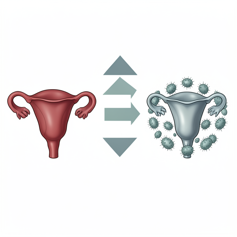
Hormonal Shift Summary
AI-generated conceptual illustration (pregnant human uterus and Toxoplasma gondii parasites). It explains the idea and does not show study data.
View source evidence ↗Figures & tables
From the original paper Table 1
 Table 2
 Table 3
 Table 5
 Table 4
 Table 6
Introduction
The arched parasite Toxoplasma gondii is considered a protozoan belonging to the primary animals (Protozoa) and is an obligate intracellular parasite. It infects mammals and birds as intermediate hosts, including humans, causing the disease Toxoplasmosis. Many members of the feline family (Felidae), which includes cats, serve as definitive hosts for the parasite [1] . The parasite affects humans through the ingestion of contaminated food or water containing expelled Oocysts in the feces of infected cats. Additionally, it can be transmitted through the consumption of undercooked or poorly cooked meat containing tissue cysts. A crucial means of transmission is through transplacental [2] .
Congenital toxoplasmosis in humans can lead to diverse clinical symptoms, including abortion, mental retardation, blindness, hydrocephalus, and congenital anomalies [5] . The parasite has varied effects on sexual hormones, with progesterone showing diverse impacts on Toxoplasmosis. Progesterone, produced in the ovaries, uterus, and brain, plays crucial roles in the body [6] , Progesterone, when present in cells infected with this parasite, impedes or prevents the regulation of the parasite's proliferation [7] , Additionally, the parasite affects Anti-Müllerian Hormone (AMH), as studies indicate fluctuations in AMH levels depending on the patient's age. The reason for increased levels may be attributed to the fact that AMH is present only in the ovaries until menopause, produced by granulosa cells [8] . Furthermore, the parasite causes an increase in oxytocin levels in the Paraventricular Nuclei (PVN) adjacent to the ventricles in the hypothalamus. Oxytocin released within the PVN leads to interconnected signals in distant brain regions, suggesting that Toxoplasma gondii infection enhances oxytocin signals in the brain [9] . T. gondii also stimulates the biosynthesis of Prostaglandin E2 (PGE2) in epithelial cells by regulating the production of arachidonic acid [10] .
Materials and Methods
The current study was conducted in Mosul and involved 150 blood samples collected randomly from women aged 20 to 35, representing the ideal period for pregnancy. Samples were gathered from Women's Health Hospital, Doctors without Borders Hospital, Al-Khansa General Hospital, outpatient clinics, and research laboratories between March, 2023 and November, 2023. For women suspected of parasitic infection, 5 ml of venous blood was withdrawn using a 5 ml medical syringe. The blood was placed in tubes containing gel for blood separation and centrifuged at 3000 rpm for 5 minutes to obtain serum. The separated serum was then distributed into 1.5 ml Eppendorf tubes and stored at -200 degrees until IgG, IgM ELISA and hormonal testing was conducted on all 150 samples, hormones were estimated following the manufacturer's instructions.
The samples were divided into two groups 1. The control group, consisting of 60 samples from uninfected women. 2. The patient group, comprising 90 samples from women with miscarriages suspected of parasitic infection.
Results and Discussion
The ratio of IgM is morally higher in infected women (1.176±0.591 pg/ml) compared to the control group (0.282± 0.282 pg/ml). The rise in serum of women with the parasite may be due to the tendency of the antidote, to survive for a long time in the case of recent infections [11] . Also, in the case of acute infection, the IgM antigen is an indicator of a low level for many years, the factors that help increase the IgM antigen when dividing and reproducing the parasite within the body as a reaction to the occurrence of reproduction, IgM often appears during the first two weeks of infection and is usually an important indicator of acute infection and when a pregnant mother is infected during pregnancy it threatens the life of the fetus causing congenital infection [12] . The current study shown in Table 2 also showed that the IgG antibodies increased to a ratio of (0.776±0.175 pg/ml) compared to control group (0.312 ±0.040 pg/ml) and its height may be due to the persistence of the anti-IgG, It is one of the most important components of the congenital immune response in control in a parasitic outbreak, which often begins one week after the two weeks of infection and may peak at 6-8 weeks, then begins to decline gradually for 1-2 years, and the low calibre may survive [13] .
| P-Value | IgG Mean ± SD | IgG |
|---|---|---|
| A | 0.776±0.175 | Patients |
| A | 0.312±0.040 | Controls |
Table text extracted from the original PDF; superscript letters are shown inline. The original table image is below.
View original table image
Concentration of prostaglandin hormone
The results, as per Table (3), demonstrate a significant increase (p≤0.001) in prostaglandin hormone levels in infected women (1508±243 pg/ml), compared to the control group (245±181 pg/ml). These findings align with studies by [14] , [15] , and [16] , but differ from [17] . The elevated levels may be attributed to intracellular signaling pathways in macrophage cells induced by Toxoplasma gondii. PGE2 production in macrophages exhibited a time-dependent pattern, initiating at 4 hours post-infection, peaking at 12 hours, stabilizing at a high level, and reaching another peak at 16 hours [18,19,20,21] . The upregulation of PGE2 in pathological conditions is significantly regulated by COX-2 gene induction, analyzed using qRT-PCR [22] . COX-2 gene expression analysis reveals regulated expression after 3 and 24 hours of T. gondii interaction. The COX-2 promoter region contains potential regulatory elements influencing gene transcription. Studies also link increased lipid droplet numbers during infection, caused by various pathogens, to enhanced eicosanoid production due to arachidonic acid cleavage and enzyme activity within lipid droplets [23] . Results indicate an increase in PGE2 synthesis from 6 to 48 hours post-infection with the parasite, positively correlating with lipid droplet formation. Enzymatic conversion of free arachidonic acid to PGE2 plays a crucial role in cellular responses to pathogens and host survival [15] . High PGE2 concentrations effectively inhibit Th1 response, Tumor Necrosis Factor (TNF), and Nitric Oxide (NO) production, favoring parasite growth. Overall, infected females treated with COX-2 inhibitors exhibited higher body weight changes and lower disease severity compared to untreated counterparts. This suggests that all COX-2 inhibitors can inhibit Toxoplasma gondii proliferation in vivo and in vitro, irrespective of strain or cell type [18,19,20] .
Concentration of progesterone hormone:
The results, as per Table (4), indicate a significant decrease (p≤0.001) in progesterone hormone levels in infected women (3.08±1.59 pg/ml) compared to the control group (28.3±12.6 pg/ml). These findings align with studies by [24, 25 26, 27] , but differ from studies by [28,29,30] . The observed substantial increase in hormone levels in infected pregnant women is attributed to hormonal fluctuations during pregnancy, impacting women's immune responses. This diminishes the inflammatory response, particularly the production of antibodies against parasites, increasing susceptibility to infections and undesirable developments in toxoplasmosis, during the second and third weeks of pregnancy, there is a noted increase in progesterone levels during Toxoplasma gondii spread, as observed by [31] . The decline in cellular immunity is linked to elevated levels of steroid hormones, enhancing the parasite's ability to survive in the body [7] . The decrease in hormone levels in pregnant women with toxoplasmosis may be due to the infection being in a nonacute stage [32] , Changes in pituitary gland activities during pregnancy, as well as placental size, maturity, and fetal immune responses, influence hormone levels during infection [33] , However, the mechanism remains unknown [32] . Studies on progesterone in mice, sheep, goats, and bone marrow-derived stem cells revealed low progesterone levels during pregnancy after T. gondii infection [24] . Pregnant mice infected with T. gondii showed clinical signs of infection, inflammation, and necrosis at the maternal-fetal interface, accompanied by reduced blood progesterone levels. Interestingly, direct progesterone treatment inhibited parasite invasion and stimulated autophagy. However, concentrations of many hormones, including progesterone, significantly increase during pregnancy [34] .
Concentration of oxytocin hormone
The results from Table (5) indicate a significant increase (p≤0.001) in oxytocin hormone concentration in infected women (644±205 pg/ml) compared to the control group (115±114 pg/ml). These findings are consistent with studies by [36,28,9] , which employed infected rat females and observed elevated oxytocin levels in the Paraventricular Nucleus (PVN) adjacent to the ventricles. The observed rise is attributed to the release of oxytocin within the PVN, initiating complex signals in distant brain regions. These observations suggest that Toxoplasma gondii infection enhances oxytocin signals in the brain. Oxytocin is crucial for social sexual behaviors in rodent females, and disrupting its signals in the brain leads to the loss of sexual approach in female mice towards specific males [36] . The study indicates that Toxoplasma gondii infection strengthens oxytocin signals from PVN to the Posterodorsal Medial Amygdala (MePD). The heightened oxytocin levels in pregnant infected women may be due to T. gondii influence, despite oxytocin naturally increasing during early labor in pregnant women [37] , Oxytocin stimulates uterine contractions, making it relevant for managing labor. Oxytocin is administered intravenously during labor induction, and its receptors' regulation increases sensitivity to oxytocin-induced contractions significantly in nonpregnant women. The study found a highly statistically significant difference (p< 0.001) between the two groups. In summary, the study suggests a significant correlation between immune markers and hormonal changes in pregnant women infected with Toxoplasma.
Concentration of anti-müllerian hormone
The recent study, detailed in Table (6), highlights a significant increase in Anti-Mullerian Hormone (AMH) concentration among infected women (9293±8324 pg/ml) compared to the control group (8324±1704 pg/ml). This contrasts with the findings of previous studies by [38,39] . The notable elevation can be attributed to the fact that AMH is exclusively produced in the ovary until menopause. It acts as a product of granulosa cells enveloping each egg, serving as a molecular marker for the relative ovarian reserve in women. The hormone's blood levels fluctuate depending on age and gender [8,40] . Considering the study's age group spanning 20 to 35 years, a higher AMH concentration can be deemed normal, especially within this age range optimal for fertility. The study implies that Toxoplasma gondii infection might impact AMH levels differently, depending on the patient's condition. This variation arises from individual factors and the patient's age, which influences the hormone's levels.
Acknowledgement
AcknowledgmentI extend my sincere thanks and gratitude to Dr. Ohood Mozahim Shaker for the effort and assistance she gave me in completing the research. May God grant her success as she loves and is satisfied.
Figures and tables from the paper
| Significant Differences | IgM Mean ± SD | IgM |
|---|---|---|
| A | 0.591 pg/ml±1.176 | Patients |
| A | 0.282±0.034 pg/ml | Controls |
Table text extracted from the original PDF; superscript letters are shown inline. The original table image is below.
View original table image
| P-Value | Prostaglandin Mean ± SD | prostaglandin |
|---|---|---|
| A | 1508±243 pg/ml | patients |
| A | 245±181 pg/ml | controls |
Table text extracted from the original PDF; superscript letters are shown inline. The original table image is below.
View original table image
| P-Value | Oxytocin mean ± SD | Oxytocin |
|---|---|---|
| A | 644±205 pg/ml | Patients |
| A | 115±114 pg/ml | Controls |
Table text extracted from the original PDF; superscript letters are shown inline. The original table image is below.
View original table image
| P-Value | Progesterone mean ± SD | Progesterone |
|---|---|---|
| A | 3.08±1.59 pg/ml | patients |
| A | 28.3±12.6 pg/ml | controls |
Table text extracted from the original PDF; superscript letters are shown inline. The original table image is below.
View original table image
| P-Value | Anti-Mullerian Mean ± SD | Anti-Mullerian |
|---|---|---|
| A | 9293±8324 pg/ml | Patients |
| A | 8324±1704 pg/ml | Controls |
Table text extracted from the original PDF; superscript letters are shown inline. The original table image is below.
View original table image
References
32 in the original paper- Mahmood SI, Saeed NAHA, Ali LQ. Rubella virus and Toxoplasma gondii infection with immune antibodies diagnosis among bad obstetric and Primigravida pregnant women in Baghdad City. World Bulletin of Public Health. 2022;12:78-85.
- Addo SO, Asare TO, Arthur C, Amoateng K, Addo K, Larbi JA. Toxoplasma gondii among pregnant women attending antenatal care in a district hospital in Ghana. Public Health Challenges. 2023;2(2):e82.
- Fallahi S, Rostami A, Shiadeh MN, Behniafar H, Paktinat S. An updated literature review on maternalfetal and reproductive disorders of Toxoplasma gondii infection. Journal of Gynecology Obstetrics and Human Reproduction. 2018;47(3):133-140.
- Castro-Filice LS, Barbosa BF, Angeloni MB, Silva NM, Gomes AO, Alves CMOS, et al. Azithromycin is able to control Toxoplasma gondii infection in human villous explants. Journal of Translational Medicine. 2014;12(1):132.
- Pellatt L, Rice S, Mason HD. Anti-Müllerian hormone and polycystic ovary syndrome: A mountain too high. Reproduction. 2010;139(5):825-833.
- Abdulai-Saiku S, Vyas A. Toxoplasma gondii infection causes an atypical abundance of oxytocin and its receptor in the female rat brain. Pathogens. 2021;10(11):1495.
- Peng BW, Lin JY, Zhang T. Toxoplasma gondii induces prostaglandin E2 synthesis in macrophages via signal pathways for calcium-dependent arachidonic acid production and PKC-dependent induction of cyclooxygenase-2. Parasitology Research. 2008;102:1043-1050.
- Enitan S, Adejumo A, Oluremi A, Ileoma E, Adejumo E, Oyedele T, et al. Sero-prevalence of antiToxoplasma gondii IgM and IgG antibodies among young adults with history of ocular infection in SouthWestern Nigeria. EAS Journal of Parasitology and Infectious Diseases. 2019;1(6):133-143.
- Quan L, Ze-Dong W, Si-Yang H, Xing-Quan Z. Toxoplasmosis and typing of Toxoplasma gondii. Parasit Vectors. 2015;8:292.
- Wang X, Chen J, Zheng J. The roles of COX-2 in protozoan infection. Frontiers in Immunology. 2023;14:955616.
- Peng BW, Lin JY, Zhang T. Toxoplasma gondii induces prostaglandin E2 synthesis in macrophages via signal pathways for calcium-dependent arachidonic acid production and PKC-dependent induction of cyclooxygenase-2. Parasitology Research. 2008;102:1043-1050.
- Gomes AF, Magalhães KG, Rodrigues RM, de Carvalho L, Molinaro R, Bozza PT, et al. Toxoplasma gondii-skeletal muscle cells interaction increases lipid droplet biogenesis and positively modulates the production of IL-12, IFN-g and PGE 2. Parasit Vectors. 2014;7:1-13.
- De Souza G, Silva RJ, Milián ICB, Rosini AM, de Araújo TE, Teixeira SC, et al. Cyclooxygenase (COX)- 2 modulates Toxoplasma gondii infection, immune response and lipid droplets formation in human trophoblast cells and villous explants. Scientific Reports. 2021;11(1):12709.
- Michelin MA, Silva JS, Cunha FQ. Inducible cyclooxygenase released prostaglandin mediates immunosuppression in acute phase of experimental Trypanosoma cruzi. Infection. Experimental Parasitology. 2005;111(2):71-79.
- Abdalla GK, Faria GE, Silva KT, Castro EC, Reis MA, Michelin MA, et al. Trypanosoma cruzi: The role of PGE2 in immune response during the acute phase of experimental infection. Experimental Parasitology. 2008;118(4):514-521.
- Tatakihara VLH, Cecchini R, Borges CL, Malvezi AD, Graça-de Souza VK, Yamada-Ogatta SF, et al. Effects of cyclooxygenase inhibitors on parasite burden, anemia and oxidative stress in murine Trypanosoma cruzi infection. FEMS Immunology & Medical Microbiology. 2008;52(1):47-58.
- Moraes KC, Diniz LF, Bahia MT. Role of cyclooxygenase-2 in Trypanosoma cruzi survival in the early stages of parasite host-cell interaction. Memórias do Instituto Oswaldo Cruz. 2015;110(2):181-191.
- Zhang X, Zhang J, Yang X, Han X. Several transcription factors regulate COX-2 gene expression in pancreatic beta-cells. Molecular Biology Reports. 2007;34(3):199-206.
- Bozza PT, Magalhães KG, Weller PF. Leukocyte lipid bodies -Biogenesis and functions in inflammation. Biochimica et Biophysica Acta (BBA)-Molecular and Cell Biology of Lipids. 2009;1791(6):540-551.
- Jones LA, Jean-Paul A, Henriquez FL, Lyons RE, Nickdel MB, Carter KC, et al. Toll-like receptor-4mediated macrophage activation is differentially regulated by progesterone via the glucocorticoid and progesterone receptors. Immunology Letter. 2008;125(1):59-69.
- Brito C, Silva MT, Castro MM, Wyrwas W, Oliveira BM, Fonseca BM, et al. Toxoplasma gondii infection reduces serum progesterone levels and adverse effects at the maternal-foetal interface. Parasite Immunology. 2020;42(2):e12690.
- Galvan-Ramirez MDLL, Gutiérrez-Maldonado AF, Verduzco-Grijalva F, Jiménez JMD. The role of hormones on Toxoplasma gondii infection: A systematic review. Frontiers in Microbiology. 2014;5:503.
- Wu Y, Zhang X, Fu Y, Liu J, Xue Y, Liu Q, et al. Progesterone can directly inhibit the life activities of Toxoplasma gondii in vitro through the progesterone receptor membrane component (PGRMC). International Journal of Molecular Sciences. 2022;23(7):3843.
- Erzaiq ZS. The correlation between some immunological markers and hormonal changes among patients with toxoplasmosis in Tikrit city. Journal of Population Therapeutics and Clinical Pharmacology. 2023;30(5):451-458.
- Zamil A. Study of the relationship between toxoplasmosis disease and progesterone, testosterone, estradiol and hormone prolactin among for abortion women in Diyala government. Diyala Journal for Pure Science. 2017;13(1-1):1-12.
- Galvan-Ramirez MDLL, Gutierrez-Maldonado AF, Verduzco-Grijalva F, Duenas Jimenez JM. The role of hormone on Toxoplasma gondii infection: A systematic review. Frontiers in Microbiology, 2014;5(503):92970.
- Al-warid HS, Al-qadhi BN. Evaluation of progesterone and estrogen hormonal levels in pregnant women with toxoplasmosis. European Journal of Scientific Research. 2012;91(4):515-519.
- Macdonald PC, Gant NF, Duenhoelter JH, Porter JC. Physiology of hormones during pregnancy and hormone monitoring. Progress in Clinical and Biological Research. 1976;10:249-257.
- Abdulai-Saiku S, Vyas A. Toxoplasma gondii infection causes an atypical abundance of oxytocin and its receptor in the female rat brain. Pathogens. 2021;10(11):1495.
- Nakajima M, Görlich A, Heintz N. Oxytocin modulates female sociosexuality behavior through a specific Class of Prefrontal Cortical Interneurons. Cell. 2014;159(2):295-305.
- Salman YJ. Correlation between Toxoplasma gondii and Anti-Mullerian hormone levels in sera of women in Kirkuk City using ELISA method. International Journal of Current Microbiology and Applied Sciences. 2014;3(10):85-92.
- Kollmann Z, Bersinger NA, McKinnon BD, Schneider S, Mueller MD, von Wolff M, et al. Anti-Müllerian hormone and progesterone levels produced by granulosa cells are higher when derived from natural cycle IVF than from conventional gonadotropinstimulated IVF. Reproductive Biology and Endocrinology. 2015;13:21.
Cited by 1
From OpenAlex, OpenCitations and reference lists read by Scinovex
- Beyond Latency: Chronic Toxoplasma Infection and Its Unveiled Behavioral and Clinical Manifestations—A 30-Year Research Perspective ↗
Ashkan Latifi, Jaroslav Flegr A · Biomedicines · 2025
Over the past three turbulent decades, research has profoundly reshaped our understanding of chronic Toxoplasma gondii infection—traditionally regarded as harmless in immunocompetent individuals—unveiling its surprising impact on human health, performance, and behavior. This review emphasizes the effects of chronic Toxoplasma infection on physical and mental health, cognitive performance, and behavioral changes, highlighting key findings from studies investigating these domains, with…
❝ 12PDF