
Research article · 2024 · Full text
International Journal of Advanced Biochemistry ResearchSymptomatology and pathogenicity test of the entomopathogenic fungus, Beauveria bassiana (Bals. -Criv.) Vuill. on Spodoptera litura (Fabricius)
Volume 8(1S) · Pages 982–985 · DOI: 10.33545/26174693.2024.v8.i1sm.495 ↗
Research areasEntomopathogenic Microorganisms in Pest ControlInsect Utilization and EffectsInsect Resistance and Genetics
Abstract
Author-writtenThe experiment on symptomatology and pathogenicity of the entomopathogenic fungus, Beauveria bassiana (Bals. -Criv.) Vuill. on Spodoptera litura Fabricius which was conducted at the Department of Entomology and Department of Plant Pathology, College of Agriculture, Navsari Agricultural University, Waghai, Gujarat, India during the year 2020-22. The entomopathogenic fungus B. bassiana was isolated from The Dangs District. Symptoms produced by B. bassiana after application on Spodoptera litura noted fungi started appearing on the integument of the larva, especially on the abdomen. These progressed with the death of S. litura larvae and the larval body was covered entirely by mycelia of B. bassiana fungus. B. bassiana was found to be pathogenic to S. litura which recorded 80.00 percent mortality on the 12th day after treatment.
Summary
Source-checkedPlain-language reading of the full paper, prepared with AI and checked against the text. Each point links to the authors' own words.
Why it matters
- The tobacco caterpillar, Spodoptera litura, is a global polyphagous pest that impacts over 112 different crop species. View evidence ↗
- In India, S. litura can cause significant crop losses between 25.8% and 100% due to having 5-6 overlapping generations annually. View evidence ↗
- Frequent use of insecticides to manage S. litura has led to the potential development of pesticide resistance. View evidence ↗
The question
- The study aimed to evaluate the symptoms produced and the pathogenicity of the fungus Beauveria bassiana against S. litura in a laboratory setting. View evidence ↗
What they did
- The B. bassiana fungus used in the study was isolated from the Dangs District of Gujarat and cultured on potato dextrose agar. View evidence ↗
- The researchers used third-instar larvae of S. litura for both the symptomatology and pathogenicity experiments. View evidence ↗
- Treated larvae were maintained in conditions of 25±2 °C and 90% relative humidity. View evidence ↗
What they found
- On the first day after exposure to B. bassiana, the movement of the S. litura larvae became slow. View evidence ↗
- Feeding activity decreased on the third and fourth days, and no larval deaths occurred until the fifth day. View evidence ↗
- Larval mortality began on the 6th day at a rate of 13.33%, although fungal mycelium was not yet visible on the larvae's exterior. View evidence ↗
- Fungal growth became visible on the larvae's integument, particularly on the abdomen, during the 7th and 8th days. View evidence ↗
- By the 9th day, larvae turned dark in color, the final abdominal segment began to crease, and mycelial growth became denser. View evidence ↗
- On the 10th day, the larvae became dry and stiff, and their bodies shrank, especially around the abdomen. View evidence ↗
- By the 11th and 12th days, the larvae were completely covered in mycelia, appearing dry, black, and deformed. View evidence ↗
- Corrected mortality increased steadily from 20.00% on day 7 to 80.00% by day 12. View evidence ↗
What it means
- The authors conclude that B. bassiana is pathogenic to S. litura, achieving 80.00% mortality by the 12th day of treatment. View evidence ↗
Visual summary
2 visualsIllustrations explain the paper; charts are replotted from the paper's own table values.
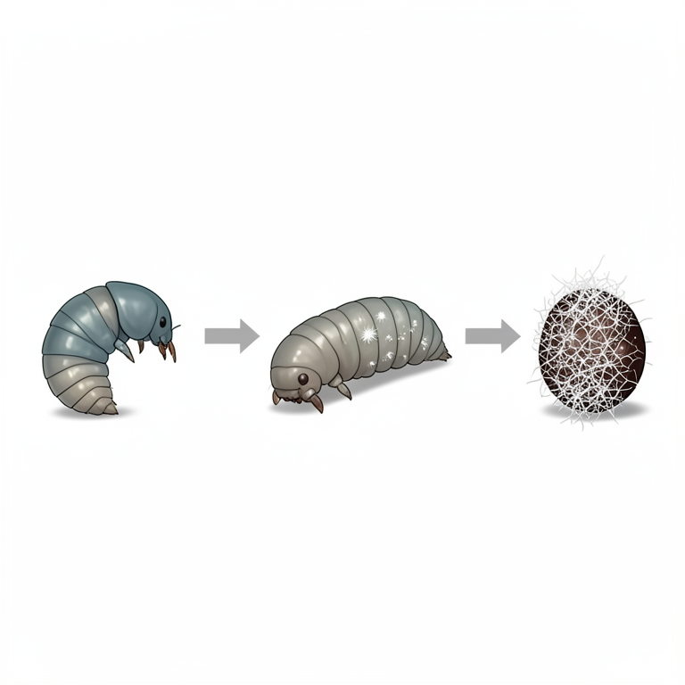
Timeline of B. bassiana Infection
AI-generated conceptual illustration (Spodoptera litura larvae (tobacco cutworm caterpillars)). It explains the idea and does not show study data.
View source evidence ↗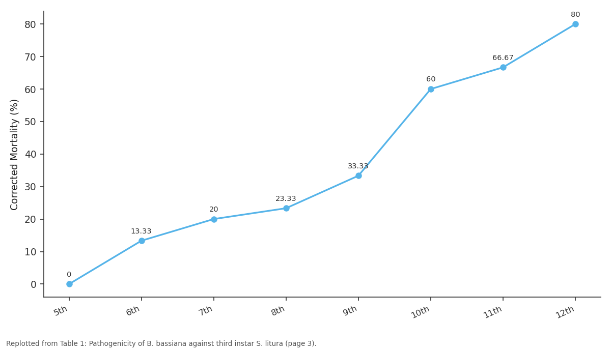
Corrected Mortality (%): 5th vs 6th vs 7th vs 8th vs 9th
Replotted from Table 1 (page 3) of the original paper. Every bar is the paper's own table value.
View source evidence ↗Figures & tables
From the original paper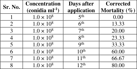 Table 1
Introduction
The tobacco caterpillar (Spodoptera litura), a polyphagous pest, affects a wide variety of cultivated crops (more than 112 species of crops) globally (Ramaiah and Maheshwari, 2018) [19] . It undergoes 5-6 overlapping generations per year, resulting in enormous crop losses ranging from 25.8-100% in different parts of India (Sasidharan and Varma, 2005) [20] . Insecticides have been used more frequently recently to manage S. litura, which has given rise to the possibility of resistance. According to reports, S. litura is now resistant to various classes of pesticides, making management more challenging. It's encouraging to see that more people are becoming aware of how using pesticides harms both the environment and human health. Alternative, more environmentally friendly management tactics have emerged as a result of this greater knowledge. Entomopathogenic fungi (EPF) is a long-lasting pestrepelling technique that restores environmental balance. The entomopathogenic fungus Beauveria bassiana has a wide range of hosts. According to De La Rosa et al. (2000) [2] , hosts have been identified as more than 200 insect species from nine insect orders, primarily Lepidoptera and Coleoptera. B. bassiana spore is a naturally occurring biopesticide that can be detected on some plants and soils (Uma Devi et al., 2008) [24] . With a concentration of 2.4×10 7 spore ml -1 , Malarvannan et al. (2010) [14] discovered a 56.67% pupation reduction in S. litura. In light of this, the present study was designed to study the symptomatology and pathogenicity test of the entomopathogenic fungi, B. bassiana on S. litura under laboratory conditions.
Materials and Methods
B. bassiana isolate used in the investigation was isolated from The Dangs District, Gujarat and routinely grown on potato dextrose agar (PDA). The plates were incubated at 27±2 °C for 10-15 days and stored in a refrigerator. The procedure followed for the rearing of S. litura larvae and proved the pathogenicity of entomopathogenic fungus was presented as follows.
collected from the Rambhas farm of Hill Millet Research Station, Waghai and brought to the laboratory. The larvae were reared in a plastic tray on castor leaves until pupa formation. The basal cut portion of the castor leaves was covered with a cotton swab dipped in water to keep them fresh and turgid for a long duration. The pupae when formed were transferred into a glass jar for the emergence of adults. The adults that emerged were paired (male and female) separately and released in a glass jar (10×20 cm). The top of the glass jar was covered with muslin cloth secured firmly by a rubber band to prevent the escape of the adults. Fresh castor leaves were provided for egg laying to the female in the jar. The cut ends of castor leaves were dipped in water to keep them fresh and turgid for long. Ten percent honey solution was provided as food to the adults by dipping cotton swabs in the solution. These swabs were changed daily. The eggs laid by each female on leaves, walls of the glass jars and on muslin cloth were collected daily with the help of a moist camel hair brush and observed for hatching. The newly emerged larvae were reared in groups in the plastic tray provided with castor leaves. The bigger larvae were reared separately in plastic vials. Thus, uniformly aged third-instar larvae were used for the experiment.
In vitro studies of symptoms produced by B. bassiana on S. litura
Third instar larvae of S. litura obtained from laboratory culture were used for symptomatological studies. The larvae were surface sterilized with 0.1 percent sodium hypochlorite solution and rinsed twice with double distilled sterilized water. Excess water was removed with blotting paper. Suspension of the 10 days old culture of B. bassiana having 1.0×10 8 conidia ml -1 concentration along with 0.5 ml Tween-80 was inoculated by spraying with a hand atomizer on the larva. Inoculated larvae were transferred to separate vials and reared on fresh castor leaves. Each larva was examined critically under a microscope daily for the development of external symptoms caused by infection of inoculated entomopathogen and the symptoms produced were recorded.
Pathogenicity test of B. bassiana against S. litura
Laboratory-reared S. litura larvae (third instar) were surface sterilized with 0.1 percent sodium hypochlorite solution and rinsed twice with double distilled sterilized water. Excess water was removed with blotting paper. 30 surface sterilized larvae were taken separately in plastic vials for pathogenicity studies. Conidial suspensions were prepared by taking a fungal mass solution from the ten-day old culture of B. bassiana and mixing them with 0.5 ml Tween-80 using distilled water. The concentration of 1.0×10 8 conidia ml -1 was prepared from this suspension. The larvae were inoculated topically with two ml of fungal suspension (1.0×10 8 conidia ml -1 ) with the help of a hand atomizer (Easwaramoorthy, 2001) [4] . Inoculated larvae were kept in plastic vials with food (fresh castor leaves). The food was replaced daily. The treated larvae were reared at 25±2 °C temperature and 90 percent relative humidity. Humidity was maintained by placing all plastic vials containing treated larvae in trays with moist absorbent cotton at the bottom and covering them with thin polythene from the outside (Li and Yang, 1988; Khan et al., 1993; Masuka and Manjonjo, 1996; Ekesi et al., 1999) [13,12,15,5] . The larval mortality on the 5 th , 6 th , 7 th , 8 th , 9 th , 10 th , 11 th and 12 th day after treatments were recorded and percent larval mortality due to observable mycosis (shows symptoms of disease development and finally death due to B. bassiana) was calculated.
Larval mortality (%) = Number of dead larvae Total number of treated larvae × 100
Results and Discussion
The visual observation on the first day after the exposure of the S. litura to B. bassiana revealed that movement of larva became slow. This might be due to penetration of hypha of B. bassiana into the tissues of the host body and it interferes with the physiological processes of the larvae. According to Neves and Alves (2004) [16] , the B. bassiana fungi require several stages of the process to infect the host, i.e., inoculation, conidia attachment and penetration. Infected hosts will become weak, inactive and eventually die (Tanada and Kaya, 1993) [23] .
The observation on second day revealed that the movements of the larva became slow and freeze. On the third and fourth days, there was a decrease in feeding activity of larvae. No mortality was observed up to fifth day. It was incubation phase of B. bassiana. Fungus needed a few days to infect and grow on the S. litura body.
The on the 6 th day, the death of larvae noticed, but mycelium was not visible on the integument of S. litura larva. It might be due to B. bassiana fungus was still in the invasion stage to damage the internal tissue of the host body. According to Desyanti (2007) [3] at the beginning of the host's death, mycelium was not seen clearly.
Photo 1: Mycelium covered on the abdomen of S. litura larvae
On the 7th and 8th days, B. bassiana fungi started appearing on the integument of the larva, especially on the abdomen. The mycelia began to grow densely inside the larval body and the food source began to decrease and it penetrated the outside of the larval body to get a new food source. In accordance with the opinion of Steinhaus (1975) [22] , the mycelia passed through the host's integument between 24-48 hours after the host died and the fungus began to damage other tissues by forming the reproductive organs. According to Sayuthi (2012) [21] , the mycelia passed through the host's integument by using penetrant hyphae.
The visual observation on the 9th day revealed that the colour of the larva appeared dark. The last segment of the abdomen started to crease and the mycelia appeared denser. It was presumably due to the crimp on the abdominal end segment of larvae's limbs that was absorbed by the B.
bassiana fungus, and larvae body began to change which was due to fluid loss and the effects of toxins and B. bassiana fungi. On the 10 th day, the larva appeared dry and stiff with the body getting smaller, especially near the abdomen because its body fluid was absorbed by B. bassiana fungus.
According to Feron (1981) [7] the cadaver became hard because the fluid of the host's body was absorbed by the entomopathogenic fungus.
The observation on the 11 th and 12 th days revealed that the larva body was covered entirely by mycelia of B. bassiana fungus became dry, black coloured, smaller and not in its proper shape. According to Haris (2005) [10] the hardening (mummification) of larva was observed on infection by B. bassiana fungus in the final stages and mycelia of B. bassiana covered the entire body of the host.
As mycelium covered the whole body of S. litura larvae it looked like cottony white coloured followed by creamy white coloured sporulation. Sporulation occurred within one to two days. Sporulation expands progressively and at last whole larva was covered with thick white sporulated fungal mass.
The systematic studies on symptoms produced by B. bassiana on S. litura were similar to the description provided by the earlier workers viz., Gloriana et al. (2000) [8] , Patel (2013) [17] , Vega et al. (2015) [25] and Yasmin and Refaei (2021) [28] . They observed white coloured growth on the surface of the test insect body. The results of the pathogenicity test of B. bassiana against third instar S. litura larvae are presented in Table 1. The data presented in Table 1 revealed there was no mortality observed up to the fifth day of exposure. It was the incubation phase of B. bassiana in the insect body. Fungus needed a few days to infect and grow on the S. litura body. Sixth day onward, the mortality of the larva was observed (13.33%). The larval mortality on the 7 th , 8 th , 9 th , 10 th , 11 th and 12 th days after the application was recorded as 20.00, 23.33, 33.33, 60.00, 66.67 and 80.00 percent, respectively. The above results indicated that the mortality of S. litura larvae started on 6 th day after application (13.33%), gradually increased and reached 80.00 percent on 12 th day after application. Petlamul and Prasertsan (2012) [18] evaluated the different strains of B. bassiana against S. litura with a concentration of 10 8 conidia ml -1 and found that mortality started after 2 nd day of inoculation and reached 50 percent on 4 th day and cent percent at 5 th day in BNBCRC and BPMC strains and at 6 th day in B14841, B16041 and B14532 strains. Gloriana et al. (2000) [8] , Anand and Tiwary (2009) [1] , Vijayavani et al. (2009) [26] , Joseph et al. (2010) [11] , Malarvannan et al.
| Sr. No. | Concentration (conidia ml-1) | Days after application | Corrected Mortality (%) |
|---|---|---|---|
| 1 | 1.0 × 10⁸ | 5th | 0.00 |
| 2 | 1.0 × 10⁸ | 6th | 13.33 |
| 3 | 1.0 × 10⁸ | 7th | 20.00 |
| 4 | 1.0 × 10⁸ | 8th | 23.33 |
| 5 | 1.0 × 10⁸ | 9th | 33.33 |
| 6 | 1.0 × 10⁸ | 10th | 60.00 |
| 7 | 1.0 × 10⁸ | 11th | 66.67 |
| 8 | 1.0 × 10⁸ | 12th | 80.00 |
Table text extracted from the original PDF; superscript letters are shown inline. The original table image is below.
View original table image
(2010) [14] , Patel (2013) [17] , Gupta and Kumar (2014) [9] , Fatima et al. (2017) [6] and Wargane et al. (2020) [27] proved pathogenicity of B. bassiana against S. litura.
Conclusion
From the present findings, it can be concluded that the S. litura larvae sprayed with B. bassiana showed the following symptoms: on the first day after the exposure of the S. litura to B. bassiana revealed that the movement of larvae became slow. On the third and fourth days, there was a decrease in the feeding activity of larvae. No mortality was observed up to fifth day. It was the incubation phase of B. bassiana. Fungus needed a few days to infect and grow on the S. litura body. On the 6 th day, the death of larvae was noticed, but mycelium was not visible on the integument of S. litura larva. On the 7 th and 8 th days, B. bassiana fungi started appearing on the integument of the larva, especially on the abdomen. The mycelia began to grow densely inside the larval body and the food source began to decrease and it penetrated the outside of the larval body. The visual observation on the 9 th day revealed that the colour of the larva appeared dark. The last segment of the abdomen started to crease and the mycelia appeared denser. On the 10 th day, the larva appeared dry and stiff with the body getting smaller, especially near the abdomen. The observation on the 11 th and 12 th days revealed that the larva body was covered entirely by mycelia of B. bassiana fungus and became dry, smaller and not in its proper shape. The pathogenicity test was carried out with topical application of two ml fungal suspension of B. bassiana to S. litura gave 13.33, 20.00, 23.33, 33.33, 60.00, 66.67 and 80.00 percent mortality on the 6 th , 7 th , 8 th , 9 th , 10 th , 11 th and 12 th days after application, respectively.
References
24 in the original paper- Anand R, Tiwary BN. Pathogenicity of entomopathogenic fungi to eggs and larvae of Spodoptera litura, the common cutworm. Biocontrol Sci Technol. 2009;19(9):919-929.
- De La Rosa W, Alatorre R, Barrera JF, Toriello C. Effect of Beauveria bassiana and Metarhizium anisopliae (Deuteromycetes) upon the coffee berry borer (Coleoptera: Scolytidae) under field conditions. J Econ Entomol. 2000;93:1409-1414.
- Coptotermes spp. (Isoptera: Rhitomidae) dengan menggunakan cendawan entomopatogen isolate lokal (Disertasi). Bogor: Program Pasca Sarjana, Institut Pertanian Bogor. 2007.
- Easwaramoorthy S, Nirmala R, Santhalakshmi G. Occurrence of Metarhizium anisopliae Var. anisopliae on Sugarcane Internode Borer, Chilo sacchariphagus indicus (Kapur). J Biol Control. 2001;15(1):81-84.
- Ekesi S, Maniania NK, Ampong NK. Effect of temperature on germination, radial growth, and virulence of Metarhizium anisopliae and Beauveria bassiana on Megalurothrips sjostedti. Biocontrol Sci Technol. 1999;9(2):177-185.
- Fatima T, Reddy KD, Rafi MG, Umamaheswari T, Sumalatha J. Evaluation of pathogenicity of Beauveria bassiana against major lepidopteran pests, Spodoptera litura and Plutella xylostella in cabbage. Prog Res Int J. 2017;12(1):959-963.
- Feron P. Pest control by the fungi Beauveria and Metarhizium. In: H.D. Burgs (Ed). Microbial Control of Pest and Plant Diseases 1970-1980, Academic Press, New York; c1981.
- Gloriana AS, Raja N, Seshadri S, Janarthanan S, Ignacimuthu S. Pathogenicity of entomopathogenic, Bacillus thuringiensis sub sp. kurstaki and Beauveria bassiana to the larvae of Spodoptera litura (F.) and Pericallia ricini (F.). Biol Agric Hortic. 2000;18(3):235-242.
- Gupta SD, Kumar B. Bioefficacy of Beauveria bassiana (Balsamo) against third instar larvae of Spodoptera litura (Far.). Int J Plant Sci. 2014;9(1):97100.
- Haris TA. Bioekologi Cendawan Beauveria bassiana (Balsamo) Vuillemin Balai Penelitian Tanaman Serealia, Prosiding Seminar Nasional Jagung. 2005;482-487.
- Joseph I, Chellaiah DE, Singh AR. Studies on the influence of Beauveria bassiana on survival and gut flora of groundnut caterpillar, Spodoptera litura Fab. J Biopestic. 2010;3(3):553-555.
- Khan KH, Gopalan M, Rabindra RJ. Influence of temperature on the growth, sporulation, and infectivity of mycopathogens against termites. J Biol Control. 1993;7(1):20-23.
- Malarvannan S, Murali PD, Shanthakumar SP, Prabavathy, Sudha NVR. Laboratory evaluation of the entomopathogenic fungi, Beauveria bassiana against the tobacco caterpillar, Spodoptera litura Fabricius (Noctuidae: Lepidoptera). J Biopes. 2010;3(1):126-131.
- Masuka AJ, Manjonjo V. Laboratory screening of Metarhizium flavoviride and Beauveria bassiana for the control of Mecastbus pinivorus, a Pinus patula defoliator in Zimbabwe. JASSA. 1996;2:91-95.
- Neves PMOJ, Alves SB. External events related to the infection process of Cornitermes cumulans (Kollar) (Isoptera: Termitidae) by the entomopathogenic fungi Beauveria bassiana and Metarhizium anisopliae. Neotrop Entomol. 2004;33(1):51-56.
- Patel RS. Isolation, identification, physiological studies, and evaluation of potent entomopathogenic fungus Beauveria bassiana (Bals.) Vuill. against Spodoptera litura (Feb). Ph. D. (Agri.) thesis submitted to the Navsari Agricultural University, Navsari. 2013;1-104.
- Petlamul W, Prasertsan P. Evaluation of strains of Metarhizium anisopliae and Beauveria bassiana against Spodoptera litura on the basis of their virulence, germination rate, conidia production, radial growth, and enzyme activity. Mycobiology. 2012;40(2):111-116.
- Ramaiah M, Maheshwari T. Biology studies of tobacco caterpillar, Spodoptera litura Fabricius. J Entomol Zool Sci. 2018;31(1):31-34.
- Sasidharan KR, Varma RV. Laboratory evaluation of Beauveria bassiana (Balsamo) Vuillemin against Indarbela quadrinotata Walker (Lepidoptera: Metarbelidae) -a key pest of Casuarina equisetifolia L. in Tamil Nadu. J Biol Control. 2005;19:197-200.
- Sayuthi M. Kajian cendawan entomopatogen Metarhizium brunneum Petch sebagai agens hayati rayap Macrotermes gilvus Hagen (Isoptera: Termitidae) pada tanaman jarak pagar (Jatropha curcas L.) [disertasi]. Bogor: Fakultas Pertanian, Institut Pertanian Bogor. 2012.
- Devi UK, Padmavathi J, Maheswara URC, Khan AAP, Murali MC. A study of host specificity in the entomopathogenic fungus, Beauveria bassiana (Hypocreales: Clavicipitaceae). Biocontrol Sci Technol. 2008;18(10):975-989.
- Vijayavani S, Reddy KRK, Murthy GBVN. Pathogenicity of Beauveria bassiana strains on Spodoptera litura (Fab.). J Biopes. 2009;2(2):205-207.
- Wargane VS, Parate RL, Lavhe NV, Sonune BD, Mane KK, Sharath CHR. Pathogenicity of Beauveria bassiana against second instar larvae of Spodoptera litura and compatibility with insecticides. J Pharmacogn Phytochem. 2020;9(1):576-578.
- Yasmin AF, Refaei EA. Pathogenicity induced by indigenous Beauveria bassiana isolate in different life stages of the cotton leafworm, Spodoptera littoralis (Boisduval) (Lepidoptera: Noctuidae) under laboratory conditions. Egypt J Biol Pest Control. 2021;31:64.29.
Cited by 1
From OpenAlex, OpenCitations and reference lists read by Scinovex
- Unveiling the endophytic abilities and pathogenicity of native Beauveria bassiana isolates against rice brown planthopper ( Nilaparvata lugens ) in rice ( Oryza sativa ): a detailed exploration ↗
S. S. Monica, P. Rajanikanth, P. Duraimurugan, S. Ameer Basha, Darshanoju Srinivasa Chary · International Journal of Pest Management · 2025
This study extensively explores indigenous B. bassiana isolates for controlling the rice brown planthopper (BPH). It investigates endophytic colonization dynamics, virulence, and molecular confirmation within rice tissues. Various inoculation methods were evaluated, with a combination showing the highest efficacy. Colonization rates of ChBb and NaBb reached 86.67% and 80.00%, respectively, at 14 d post-inoculation (DPI). Nested PCR confirmed the presence…
❝ 1