
Research article · 2024 · Full text
International Journal of Advanced Biochemistry ResearchTherapeutic management of theileriosis in young Deoni calves
Volume 8(1) · Pages 628–630 · DOI: 10.33545/26174693.2024.v8.i1i.467 ↗
Research areasToxin Mechanisms and ImmunotoxinsAnimal Virus Infections StudiesVector-borne infectious diseases
Abstract
Author-writtenBovine theileriosis is a tick-borne disease caused by protozoan parasite of the genus Theileria. Seven deoni calves affected with Theileriosis. The calves shows clinical signs such as pyrexia, anorexia, anemia, pale mucus membrane, enlarged lymph node, general weakness, lameness and difficulty in getting up etc. The diagnosis was done by blood smear examination and lymph node smear examination. Animals were treated with buparvaquone injection along with symptomatic and supportive therapy. The infection can be fully cured with early detection, appropriate medication and control of ticks.
Summary
Source-checkedPlain-language reading of the full paper, prepared with AI and checked against the text. Each point links to the authors' own words.
Why it matters
- Bovine theileriosis is a disease in cattle caused by protozoan parasites of the genus Theileria and is transmitted by ticks. View evidence ↗
- Theileriosis is a significant problem in the Indian subcontinent because it causes calf mortality, hindering livestock upgrading projects. View evidence ↗
- Diseases caused by blood protozoans create major constraints for the global dairy industry and cause severe livestock losses. View evidence ↗
The question
- The study aims to demonstrate the therapeutic management of theileriosis in calves within field conditions. View evidence ↗
What they did
- The study involved 7 calves from various localities in Aurad, Bidar, Karnataka. View evidence ↗
- Diagnosis was performed using Giemsa-stained blood and lymph smears collected on day 0, day 5, and day 15 post-treatment. View evidence ↗
- Treatment consisted of a 2.5 mg/kg BW deep intramuscular injection of Buparvaquone on day 0, supplemented with Melonex plus (0.5mg/kg BW), Avil (1ml), and Tribivet (1.5 ml) for 5 days. View evidence ↗
What they found
- Over 75% of the infected calves exhibited poor appetite, pale mucous membranes, enlarged superficial lymph nodes, and fever between 104-107 °F. View evidence ↗
- Blood smears confirmed the presence of piroplasms in the red blood cells of 42.85% (3 out of 7) of the calves. View evidence ↗
- Lymph node aspirate smears showed schizonts in the mononuclear cells of 71.42% (5 out of 7) of the calves. View evidence ↗
- A total of 57.14% (4 out of 7) of the calves tested positive for both piroplasms in blood and schizonts in lymph node aspirates. View evidence ↗
- Four of the calves recovered within five days after receiving a single dose of buparvaquone. View evidence ↗
- Three calves remained positive after five days and required a second dose of buparvaquone, recovering within 15 days. View evidence ↗
- The authors found that lymph smears are the most effective rapid diagnostic test for calves presenting with enlarged lymph nodes. View evidence ↗
What it means
- The authors conclude that early treatment with a single dose of buparvaquone and supportive therapy is effective for theileriosis. View evidence ↗
- The authors state that full cure of the infection is possible through early detection, correct medication, and tick control. View evidence ↗
Visual summary
1 visualsIllustrations explain the paper; they do not show study data.
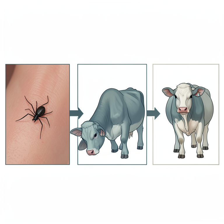
Theileriosis Transmission and Treatment Cycle
AI-generated conceptual illustration (Deoni calves and ticks). It explains the idea and does not show study data.
View source evidence ↗Figures & tables
From the original paper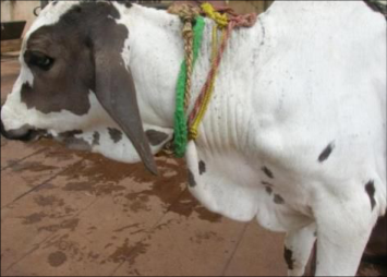 Figure 1
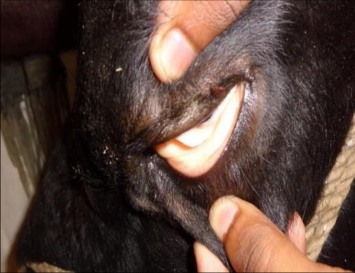 Figure 2
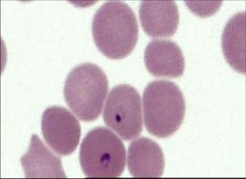 Figure 3
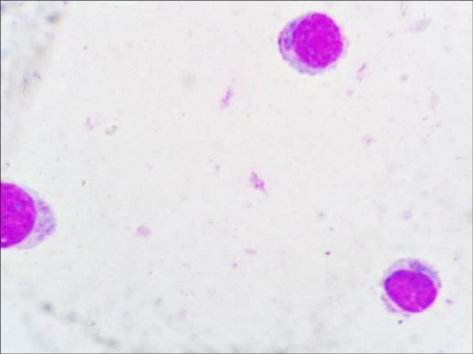 Figure 4
Introduction
Theileria annulata, a protozoan parasite of cattle and domestic buffaloes, is transmitted by ticks of the genus Hyalomma and causes a disease named Mediterranean or tropical theileriosis (Mirzaei, 2007) [6] . On the pasture, ticks can remain infected for upto two years. Animals that are vulnerable to sporozoites are exposed through the saliva of feeding ticks.
Once the tick clings to its host, theileria develop and emerge into the saliva. Theileriainfected animal's spleen, lymph nodes, liver and entire blood contain schizonts; injected schizonts can spread easily among cattle. One of the main obstacles to livestock upgrading projects in the Indian subcontinent is calf mortality from theileriosis. (Godara et al., 2009; Sudan et al., 2012) [9,10] . In neonates, theileriosis can present with a variety of clinical presentations, ranging from subacute to chronic or acute to peracute. It mainly depends upon the damaging effect of the pathogen on lymphoid tissues and the host's susceptibility. A marked rise in body temperature, reaching 104-107°F is followed by lethargy, lacrimation, nasal discharge, exophthalmia and swelling of superficial lymph nodes; hemolytic anemia is among the characteristic features of tropical theileriosis (Fartashvand et al., 2013; Singh et al., 2013) [3,11] . Haemoprotozoan animal diseases are causing severe losses to the livestock industry and thus pose major constraints to the dairy industry worldwide (Kohli et al., 2014) [5] . This case study illustrates how theileriosis in calves can be treated therapeutically under field conditions.
Materials and Methods
Materials and Methods
Animals
A total number of 07 calves belong to different localities in Aurad, taluka of Bidar Karnataka, were subjected to study. All animals showed clinical signs of theileria infection, out of them only 04 cows showed enlargement of superficial lymph nodes.
Clinical examination
A thorough clinical examination was carried out on all the calves. The signs in clinical cases of Theileria annulata infection were reported and recorded. From each calf ear vein, thin blood smears were made. Aspirates of lymph nodes were taken from suspected instances with enlarged superficial lymph nodes.
Samples
Blood and lymph aspiration were collected from each calf. Blood was drawn from the vein in the ear and utilized to create blood film. Lymph node aspirates were collected from enlarged lymph nodes and used for lymph smear. Blood and lymph smears were prepared directly after collections, fixed with absolute methanol, and stained with Giemsa. Samples were collected on day '0', 5 th day after treatment and 15 th day after treatment.
Diagnosis and Treatment
Out of 07 presented calves, four were female and three were male. More than 75% of the infected calves displayed clinical signs of pyrexia reaching 104-107 °F, poor appetite and superficial lymph node enlargement (Fig. 1) and pallor mucous membrane (Fig. 2). Blood smear examination revealed presence of piroplasms in the RBCs of 42.85% (3/07) calves (Fig. 3) While, Lymph node aspirate smear examination revealed presence of schizonts in mononuclear cells of 71.42% (05/07) calves (Fig. 4). Moreover, 57.14% (4/07) of calves were positive for the both piroplasms in blood smear and presence of schizonts in mononuclear cells of lymph nodes aspirate smear. In present investigation, all the infected calves were treated with Buparvaquone (Zubion) at the dose rate of 2.5 mg/kg BW deep intramuscular injection at neck region on day '0' along with Melonex plus at a dose rate of 0.5mg/kg BW intramuscular, Avil 1ml intramuscular and Tribivet 1.5 ml intramuscular for 5 days. After the 5 th day of treatment, three (03) were positive and those were repeated 2 nd dose of buparvoquone (Zubion). These calves recovered with 2 nd dose of buparvaquone within 15 days. After receiving a single dosage of buparvaquone, four calves recovered in five days. In the current instance, it was discovered that, when treated as soon as possible, a single dosage of buparvaquone combined with supportive therapy is effective against cases of theileriosis.
Discussion
The findings of this investigation indicate that some of the most prevalent clinical signs that spontaneously infected calves with T. annulata exhibit are lymph node enlargement, pallor mucous membrane, decreased appetite, a noticeable rise in body temperature and exophthalmia. Anemia caused by cytokine overproduction, immune-mediated hemolysis and reactive oxygen species is a significant aspect of the disease (Saleh et al., 2012) [7] . Pallid mucous membranes are a sign of anemia in calves who are affected. Since it is not confined by a parasitophorous vacuole, the changing schizont can easily disrupt the signaling pathways of host cells that control cell division and survival (Shiels et al., 2006) [8] . According to Woods et al. (2013) [12] , T. annulata primarily infects macrophages/monocytes and B-cells. It then transforms the infected host cells, causing unchecked proliferation and clonal expansion of the parasitized cell population, which causes generalized lymphadenopathy and lympho-proliferative disease in cattle. T. annulata infected cells also generate large amounts of inflammatory cytokines, including TNF- (Brown et al., 1995) [1] . According to Graham et al. (2001) [4] , this cytokine is a strong inducer of all the main clinical signs and symptoms of acute tropical theileriosis, including pyrexia, anemia, anorexia, muscle wasting and necrosis. Results of the current investigation revealed that 42.85% (3/07) calves were positive for Theileria infection by blood film. Furthermore, 71.42% (05/07) calves with enlarged lymph nodes were positive for theileriasis by lymph smear, this indicates that lymph smear is the best rapid test for diagnosis of theileriasis in calves with enlarged lymph nodes. Similarly, Abdel-Rady revealed that, 31 cows (25.8%) out of the examined cattle (120) were positive by blood film Theileria infection, and all the cows (18) with enlarged lymph nodes were positive by lymph smear theileriasis. The intramuscular method of buparvaquone at a dose rate of 2.5 mg/kg body weight was found to be efficacious. The outcome matched that of Devadevi et al. (2018) [2] . The present study showed that in 03 calves required doses of injections for complete recovery. In contrast to the findings of Devadevi et al. (2018) [2] , a single intramuscular dosage of buparvaquone at 2.5 mg/kg body weight demonstrated 100% recovery and removed the Theileria organism from the bloodstream. According to Singh et al. (2013) [11] , in these particular circumstances, vitamin supplementation may have helped to improve the anemia of the affected calves by reducing the oxidative damage to erythrocytes caused by Theileria.
References
11 in the original paper- Brown DJ, Campbell JD, Russell GC, Hopkins J, Glass EJ. T cell activation by Theileria annulata infected macrophages correlated with cytokine production. Clin Exp Immunol. 1995;102:507-514.
- Devadevi N, Rajkumar K, Vijayalekshmi P. Efficacy of buparvaquone in the treatment of bovine benign theileriosis. Int J Chem Stud. 2018;6(3):3110-3112.
- Fartashvand M, Nadalian MG, Sakha M, Safi S. Elevated Serum Cardiac Troponin I in Cattle with Theileriosis. J Vet Intern Med. 2013;27:194-199.
- Graham SP, Brown DJ, Vatansever Z, Waddington D, Taylor LH, Nichani AK, et al. Proinflammatory cytokine expression by Theileria annulata infected cell lines correlates with the pathology they cause in vivo. Vaccine. 2000;19:2932-2944.
- Kohli S, Atheya UK, Thapliyal A. Prevalence of theileriosis in cross-bred cattle: its detection through blood smear examination and polymerase chain reaction in Dehradun district, Uttarakhand, India. Vet World. 2014;7(3):168-171.
- Mirzaei M. Treatment of natural tropical theileriosis with the extract of the plant Peganum harmala. Korean J Parasitol. 2007;45:267-271.
- Saleh MA, Mahran OM, Al-Salahy MB. Corpuscular oxidation in newborn crossbred calves naturally infected with Theileria annulata. Vet Parasitol. 2012;182(2-4):193-200.
- Shiels B, Langsley G, Weir W, Pain A, McKellar S. Alteration of host cell phenotype by Theileria annulata and Theileria parva: mining for manipulators in the parasite genomes. Int J Parasitol. 2006;36:9-21.
- Godara R, Sharma RL, Sharma CS. Bovine tropical theileriosis in a neonate calf. Trop Anim Health Prod. 2009;42:551-553.
- Sudan V, Sharma RL, Borah MK, Mishra R. Acute bilateral proptosis in a crossbred calf naturally infected with Theileria annulate. J Parasit Dis. 2012;36:215219.
- Woods KL, Theiler R, Mühlemann M, Segiser A, Huber S, Ansari HR, et al. Recruitment of EB1, a Master Regulator of Microtubule Dynamics, to the Surface of the Theileria annulata Schizont. PLOS Pathogens. 2013;9:e1003346. Doi: 10.1371/journal.ppat.1003346.