
Research article · 2024 · Full text
International Journal of Advanced Biochemistry ResearchSynthesis, characterization and efficacy of green synthesized copper nanoparticles from eucalyptus, against Fusarium wilt of green gram
Volume 8(1) · Pages 272–277 · DOI: 10.33545/26174693.2024.v8.i1d.412 ↗
Research areasNanoparticles: synthesis and applicationsMedicinal Plants and NeuroprotectionMedicinal Plant Research
Abstract
Author-writtenGreen gram suffers heavy losses in yield due to many diseases especially wilt caused by Fusarium sp. Thus, the present study was undertaken with emphasis on development of simple and cost-effective methods for synthesis of nanomaterials and their application in the management of the pathogen. Green synthesis methods, employing plant extracts, have emerged as an eco-friendly and clean alternative to physical and chemical methods. The eucalyptus plant extract mediated copper nanoparticles were synthesized using 0.01M of CuSO4 solutions and characterized by usingUV-Visible spectrophotometry, Zeta size analysis, Zeta potential, and Nano Tracking Analysis. The synthesized nanoparticle (129.23 nm) were applied at various concentrations to determine their antifungal activities. The result showed the highest inhibition percentage of mycelial growth (80.59%) was recorded on CuNP-E @ 1000 ppm.
Summary
Source-checkedPlain-language reading of the full paper, prepared with AI and checked against the text. Each point links to the authors' own words.
Why it matters
- Green gram crops experience significant yield losses due to various diseases, particularly wilt caused by Fusarium species. View evidence ↗
- Managing Fusarium wilt with traditional fungicides can lead to environmental pollution and residual toxicity. View evidence ↗
- Green synthesis using plant extracts is considered a clean and eco-friendly alternative to chemical or physical nanoparticle synthesis methods. View evidence ↗
The question
- The study aimed to develop a simple, cost-effective method for synthesizing nanomaterials to manage the Fusarium pathogen. View evidence ↗
What they did
- Copper nanoparticles (CuNP-E) were synthesized by adding 10 ml of fresh eucalyptus leaf extract to 100 ml of 0.01 M copper sulphate solution. View evidence ↗
- The synthesized nanoparticles were characterized using UV-Visible spectrophotometry, Zeta size analysis, Zeta potential, and Nano Tracking Analysis. View evidence ↗
- The antifungal efficacy of the nanoparticles was tested at concentrations ranging from 25 to 1000 ppm using a Completely Randomized Design with three replications. View evidence ↗
- The study used the poison food technique, comparing the nanoparticles against a water control and standard fungicides: carbendazim (500 ppm), mancozeb (2500 ppm), and thiram (500 ppm). View evidence ↗
What they found
- The formation of copper nanoparticles was visually indicated by the solution changing from blue to dark brown within 2 hours. View evidence ↗
- UV-Visible spectroscopy confirmed the presence of copper nanoparticles via a sharp absorbance peak at 345 nm. View evidence ↗
- Zeta size analysis determined that the eucalyptus-mediated copper nanoparticles had an average diameter of 129.23 nm. View evidence ↗
- The zeta potential of the biosynthesized CuNP-E was measured at -2.41 mV, which the authors state indicates high stability. View evidence ↗
- Nano Tracking Analysis showed varying nanoparticle sizes based on camera level: 149.5 nm at level 12 and 196.5 nm at level 11. View evidence ↗
- The highest inhibition of mycelial growth for Fusarium sp. was 80.59%, achieved using CuNP-E at a concentration of 1000 ppm. View evidence ↗
- At 1000 ppm, CuNP-E (80.59% inhibition) was more effective than the standard fungicides Thiram (80.41%), Carbendazim (76.44%), and Mancozeb (72.15%). View evidence ↗
- CuNP-E concentrations of 500 ppm and below were less effective than the recommended standard fungicides. View evidence ↗
What it means
- The authors conclude that green-synthesized copper nanoparticles from eucalyptus are effective in vitro for inhibiting the mycelial growth of Fusarium sp., with peak efficacy at 1000 ppm. View evidence ↗
Visual summary
3 visualsIllustrations explain the paper; charts are replotted from the paper's own table values.
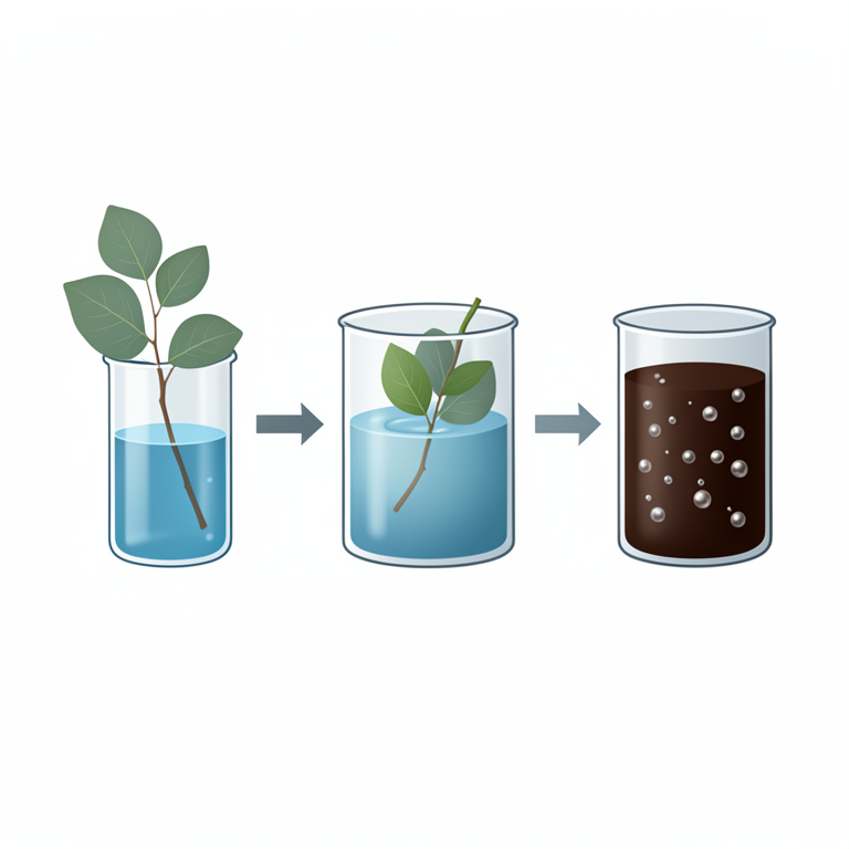
Green Synthesis Process
AI-generated conceptual illustration (eucalyptus leaves and copper sulphate solution). It explains the idea and does not show study data.
View source evidence ↗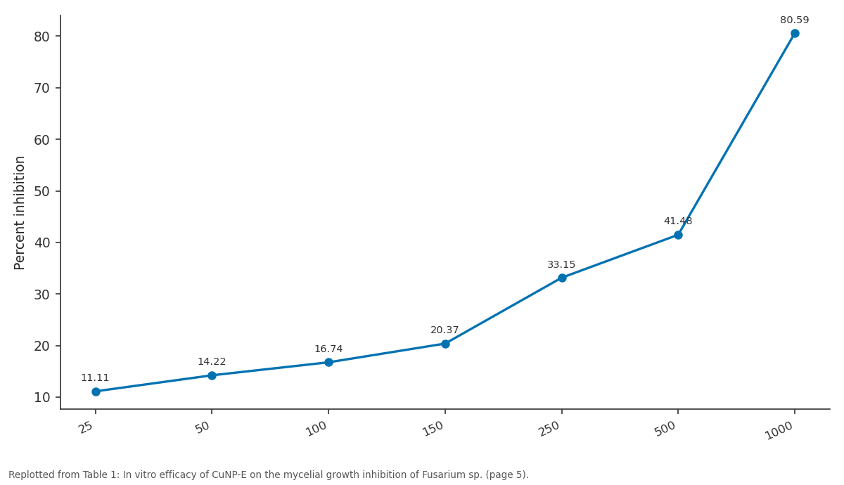
Percent inhibition: 25 vs 50 vs 100 vs 150 vs 250
Replotted from Table 1 (page 5) of the original paper. Every bar is the paper's own table value.
View source evidence ↗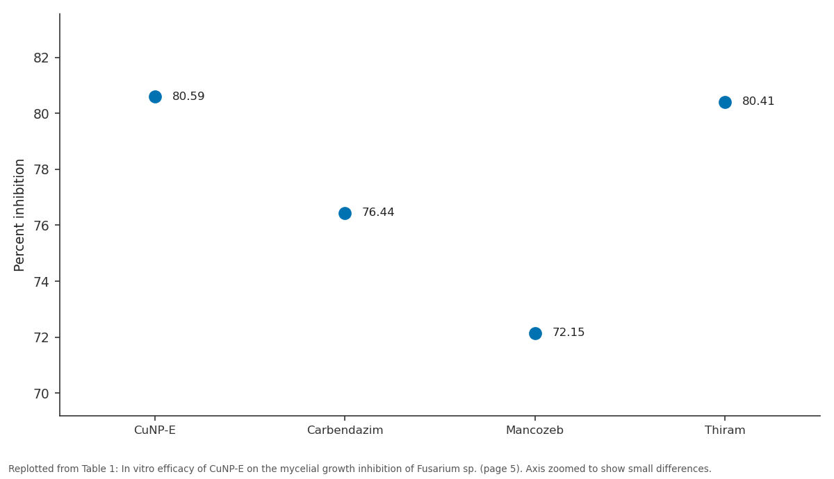
Percent inhibition: CuNP-E vs Carbendazim vs Mancozeb vs Thiram
Replotted from Table 1 (page 5) of the original paper. Every bar is the paper's own table value.
View source evidence ↗Figures & tables
From the original paper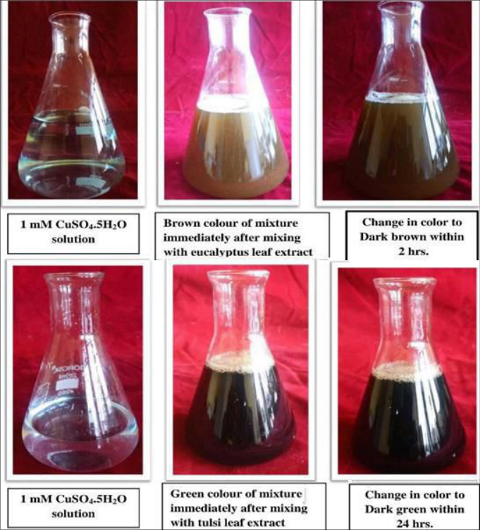 Plate 1
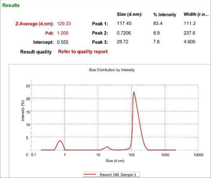 Figure 1
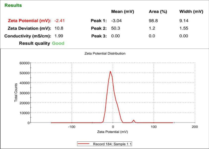 Figure 2
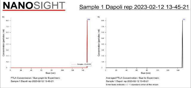 Figure 3
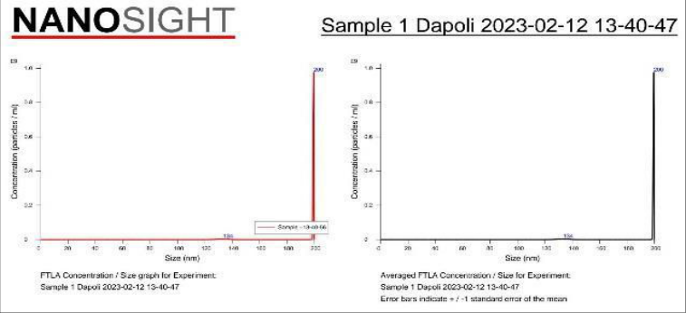 Figure 4
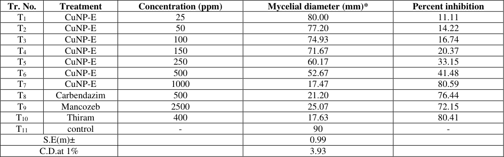 Table 1
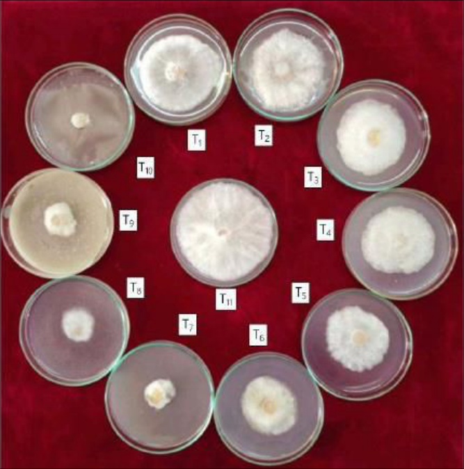 Plate 2
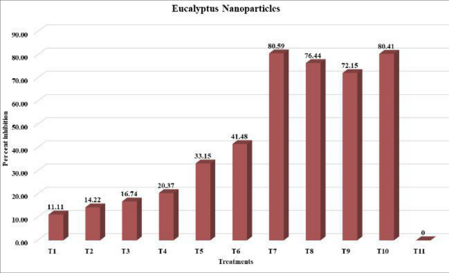 Figure 3
Introduction
In recent years, Synthesis of metal Nanoparticles using plant leaf extract has attracted attention of many researchers because of easy availability of materials, inexpensive procedure to carry out in any laboratory and use of non-toxic reagent (Kulkarni et al. 2015) [4] . Nanomaterial has completely new or improved properties based on specific characteristics such as size distribution and morphology. The two approaches were used for the synthesis of nanoparticles i.e., 'top down" approach and "bottom up" approach (Gaikwad et al. 2016) [2] .
The confirmation of synthesis and stabilization of nanoparticles were done by various characterization methods. Green gram is one of the important pulse crop and the crop is suffers from many disease among that the wilt caused by Fusarium sp. is the most common. The use of fungicide to manage this disease causes several adverse effect like residuel toxicity, pollution to environment, therefore the present study was undertaken to provide the farmer with the most reliable and advanced measures of nanotechnology for managing the incidence and losses caused by wilt of green gram.
Materials and Methods
Materials and Methods
Collection of sample and isolation of the pathogen
The green gram seed were collected and Fusarium sp. isolated from that seeds. Potato dextrose agar media is used for the isolation of pathogen.
Preparation of Eucalyptus leaves extract
Eucalyptus (Eucalyptus globulus L.) leaves were collected. These plant products were cleaned under running tap water subsequently dried under shade to remove moisture completely, and then it was powdered and stored. 10 g of powdered material were taken in a beaker along with 100 ml of de-ionized water and allowed to boil at 80 °C for 25-30 minutes under reflux condition, cooled down to room temperature. Each prepared solution was initially filtered through Whatman No.42 filter paper and then centrifuged at 3000 rpm for 10 minutes to remove heavy biomaterials and to get clear solution. Then plant extract was stored at 4 °C for further studies.
(Iliger et al. 2020) [3] .
Preparation of Copper sulphate Solution
Accurate amount of 1mm copper sulphate solution can be prepared by dissolving 0.2496 gm of CuSO 4 .5H 2 0 in 100 ml of double distilled water and stored in clean, dried reagent bottle.
Synthesis of copper nanoparticles
In a typical synthesis of copper nanoparticles from eucalyptus, 10 ml of fresh leaf extracts were added to 100 ml of 0.01 M copper sulphate (CuSO 4 .5H 2 O) aqueous solutions. Tulsi leaves extract 50 ml were mixed with 50 ml aqueous solution of 1mM copper sulphate (1:1 ratio of tulsi extract to copper solution). A prepared mixture was kept at constant stirring on a magnetic stirrer for 2 hrs. The suspended solution was autoclaved and centrifuged at 5000 rpm for 25 min and the supernatant liquids were decanted off and the residues were repeatedly washed with de-ionized water. Centrifugation-decantation-washing processes were repeatedly done several times to remove impurities if any on the surface of the copper nanoparticles. The obtained precipitate was dried in an oven at 60 °C for 24 hr. (Seeram et al. 2016 and Mali et al. 2020) [13,6] .
Characterizations of synthesized nanoparticles
UV-Visible spectrophotometry, Zeta size analysis, Zeta potential, and Nano Tracking Analysis, were used to characterize the synthesized copper nanoparticles.
In-vitro efficacy of synthesized nanoparticles
Evaluation of synthesized nano-particles were carried out under controlled conditions through poison food technique with carbendazim 50 WP (500 ppm), mancozeb (2500 ppm) and thiram(500 ppm) served as standard checks while as water served as control. CuNP-E including three standard fungicides for each were tested against the F. oxysporum f. sp. Vigni under study. The nanoparticles were evaluated at 25, 50, 100, 150, 250, 500 and 1000 ppm concentrations with three replications each in a Completely Randomized Design (CRD). The efficacy of nanoparticles prepared from eucalyptus against the test fungus was expressed as inhibition of mycelial growth over control percent calculated by using the formula given by Vincent (1947) [12] .
C -T Percent Inhibition (I) = x 100 C
Where, C = Growth (mm) of test fungus in untreated control plate. T = Growth (mm) of test fungus in treated plate.
Results and Discussion 1. Isolation, Purification, and identification of the pathogen
The seeds which showing the symptoms of disease after examination, were selected for further research work. The agar plate method was used for the isolation of the pathogen.The incubated Petri dishes at 22-25 °C were shows mycelial growth of the fungus after 3 days. The pure culture of fungi was obtained by transfer of hyphal tip of actively growing mycelium at 7 days after incubation. Sandhyarani et al. (2020) [8] and Deshmukh et al. (2019) [1] utilized similarly the agar plate method (ISTA, 1999) for the isolation of seed mycoflora associated with green gram seeds.
Green synthesis of nanoparticles
The biosynthesis of copper nanoparticles was carried out using copper sulphate pent hydrate (CuSO 4 .5H 2 O) solution mixedwith leaf extracts of eucalyptus and leaf extracts. The leaf extract act as a capping and stabilizing agent and copper sulphate pent hydrate act as a reductant chemical. The results estimated that the 100 gm. of eucalyptus leaves (powder) produce 0.12 gm. of copper nanoparticles, which was in solid form. The green synthesis of CuNP from the eucalyptus leaves was conducted by Illiger et al. (2020) [3] and Kulkarni et al. (2015) [4] , they utilize copper sulphate pent hydrate (CuSO 4 .5H 2 O) as the reducing agents.
Characterizations of synthesized nanoparticles 3.1 Visual observation
The reaction of nanoparticles synthesis started when the leaf extract of eucalyptus leaves was added into 1mM aqueous copper sulphate pent hydrate (CuSO 4 .5H 2 O) solution. The Copper nanoparticle solution was autoclaved to increase the intensity of nanoparticles in the solution. The aqueous copper sulphate solution turned blue to dark brown within 2 hours indicates the formation of copper nanoparticles in the solution. The similar results were reported by Illiger et al. (2020) [3] where Copper sulphate solution (aqueous) which was blue in colour changed to brown colour within 2 hours after addition of leaf extract of Eucalyptus shows the presence of Cu nanoparticles in the solution.
UV-Visible spectrophotometry
Absorption spectra of copper nanoparticles formed in the reaction has sharp absorbance peak at 345 nm which can be attributed to absorption by Nano size copper particle confirming the presence of copper nanoparticles by UV-Visible spectroscopy. Similar observations were reported by Shende et al. (2016) [10] , where the presence of copper nanoparticle was confirmed by UV-visible spectroscopy technique.
Zeta size Analysis
The characterization of copper nanoparticles in terms of average particle diameter was recorded in nanometer from the intensity distribution analysis by using Zeta sizer as shown in Fig. 1 The result obtained shows that sample of eucalyptus copper nanoparticle is with average particles diameter 129.23 nm size. The similar method was used by Usha et al. (2017) [11] for the characterization of nanoparticles. Zeta sizer was used by her to examine average particle diameter (nm) of biosynthesized copper nanoparticles and it revealed that three biosynthesized samples with average particles diameter were 37.61 nm. The above finding was similar to the work done by Manikandan et al. (2015) [7] in which he synthesized stable CuNP and the zeta potential was -29.0 mV. [10] , where nanoparticles tracking analyzer measurements showed average particle size of copper nanoparticles was 25 nm.
In-vitro efficacy of synthesized nanoparticles
The poison food technique was used to evaluate eucalyptus nanoparticles and the data of their effect on mycelial growth calculating percent inhibition of Fusarium sp. was presented in Table 1. Observations on mycelial diameter of Fusarium sp. was recorded after ten days of incubation. Mycelial growth of Fusarium sp. decreased significantly with increase in the concentration of nanoparticles. Perusal of the data (Table 1) reveals that mycelial growth inhibition (%) of Fusarium sp. increased significantly with increase in the concentration of nanoparticles. Highest inhibition percentage of mycelial growth (80.59%) was recorded on CuNP-E @1000 ppm, followed by (41.48%) was recorded on CuNP-E @ 500 ppm. Highest inhibition percentage of mycelial growth (80.59%) was recorded on CuNP-E @ 1000 ppm, which was significantly higher than the inhibition percent of Thirum (80.41%), Carbendazim (76.44%) and Mancozeb (72.15%) respectively. The data further reveals that the recommended fungicides used at recommended concentrations were not as effective as nanoparticles CuNP-E @1000 ppm. But the inhibition percentage of CuNP-E @ 25 ppm, 50 ppm, 100 ppm, 150 ppm, 250 ppm and 500 ppm was showing lower inhibition percentage than the reccomonded fungicide viz. Thirum (80.41%), Carbendazim (76.44%) and Mancozeb (72.15%). This data further reveals that all the CuNP-E when used at lower concentration were not as effective as the recommended fungicides at their recommended concentrations. The similar research work was conducted by Illiger et al. (2020) [3] on impact of copper nanoparticles prepared from eucalyptus on mycelial growth of C. capsici. They noted that significantly decreased inmycelial growth of C. capsici with increase in the nanoparticle concentration. Lowest growth of pathogen CuNP-E @ 1000 ppm (4.75 mm) was recorded by them.
| Tr. No. | Treatment | Concentration (ppm) | Mycelial diameter (mm)* | Percent inhibition |
|---|---|---|---|---|
| T1 | CuNP-E | 25 | 80.00 | 11.11 |
| T2 | CuNP-E | 50 | 77.20 | 14.22 |
| T3 | CuNP-E | 100 | 74.93 | 16.74 |
| T4 | CuNP-E | 150 | 71.67 | 20.37 |
| T5 | CuNP-E | 250 | 60.17 | 33.15 |
| T6 | CuNP-E | 500 | 52.67 | 41.48 |
| T7 | CuNP-E | 1000 | 17.47 | 80.59 |
| T8 | Carbendazim | 500 | 21.20 | 76.44 |
| T9 | Mancozeb | 2500 | 25.07 | 72.15 |
| T10 | Thiram | 400 | 17.63 | 80.41 |
| T11 | control | - | 90 | - |
| S.E(m)± | 0.99 | |||
| C.D.at 1% | 3.93 |
Table text extracted from the original PDF; superscript letters are shown inline. The original table image is below.
View original table image
Conclusions
Green synthesis of nanoparticles was done by using aqueous plant extracts of eucalyptus with inorganic salts of copper (CuSO 4 .5H 2 0). This method of green synthesis has merits over other methods of nanoparticles synthesis. The characterization of nanoparticles were done by various analytical techniques viz., UV visible electroscopy, Zeta sizer, Zeta potential, Nano tracking analyser. The synthesize nanoparticles were effective in inhibiting mycelial growth of Fusarium sp. in vitro. The highest inhibition 80.59% is observed @1000 ppm concentration of CuNP-E.
Acknowledgement
AcknowledgementAuthors are thankful to the Department of Plant Pathology, Dr. Balasaheb Sawant Konkan Krishi Vidyapeeth, Dapoli, (M.S.), India for providing necessary facilities.
Figures and tables from the paper
References
13 in the original paper- Deshmukh AJ, Sabalpara AN, Patil VA, Prajapati VP. Isolation and identification of seed-borne fungi in green gram. Seeds International journal for innovative research in multidisciplinary field. 2019;2(8):108-111.
- Gaikwad ND, Shende SS, Bansod S. Synthesis and evaluation of antimicrobial potential of copper nanoparticle against agriculturally important phytopathogens. Int. J of Biol. Research. 2016;1(4):4147.
- Iliger K, Sofi T, Ahager F, Sekhar C. Compare the efficacy of nanoparticle and fungicide. Saudi Journal of Biological Sciences. 2020;28(2):1477-1486.
- Kulkarni V, Suryawasanshi S, Kulkarni P. Biosynthesis of copper nanoparticles using aqueous extract of Eucalyptus sp. plant leaves. Current Sciences. 2015;109(2):255-257.
- Kulakarni VD, Kajare KK, Patoles PD, Rode SB, Kulkarni PS. Biosynthesis of copper nanoparticles using Aegle marmeloes leaf extract. Indian Streams Research Journal. 2014. ISSN:-2230-7850.
- Mali S, Dhaka A, Chanda G, Trivedi R. Green synthesis of copper nanoparticles using Celastrus paniculatus wild leaf extract and their photocatalytic and antifungal properties. Journal/biotechnologyreport. 2020;27:518.
- Manikandan A, Sathiyabama M. Green synthesis of copper-chitosan nanoparticles and study of its antibacterial activity. Journal of nanomedicine and nanotechnology. 2015;6(1):2157-7439.
- Sandhyarani N, Mishra MK, Pradhan T, Jena B, Lipilipsa P. Mycoflora associated with green gram seeds in Odisha and their effect on seed health. Int. J Curr. Microbiol. App. Sci. 2020;9(5):96-103.
- Saadaoui E, Yahia KB, Chemlali I, Belaïd S, Romdhane CB. Eucalypt in the Tunisian arid region: Diversity and valorization for honey production. Int. J Agric. Nutr. 2022;4(1):1-5. DOI: 10.33545/26646064.2022.v4.i1a.45
- Shende SS, Gaikwad ND, Bansod SD. Synthesis and evaluation of antimicrobial potential of copper nanoparticle against agriculturally important phytopathogens. Int. J. Biol. Res. 2016;1(4):41-47.
- Usha S, Ramappa KT, Hiregoudar S, Vasanthkumar GD, Ashwathanarayana DS. Biosynthesis and characterization of nanoparticle from tulsi leaves. Int. J Curr. Microbiol. App. Sci. 2017;6(11):2219-2228.
- Vincent JM. Distortion of fungal hyphae in the presence of certain inhibitors. Nature. 1947 Jun 21;159(4051):850.
- Seeram H, G Susheela, J Santhoshkumar, Madhu CH, Sravani D. Greensynthesis of copper nanoparticles by Arevalanata leaves. International Journal of Chem Tech Research. 2016:98-105.