
Research article · 2024 · Full text
International Journal of Advanced Biochemistry ResearchIdiopathic chronic hepatitis in a buffalo calf: A postmortem study
Volume 8(1S) · Pages 563–566 · DOI: 10.33545/26174693.2024.v8.i1sh.411 ↗
Research areasLiver Disease Diagnosis and TreatmentAnimal health and immunology
Abstract
Author-writtenA one-year-old Buffalo male calf carcass was brought to the Department of Veterinary Pathology, CVAS, Navania, Udaipur for postmortem examination, died with a history of anorexia, emaciation, and chronic illness. External examination of carcass revealed severe debilitation, pale mucous membranes and absence of rigor mortis. Gross examination of the organs of buffalo calf carcass revealed severe congestion in viscera, smaller and nodular liver with an enlarged distended gallbladder filled with watery consistency fluid, smaller size spleen with wrinkled capsule and gelatinization of fat was observed in fat depots and visceral organ. Microscopic examination of the liver revealed heavy infiltration of chronic inflammatory cells such as lymphocytes and macrophages, as well as an increase in fibrous tissue proliferation within and around the hepatic lobules. Pseudolobulation and in some areas eccentric position of central vein were also noted. Additionally, kidney showed degeneration of tubular epithelium, hyaline cast formation in tubules and chronic infiltration of cells and sarcocyst were observed in the myocardium of the heart, while the lungs exhibited interstitial pneumonia with congestion. Based on the findings case was identified as idiopathic chronic hepatitis.
Summary
Source-checkedPlain-language reading of the full paper, prepared with AI and checked against the text. Each point links to the authors' own words.
Why it matters
- Buffaloes are highly valued animals due to their productivity, biological performance, and use as draft power. View evidence ↗
- The liver is the largest organ in herbivores, making up about 1% of their total body weight. View evidence ↗
- Liver diseases caused by factors ranging from parasites and bacteria to idiopathic causes can lead to significant economic losses. View evidence ↗
The question
- The study aimed to identify liver pathological conditions in buffalo calf carcasses using gross and histopathological examinations. View evidence ↗
What they did
- The study was conducted on a single one-year-old male buffalo calf carcass submitted for postmortem examination. View evidence ↗
- Tissue samples from the liver and other organs were fixed in 10% neutral buffered formalin for 24 to 48 hours. View evidence ↗
- Tissue sections of 3-5 mm thickness were embedded in paraffin using an acetone and benzene technique. View evidence ↗
- Routine bacteriological analysis was performed on samples taken from the heart blood and liver. View evidence ↗
What they found
- Gross examination of the liver showed it was small, nodular, and congested, with visible necrotic foci. View evidence ↗
- The gallbladder was found to be enlarged and distended, containing watery fluid. View evidence ↗
- The spleen was smaller than normal and exhibited a wrinkled capsule. View evidence ↗
- Microscopic analysis of the liver revealed necrosis, degeneration, and the proliferation of fibrous tissue. View evidence ↗
- The liver showed significant infiltration of chronic inflammatory cells, specifically lymphocytes and macrophages, and increased fibrous tissue around hepatic lobules. View evidence ↗
- Kidney examination revealed tubular epithelium degeneration, hyaline cast formation, and chronic inflammatory cell infiltration. View evidence ↗
- Sarcocysts were detected within the heart's myocardium. View evidence ↗
- Bacteriological cultures from the samples showed no growth, ruling out common bacterial causes. View evidence ↗
What it means
- Based on the histopathological and gross findings, the authors concluded the case was idiopathic chronic hepatitis. View evidence ↗
Visual summary
1 visualsIllustrations explain the paper; they do not show study data.
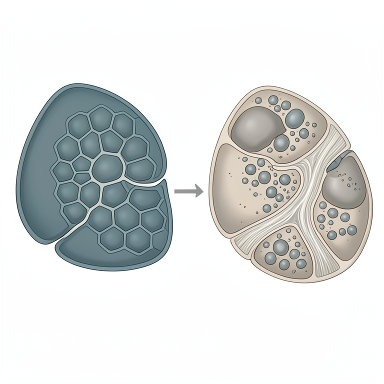
Liver Histopathology
AI-generated conceptual illustration (buffalo calf liver tissue). It explains the idea and does not show study data.
View source evidence ↗Figures & tables
From the original paper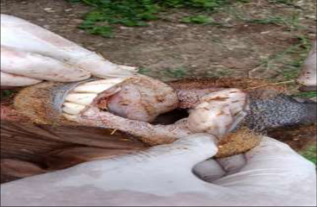 Figure 1
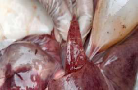 Figure 2
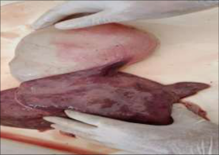 Figure 3
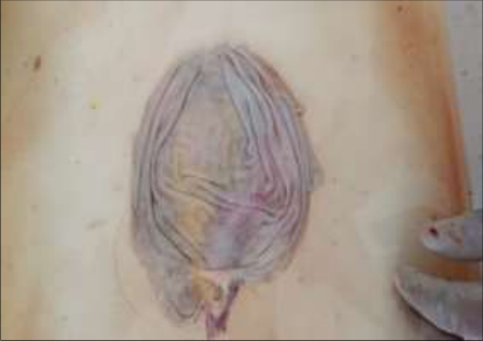 Figure 4
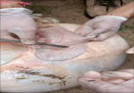 Figure 5
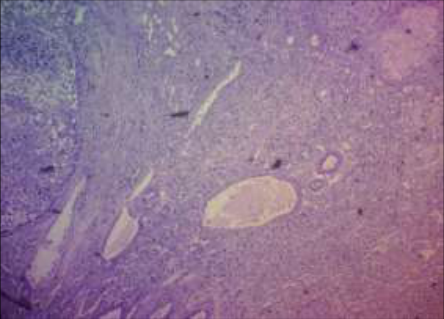 Figure 6
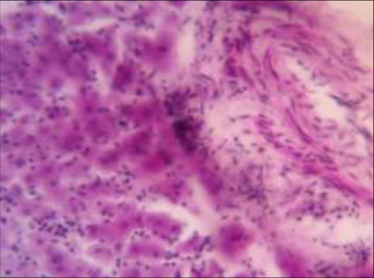 Figure 7
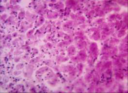 Figure 8
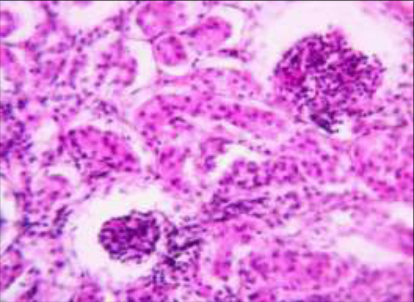 Figure 9
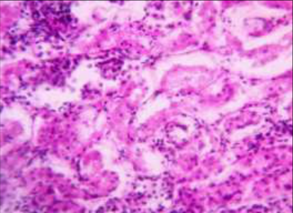 Figure 10
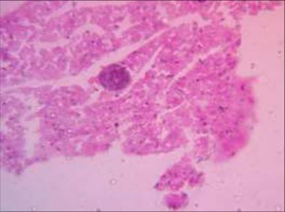 Figure 11
Introduction
Buffalo species possesses significant value in terms of its draft power, biological performances, and high ecological potential on productivity (Latif, 1994) [5]. The liver, being the largest organ in the body of herbivores, constitutes approximately 1% of their body weight. The liver plays a crucial role in various functions, including the metabolism of bilirubin, carbohydrates, xenobiotics, protein production, and immune function (Cullen et al., 2012) [2]. Furthermore, research has indicated that buffalo liver can also be commercially utilized in the preparation of acceptable comminuted meat products within the food industry (Naveena et al., 2014) [8].
Pathological conditions affecting the liver can arise from a variety of causes, such as parasites, viruses, mycoses, bacteria, genetic factors, immunological factors, and idiopathic factors, resulting in significant economic losses (Ahmed et al., 2013) [1]. Chronic hepatitis, also known as cirrhosis, is an inflammatory and necrotizing liver disease (Sterczer et al., 2001) [10]. It is characterized by fibrosis, degeneration, and hyperplasia of hepatic cells. Therefore, the present study aims to identify the pathological conditions of the liver in buffalo calf carcasses through gross and histopathological studies.
Materials And Methods
A buffalo calf carcass, one-year-old received in the Department of Veterinary Pathology at CVAS, Navania, Udaipur for postmortem examination. Following the gross examination of the entire carcass, representative tissue specimens were collected from the liver and other organs. These specimens were then fixed in 10% neutral buffered formalin for a minimum of 24-48 hours and subsequently subjected to routine processing, as described by Salmo et al. in 2014. The fixed tissue samples, measuring 3-5 mm in thickness, were further processed using the acetone and benzene technique for paraffin embedding, as outlined by Lillie in 1965.
Bacteriological samples were also taken from liver and heart blood, processed as per routine. IJABR 2024; SP-8(1): 563-566 Indu Bala PG Scholar, Department of Veterinary Pathology, College of Veterinary and animal Science, Rajasthan University of Veterinary and Animal Sciences, Bikaner, Rajasthan, India Goverdhan Singh Assistant Professor, Department of Veterinary Pathology, College of Veterinary and Animal Science, Rajasthan University of Veterinary and Animal Sciences, Bikaner, Rajasthan, India Mamta Assistant Professor, Department of Veterinary Pathology, College of Veterinary and Animal Science, Rajasthan University of Veterinary and Animal Sciences, Bikaner, Rajasthan, India Kamal Purohit Assistant Professor, Department of Veterinary Pathology, College of Veterinary and Animal Science, Rajasthan University of Veterinary and Animal Sciences, Bikaner, Rajasthan, India Bhavana Kanwar Rathor Department of Animal Nutrition, College of Veterinary and Animal Science, Rajasthan University of Veterinary and Animal Sciences, Bikaner, Rajasthan, India Mamta Kumari PG Scholar, Department of Veterinary Pathology, College of Veterinary and Animal Science, Rajasthan University of Veterinary and Animal Sciences, Bikaner, Rajasthan, India Pankaj Kanwar PG Scholar, Department of Livestock Production and Technology, College of Veterinary and Animal Science, Rajasthan University of Veterinary and Animal Sciences, Bikaner, Rajasthan, India Indu Bala PG Scholar, Department of Veterinary Pathology, College of Veterinary and animal Science, Rajasthan University of Veterinary and Animal Sciences, Bikaner, Rajasthan, India Fig 3: Gross picture of small nodular liver and enlarge distended gallbladder Fig 4: Gross picture of distended gallbladder with thinning of wall. https://www.biochemjournal.com
Results And Discussion
Upon gross examination, the conjunctival and oral mucous membranes exhibited a pale appearance (Fig 1), while congestion was evident in most of the organs. The liver displayed congestion and appeared small and nodular, with the presence of necrotic foci (Fig 2 & 3). The gallbladder was enlarged, distended, and contained fluid with a watery consistency (Fig 3). Additionally, thinning of the gallbladder wall was observed (Fig 4).
The spleen appeared smaller in size and had a wrinkled capsule (Fig 5). Gelatinization of fat was observed in fat depots and on the visceral organs. Histopathological examination of liver tissues revealed varying degrees of degeneration, necrosis, and fibrous tissue proliferation (Fig 6). These findings align with the research conducted by Talukder et al. (2010) [11] and MacGavin et al. (2001) [7].
In the liver tissues, there was a significant presence of chronic inflammatory cells, including Macrophages and lymphocytes, as well as an increase in fibrous tissue growth within and around the hepatic lobules (Fig 7 & 8). These findings align with the research conducted by Doy et al. (1984) [3] and Wiedosari et al. (1991) [12]. Additionally, certain areas of the liver lobules exhibited neovascularization and an excessive growth of bile ducts. Faccin et al. (2016) [4] also observed bile duct hyperplasia in their study.
Furthermore, there were instances of pseudolobulation and an eccentric positioning of central veins in certain areas. Upon microscopic examination of the kidney, degeneration of the tubular epithelium was observed (Fig 9). The tubules also displayed the formation of hyaline casts and infiltration of chronic inflammatory cells (Fig 9 & 10). Sarcocysts were identified in the myocardium of the heart (Fig 11).
Bacteriological analysis of the samples did not yield any growth on culture media. Based on all these findings, the case was diagnosed as idiopathic chronic hepatitis. Fig 5: Gross picture of smaller sized spleen and its wrinkled capsule Fig 1: Gross pic of oral cavity of buffalo calf showing Pale mucous membranes. Fig 6: Microphotograph of liver showing chronic hepatitis, severe atrophic changes, necrosis and proliferation of fibrotic tissue in liver parenchyma.
H&E-4X Fig 2: Gross picture of Liver showing severe congestion along with necrotic foci https://www.biochemjournal.com Fig 7: Photmicroograph of liver tissue showing, infiltration of chronic inflammatory cells around the hepatocyte and fibrous tissue proliferation. H&E, 40X Fig 8: Photomicrograph of liver tissue showing, degeneration of hepatic cells infiltration of chronic inflammatory Cell. H&E, 40X Fig 9: Photomicrograph of kidney tissue showing, infiltration of chronic inflammatory cells in glomeruli and in tubules. H&E, 40X Fig 11: Photomicrograph of heart tissue showing presence of sarcocyst, H&E, 40X
Conclusion
As per histopathological examination of tissue indicated a case of idiopathic chronic hepatitis.
Acknowledgement
The authors are highly grateful to the Dean, College of Veterinary and Animal Science, Navania, Udaipur (Rajasthan) for providing essential facilities for completing the study.
Figures and tables from the paper
References
12 in the original paper- Ahmed AM, Ismail SAS, Dessouki AA. Pathological lesions survey and economic loss for male cattle slaughtered at Ismailia abattoir. International Food Research Journal. 2013;20:857-863.
- Cullen JM, Brown DL. Pathologic basis of veterinary disease: Hepatobiliary system and exocrine pancreas. In: Zachary JF, McGavin MD, editors. Elsevier Mosby Ltd., Missouri, USA; c2012. p. 405-446.
- Doy TG, Hughes DL. Early migration of immature Fasciola hepatica and associated liver pathology in cattle. J Res Vet Sci. 1984;37:219-22.
- Faccin TC, Kommers GD, Galiza GJND, Pupin RC, Madureira RC, Lemos RAA. Chronic liver disease in cattle associated with ingestion of Brachiaria spp. Ciencia rural. 2016;46:2036-2042.
- Latif MA. Bufffalo production in Bangladesh problems and prospects. Proceedings of 1st Asian Buffalo Association Congress, January 17-21, Khon Kaen, Thailand; c1994.
- Lillie RD. Histopathological technique and practical histochemistry. McGrawHill Book Co., New York and London; c1965.
- MacGavin MD, Carlton WW, Zachary JF. Thomson's Special Veterinary Pathology. 3rd ed. St Louis: Mosby; c2001. P. 325-245.
- Naveena BM, Kirant M. Buffalo meat quality, composition, and processing characteristics: Contribution to the global economy and nutritional security. Animal Front. 2014;4:18-24. https://doi.org/10.2527/af.2014-0029
- Salmo NA, Hassan SM, Saeed AK. Histopathological study of chronic livers Fascioliasis of cattle in Sulaimani abattoir. AL-Qadisiyah Journal of Veterinary Medicine Sciences. 2014;13(2):71-80.
- Sterczer A, Gaal T, Perge E, Rothuizen J. Endocrinology: Chronic hepatitis in the dog - a review. Veterinary Quarterly. 2001;23(4):148-152. Fig 10: Photomicrograph of kidney tissue showing, degeneration of tubular epithelium & chronic infiltration of inflammatory cell. H&E, 40X https://www.biochemjournal.com
- Talukder S, Bhuiyan MJ, Hossain MM, Uddin MM, Paul S, Howlader MMR. Pathological investigation of liver fluke infection of slaughtered black Bengal goat in a selected area of Bangladesh. Bangl J Vet Med. 2010;8(1):35-41.
- Wiedosari E, Graydon R, Copeman DB. Comparative pathological study of hepatic changes induced by Fasciola gigantica and Gigantocotyle explanatum in Javanese thin-tailed sheep. J Comp Path. 1991;105:147- 155.