
Research article · 2024 · Full text
International Journal of Veterinary Sciences and Animal HusbandryEpidemiological and haemato-biochemical studies in parvo viral infected dogs
Volume 9(1) · Pages 656–659 · DOI: 10.22271/veterinary.2024.v9.i1i.992 ↗
Research areasVirus-based gene therapy researchAnimal Virus Infections StudiesParvovirus B19 Infection Studies
Abstract
Author-writtenThe present study was carried out at Department of Veterinary Clinical Complex, Veterinary College, Gadag, for the period of 1 year (January-2021 to April 2022). Total of 101 dogs were presented with history of vomition, diarrhea, depression & dehydration to the Department of Veterinary Clinical Complex, Veterinary College, Gadag during the study period. Out of 101 cases 57 cases were screened positive for Parvoviral infection by using “Anigen Rapid” CPV Ag test kits. Out of these 57 cases 14 cases were selected for the hemato-biochemical study. The pups below the age of 6 months were highly susceptible compare to the adult ones and the more number of cases reported were of Labrador breed. The case susceptibility was comparatively more in male dogs than female ones and the highest incidence was recorded in post monsoon season (October-December). Reduced HB, PCV, Leukocytopemia, lymphocytopenia, thrombocytopenia, neutrophilia and elevated levels of ALP and ALT enzymes were observed in the CPV infected cases.
Summary
Source-checkedPlain-language reading of the full paper, prepared with AI and checked against the text. Each point links to the authors' own words.
Why it matters
- Canine parvovirus infection (CPV) is a highly contagious disease in dogs globally that is frequently fatal. View evidence ↗
- CPV gastroenteritis typically manifests as severe vomiting, loss of appetite, and foul-smelling diarrhea, which may contain blood. View evidence ↗
- Puppies are predisposed to CPV due to factors such as lack of immunity, intestinal parasites, and poor environmental conditions like overcrowding and lack of sanitation. View evidence ↗
The question
- The study aimed to investigate the epidemiological patterns and the changes in blood (hematological) and chemical (biochemical) parameters associated with canine parvovirus infection. View evidence ↗
What they did
- Out of 101 dogs presenting with symptoms like vomiting and diarrhea, 57 were confirmed positive for CPV using Anigen Rapid CPV Ag test kits. View evidence ↗
- For the blood analysis, 14 infected dogs and 6 healthy control dogs were selected for sampling. View evidence ↗
- Hematological parameters (Hb, RBC, TLC, PCV, Platelets) and biochemical markers (ALT, ALP) were measured using automated analyzers, with data analyzed via unpaired t-tests. View evidence ↗
What they found
- Puppies under 6 months were the most susceptible to CPV (54.39%), followed by those aged 6-12 months (31.58%) and adults (14.03%). View evidence ↗
- Labradors showed the highest susceptibility to the infection (36.84%), while Golden Retrievers showed the lowest (7.01%). View evidence ↗
- The highest incidence of CPV occurred during the post-monsoon season (October-December) at 42.10%, with the lowest in the pre-monsoon season (8.77%). View evidence ↗
- Infected dogs exhibited marked anemia, characterized by significantly reduced hemoglobin (9.75±0.2 g/dl) and hematocrit (28.9±0.82%). View evidence ↗
- A majority of infected dogs suffered from leukocytopenia (78.57%, mean 3.76±0.17 x10³/μl) and lymphocytopenia (57.14%, mean 18.86±0.73%). View evidence ↗
- Thrombocytopenia was observed in 85.71% of the infected cases, with a mean platelet count of 98.57±3.26 x10³/μl. View evidence ↗
- All 14 infected dogs showed a significant increase in the liver enzymes ALP and ALT. View evidence ↗
What it means
- The authors conclude that CPV is most common in young dogs and during the monsoon season, and that blood and biochemical tests are vital for diagnosis and prognosis. View evidence ↗
Visual summary
3 visualsIllustrations explain the paper; charts are replotted from the paper's own table values.
Epidemiological Risk Factors
AI-generated conceptual illustration (domestic dogs (puppies and adult Labradors)). It explains the idea and does not show study data.
View source evidence ↗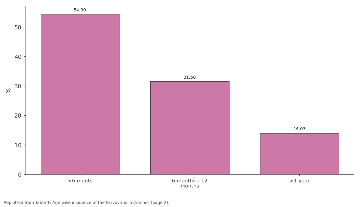
%: <6 monts vs 6 months – 12 months vs >1 year
Replotted from Table 1 (page 2) of the original paper. Every bar is the paper's own table value.
View source evidence ↗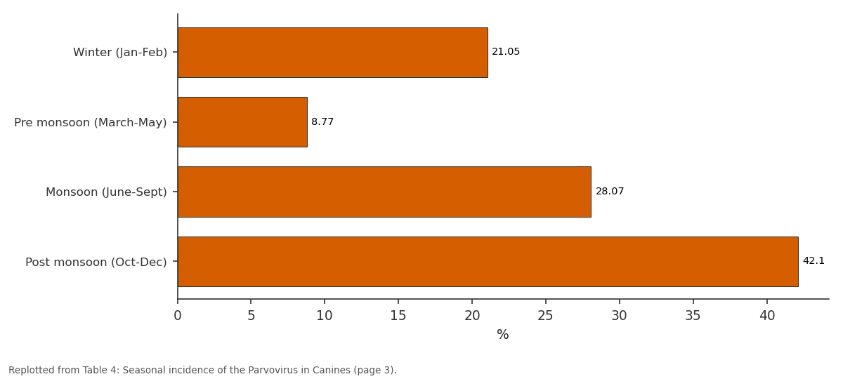
%: Winter (Jan-Feb) vs Pre monsoon (March-May) vs Monsoon (June-Sept) vs Post monsoon (Oct-Dec)
Replotted from Table 4 (page 3) of the original paper. Every bar is the paper's own table value.
View source evidence ↗Figures & tables
From the original paper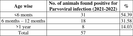 Table 1
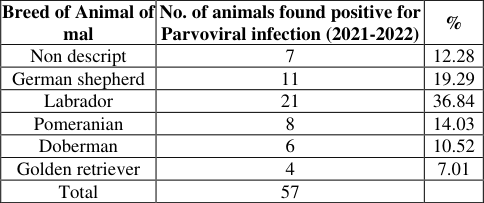 Table 2
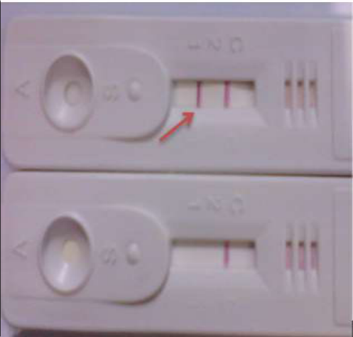 Figure 1
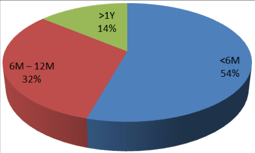 Figure 1
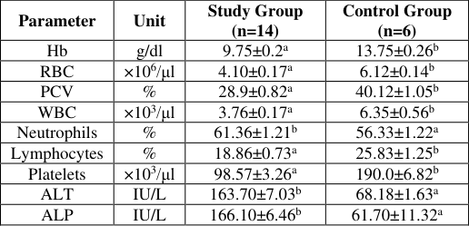 Table 5
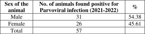 Table 3
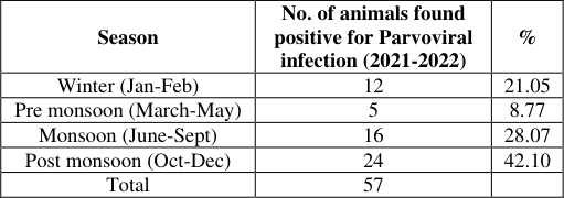 Table 4
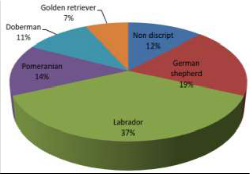 Figure 2
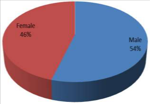 Figure 3
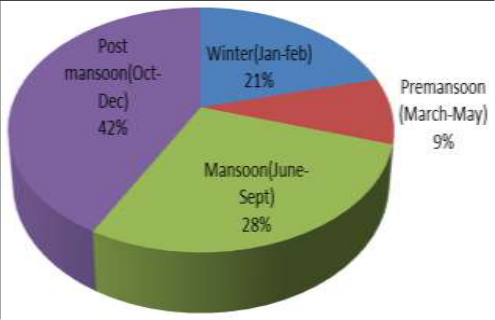 Figure 4
Introduction
Canine parvovirus infection (CPV) is a highly contagious and often fatal disease of dogs in many parts of the world. Clinical manifestation of CPV gastroenteritis are severe emesis, inappetance, diarrhea, with or without presence of blood and foul smell (Castro et al., 2007 and Sagar et al., 2008) [4,16] . Parvoviral enteritis can be seen in dogs of any breed, age or sex but puppies between 6 weeks and 6 months of age are more prone to the disease (Quinn et al., 2002 and Gaddard et al., 2008) [14,6] . Factors that predispose to parvoviral infection in puppies are lack of protective immunity, intestinal parasites and overcrowded, unsanitary and stressful environmental conditions (Greene, 2006) [5] . The present study was an attempt to study the Epidemiological and hemato-biochemical alterations in canine parvovirus infection.
Materials and Methods
Total of 101 dogs were presented with history of vomition, diarrhea, depression & dehydration to the Department of Veterinary Clinical Complex, Veterinary College, Gadag during the study period. Out of 101 cases 57 cases were screened positive for Parvoviral infection by using "Anigen Rapid" CPV Ag test kits. Out of these 57 cases 14 cases were selected for the hemato-biochemical study. 4 ml Blood sample was collected from all the infected dogs (n=14) and also from the healthy control group dogs (n=6) using new syringes and then transferred 2ml each to EDTA vials and serum clot activator vials. The serum was separated through centrifugation for further analysis. The Hematological Parameters (Hb, RBC, TLC, PCV and Platelets) were determined using automated blood cell counter (Beckman Coulter, Coulter diff Ac.T, USA) and biochemical parameters (ALT and ALP) were determined using automated clinical chemistry analyzer (Adiduoplus Biochemestry Analyzer). The differential leucocyte count was performed manually on blood smear stained by Wright-Giemsa staining method. Statistical analysis is done by unpaired "t" test.
Results and Discussion
Total of 101 dogs were presented with history of vomition, diarrhea, depression & dehydration to the Department of Veterinary Clinical Complex, Veterinary College, Gadag during the study period. Out of 101 cases 57 cases were screened positive for Parvoviral infection by using "Anigen Rapid" CPV Ag test kits. The pups below the age of 6 months (54.39%) were found to be more susceptible for parvoviral infection followed by 6-12 months (31.58%) age groups and the adult ones (14.03%). similar observations are recorded by Quinn et al. (2002) [14] , Mohammed et al. (2005) [13] , Goddard et al. (2008) [6] . This could be due to due to lack of immunity (Greene, 2006) [5] .
The breed susceptibility was seen more in Labrador breed (36.84%) of dogs followed by German Shepherd (19.29%), Pomeranian (14.03%), Non-descript (12.28%), Doberman (10.52%) and Golden Retriever (7.01%). These changes could be due to the more number of Labrador population in the Gadag district of Karnataka. The sex wise incidence study revealed, male dogs were more prone to the parvoviral infection compare to female. The similar observations are recorded by Haque and Arfa (2012) [7] . This variation in sex wise incidence could be due to more population of male dogs compare to female ones in the district. Seasonal incidence of parvoviral infection was seen more in Post monsoon during October-December (42.10%) season followed by monsoon during the month of June-September (28.07%), Winter during the months of January-February (21.05%) and Pre monsoon during the months of March-May (8.77%). Similar findings were reported by Al-Bayati et al. (2010) [1] who reported high prevalence during September-December months. This could be due to the survivability and growth of virus in cold climate as opined by Mehta et al. (2017) [12] . However contradictory to these findings reported by Mohammed et al. (2005) [13] who reported highest prevalence in May to June months. Hematological examination showed marked anemia with significantly reduced hemoglobin levels of (9.75±0.2) and reduced hematocrit (28.9±0.82). Similar observations were also recorded by Sagar (2005) [17] and Sujata et al. (2012) [19] . The reduced hemoglobin, hematocrit could be due to damages to the capillaries of the intestinal villi (Hoskins and McCaw 1998) [8] , decreased erythropoiesis as CPV directly effects the bone marrow (Boosinger et al., 1982) [3] and loss of blood due to emesis and diarrhea as it's the most common sing of CPV infection (Yadav et al., 2011) [21] . Leukocytopenia (3.76±0.17) was observed in 11 cases (78.57%) and lymphocytopenia (18.86±0.73) was seen in 8 cases (57.14%). Similar observations were also recorded by Goddard et al. (2008) [6] , Sagar et al. (2008) [16] and Sujata et al. (2012) [19] . This could be due to the virus replication in the lymphoid organs resulting in lymphocytolysis (Biswas et al.
2005) [2] and which indicates the presence of more severe form of disease (Mason et al., 1987) [11] . Thrombocytopenia (98.57±3.26 ×10 3 /μl) was observed in 12 cases (85.71%). This was in resemblance to Sujata et al.
(2012) [19] . The Virus-induced thrombocytopenia can occur because of decreased platelet production or as a result of direct action of viruses and/or immuno-logic components on platelets or endothelium (Wilson et al., 1982) [20] . Neutrophilia (61.36±1.21) was recorded in all 14 cases. These observations were similar to reports of Shah et al. (2013) [18] . This could be due to concurrent bacterial infections with parvoviral enteritis as opined by Ramprabhu et al. (2002) [15] . Biochemical evaluation revealed significant rise in the values of ALP and ALT in all 14 cases. These observations were similar to reports of Jacobs et al. (1980) [9] , Macintire and Smith, (1997) [10] and Shah et al. (2013) [18] . Significant rise in alkaline phosphatase and alanine transaminase could be a result of hepatic hypoxia secondary to severe hypovolemia or the absorption of toxic substances due to loss of the gut barrier as opined by Shah et al. (2013) [18] .
Conclusion
From the above study it is concluded that, the epidemiological data reveals the outbreak of CPV infection in dogs is very common in young dogs and in monsoon season and the hemato-biochemical study plays a vital role in diagnosis of the CPV infected dogs and helps in assessing the possible prognosis of the disease.
Figures and tables from the paper
| Age wise | No. of animals found positive for Parvoviral infection (2021-2022) | % |
|---|---|---|
| <6 monts | 31 | 54.39 |
| 6 months – 12 months | 18 | 31.58 |
| >1 year | 8 | 14.03 |
| Total | 57 |
Table text extracted from the original PDF; superscript letters are shown inline. The original table image is below.
View original table image
| Breed of Animal of mal | No. of animals found positive for Parvoviral infection (2021-2022) | % |
|---|---|---|
| Non descript | 7 | 12.28 |
| German shepherd | 11 | 19.29 |
| Labrador | 21 | 36.84 |
| Pomeranian | 8 | 14.03 |
| Doberman | 6 | 10.52 |
| Golden retriever | 4 | 7.01 |
| Total | 57 |
Table text extracted from the original PDF; superscript letters are shown inline. The original table image is below.
View original table image
| Parameter | Unit | Study Group (n=14) | Control Group (n=6) |
|---|---|---|---|
| Hb | g/dl | 9.75±0.2a | 13.75±0.26b |
| RBC | ×106/μl | 4.10±0.17a | 6.12±0.14b |
| PCV | % | 28.9±0.82a | 40.12±1.05b |
| WBC | ×103/μl | 3.76±0.17a | 6.35±0.56b |
| Neutrophils | % | 61.36±1.21b | 56.33±1.22a |
| Lymphocytes | % | 18.86±0.73a | 25.83±1.25b |
| Platelets | ×103/μl | 98.57±3.26a | 190.0±6.82b |
| ALT | IU/L | 163.70±7.03b | 68.18±1.63a |
| ALP | IU/L | 166.10±6.46b | 61.70±11.32a |
Table text extracted from the original PDF; superscript letters are shown inline. The original table image is below.
View original table image
References
18 in the original paper- Al-Bayati HA, Odhiso SHM, Majeed HA. Detection of Canine Parvo Virus in Iraq by using rapid antigen test kit and Haemagglutination-inhibition test. Al-Anbar J Vet Sci. 2010;3(2):17-23.
- Biswas S, Charavorty D, Pradhan NR. Clinical and haemato-biochemical changes in Parvo virus infection in dogs. Indian J Vet Med. 2005;25:16-18.
- Boosinger TR, Rebar AH, Denicola DB, Boon GD. Bone marrow alterations associated with Canine Parvo viral enteritis. Vet Pathol. 1982;19(5):558-561.
- Castro TX, Miranda SC, Labarthe NV, Silva LE, Cubel G. Clinical and epidemiological aspects of Canine Parvo virus (CPV) enteritis in the state of Rio de Janeiro. Arq Bras Med Vet Zootec. 2007;59(2):333-339.
- Goddard A, Leisewitz AL, Christopher MM, Ducan NM, Becker PJ. Prognostic usefulness of blood leukocyte changes in canine Parvo viral enteritis. J Vet Intern Med. 2008;22(2):309-316.
- Haque S, Arfa T. Epidemiological study of Parvo gastroenteritis and its management by different concentrations of fluid in pups. Indian J Canine Pract. 2012;4(2):83-85.
- Hoskins JD, McCaw DL. Canine Viral enteritis. In Greene, C.E. (Ed), Infectious diseases of dogs and cats. 2nd Edn. W.B a Sunders co, Philadelphia. 1998;208(4):542-546.
- Jacobs RM, Weiser MG, Hall RL, Kowalski JJ. Clinicopathologic features of Canine Parvo viral enteritis. J Am Anim Hosp Assoc. 1980;16:809-814.
- Macintire DK, Smith-Carr S. Canine parvovirus. Part II. Clinical signs, diagnosis and treatment. Comp. The Compendium on continuing education for the practicing veterinarian. 1997;19(3):291-302.
- Mason MJ, Gillet NA, Muggenberg BA. Pathological and epidemiological aspects of canine Parvo viral enteritis in an unvaccinated closed beagle colony. J Am Anim Hosp Assoc. 1987;23:183-192.
- Mehta SA, Patel RM, Vagh AA, Mavadiya SV, Patel MD, Vala JA, et al. Prevalence of Canine Parvo Viral Infection in Dogs in and around Navasari District of Gujarat state, India. Indian J Vet Sci Biotechnol. 2017;13(2):67-72.
- Mohammed, Gisilanbe J, Ogbe, Okuwa A, Zwandor, Joseph N, Umoh, et al. Risk factors associated with Canine Parvo virus enteritis in vom and environs. Anim Res Int. 2005;2(3):366-368.
- Ramprabhu R, Prathaban S, Nambi AP, Dhanapalan P. Haemorrhagic gastroenteritis in dogs -A clinical profile. Indian Vet J. 2002;79:374-376.
- Sagar A, Roy S, Roy M. Clinico, haemato-biochemical changes and diagnosis of canine Parvo viral enteritis. Intas Polivet. 2008;9(II):262-265.
- Shah SA, Sood NK, Wani N, Gupta K, Singh A. Haemato-biochemical changes in canine parvoviral infection. Indian J Vet Pathol. 2013;37(2):131-133.
- Sujata T, Ranjan R, Amol S, Kumar D, Bhutia C. Acute anaemia and its therapeutic management by blood transfusion in a pup. Intas Polivet. 2012;13(1):92-93.
- Wilson JJ, Neame PB, Kelton JG. Infection-induced thrombocytopenia. Semin Thromb Hemost. 1982;8(3):217-233.
- Yadav R, Gupta SR, Sharma CS. Clinical haematology in dogs affected with haemorrhagic gastroenteritis. Vet Practitioner. 2011;12(1):60-62.
Cited by 4
From OpenAlex, OpenCitations and reference lists read by Scinovex
- Molecular Occurrence of CPV-2 Infection in HGE Affected Dogs ↗
Anuja Jayaprasad, Brejesh Singh, Kajal Kumar Jadav, Yogendra Kumar Sinha, Diksha Lade, Somesh Singh · Indian Journal of Animal Research · 2025
Background: Canine parvovirus is the most prevalent infectious disease affecting dogs globally. Canine parvovirus type-2 is regarded as the causal factor of canine parvoviral enteritis (PVE). The objective of the study was to determine the occurrence of parvovirus in dogs in Jabalpur from June, 2023 to December, 2023. Methods: Faecal samples form suspected cases were handled using sterile swab and…
❝ 2PDF - OCCURRENCE OF CANINE PARVOVIRAL GASTRO ENTERITIS IN AND AROUND BENGALURU ↗
H.V. Sanjay, G.K. Chetan Kumar, G. Leena · Indian journal of veterinary and animal sciences research. · 2026
Canine parvoviral gastroenteritis is one of the most common viral diseases found in dogs and caused by canine parvovirus (CPV) type 2. Young dogs are highly susceptible to the disease with high morbidity and mortality. The present study was conducted at the Department of Veterinary Medicine, Veterinary College, Bengaluru to know the occurrence pattern of CPV infection among PCR positive…
❝ 0PDF - Anti-inflammatory effects of Saccharomyces boulardii treatment in dogs naturally infected with canine parvovirus ↗
Selin Sinem SÜMBÜL, Başak Hanedan · Veterinary Immunology and Immunopathology · 2026
❝ 0 - Detection and Epidemiology of canine parvovirus infection in dogs
Khushi Saini, Sarita Devi, Vidhi P Patel, Sandip S Patel · International Journal of Veterinary Sciences and Animal Husbandry · 2025
A total of 335 dogs suspected of Canine Parvovirus (CPV) infection presented during the nine-month study period were clinically evaluated and tested by Polymerase Chain Reaction (PCR). Of these, 232 samples (69.25%) tested positive for CPV. The highest incidence was observed in dogs aged 3-6 months (38.79%), followed by < 3 months (25.43%), 6-9 months (24.14%), and > 9 months (11.64%). Males showed higher prevalence (73.28%) than females (26.72%). Non-descript dogs were most affected (58.19%), and winter recorded the highest seasonal prevalence (46.12%). A greater incidence was noted in non-va