
Research article · 2024 · Full text
International Journal of Advanced Biochemistry ResearchCanine demodicosis: A case report and successful recovery at Izatnagar, Bareilly
Volume 8(1) · Pages 519–521 · DOI: 10.33545/26174693.2024.v8.i1g.431 ↗
Research areasDermatological diseases and infestationsYersinia bacterium, plague, ectoparasites researchPoxvirus research and outbreaks
Abstract
Author-writtenCanine demodicosis, also known as demodectic mange, is mainly caused by Demodex canis. It causes both localized and widespread (generalised) skin infections in dogs all throughout the world. An adult male dog (one-year-old), weighing 15 kg was presented to Veterinary clinical complex, Izatnagar, Bareilly. The animal had history of anorexia, alopecia, dehydrated, erythema, itchy skin, crust formation, hyperpigmentation, and dullness. On clinical examination and skin scrapping examination it was diagnosed as demodicosis (Demodex canis). The case was treated with tablet Bravecto (fluralaner 1000 mg) once a day orally for two weeks, along with antibacterial, antifungal and supportive nutritional therapy. The dog reacted well to medication, and no further notable side effects were observed. The third week of treatment marked the full recovery. The current report's goal is to educate pet owners about the prevalence of Demodex canis and its therapeutic care.
Summary
Source-checkedPlain-language reading of the full paper, prepared with AI and checked against the text. Each point links to the authors' own words.
Why it matters
- Canine demodicosis is considered one of the most significant skin diseases encountered in veterinary practice. View evidence ↗
- The primary cause of this condition is the mite Demodex canis, which lives in the sebaceous glands and hair follicles of dogs. View evidence ↗
- Infected animals often experience persistent itching (pruritus), which can lead to the development of wounds on the body. View evidence ↗
The question
- The goal of this report is to educate pet owners regarding the prevalence of Demodex canis and how to provide therapeutic care. View evidence ↗
What they did
- The study focuses on a single case involving a one-year-old male dog weighing 15 kg at the Veterinary clinical complex in Izatnagar, Bareilly. View evidence ↗
- Confirmatory diagnosis was achieved using deep skin scrapings performed with a scalpel and surgical blades. View evidence ↗
- The skin scrapings were processed by digestion in 3-5 ml of 10% potassium hydroxide solution to dissolve tissues and hairs. View evidence ↗
- The processed solution was cooled and then centrifuged at 1500rpm for 5 minutes before microscopic examination. View evidence ↗
What they found
- Microscopic examination of skin scrapings confirmed the presence of Demodex canis, identified by its elongated, cylindrical, cigar-shaped appearance. View evidence ↗
- The dog exhibited widespread clinical signs, including hair loss (alopecia), redness (erythema), and crusts across the face, ears, limbs, abdomen, thigh, loin, and tail. View evidence ↗
- The affected dog's body temperature was recorded at 101.8°F. View evidence ↗
- The primary treatment consisted of oral Bravecto (Fluralaner 1000 mg) administered once daily for two weeks. View evidence ↗
- To manage secondary bacterial infections, a 10 mg/kg combination of clindamycin hydrochloride and nicotinamide was given for ten days. View evidence ↗
- Fungal infections were treated with 5 mg/kg of itraconazole tablets for 15 days. View evidence ↗
- After two weeks of treatment, the skin texture returned to almost normal and skin scrapings no longer showed any mites. View evidence ↗
- Full recovery of the animal was achieved by the third week of treatment. View evidence ↗
What it means
- The authors conclude that the case of demodicosis was successfully and completely cured using a combination of fluralaner, antibacterial/antifungal medications, and specialized shampoo. View evidence ↗
- The authors emphasize that pet owners must provide appropriate treatment and intensive care to prevent the disease from becoming widespread. View evidence ↗
Visual summary
1 visualsIllustrations explain the paper; they do not show study data.
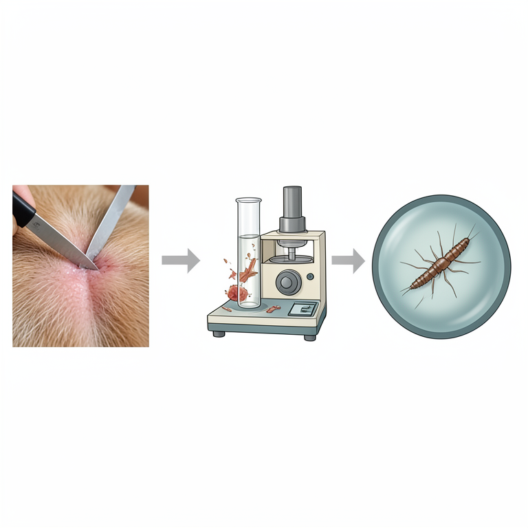
Skin Scraping Diagnostic Process
AI-generated conceptual illustration (Canine skin tissue and Demodex mites). It explains the idea and does not show study data.
View source evidence ↗Figures & tables
From the original paper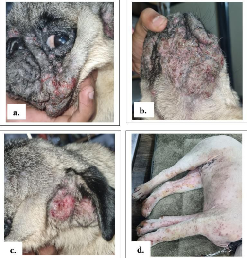 Figure 1
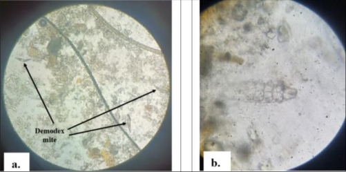 Figure 2
Introduction
Dogs are the mostly reared pet animals throughout the world. A wide range of parasites with zoonotic potential are infecting the canines, which are of human health concerns. Among that, canine demodicosis is most important cutaneous disease of dogs that is encountered across veterinary practices. Demodicosis is very commonly reported in the immuno-deficient young, adult and old dogs. There are three documented species of canine Demodex mites and they are Demodex canis, D. injai, and, D. cornei. The first species to be identified was Demodex canis; two other Demodex mite species may be either mutations of Demodex canis or distinct species (Sivajothi et al., 2015) [9] . Demodex injai is a long-bodied demodecid mites with males that are more than twice as long as male D. canis mites (Hillier and Desch, 1997) [3] . Scarff (1988) [5] reported a short and stubby form of the Demodex and described being about half the length of the female D. canis (Scarff, 1988; Chesney, 1999) [5,2] . However, Demodex canis is the major causative agent of canine demodicosis and which colonize in the hair follicles, sebaceous glands of dogs. It is characterized by significant changes in skin such as alopecia, erythema, follicular hyperkeratosis, comedones, pustules, seborrhoea, crusts etc. A secondary pyoderma can also be frequently reported as complications of demodicosis. Demodex canis is a follicular effecting, most common skin infection in dogs and, two types of infections are documented such as, a localized and a generalized form. The localized form occurs mainly in younger dogs less than a year old. But the cutaneous infection may become generalized due to proliferation of mite population. Generalized demodicosis in dog might take several months to years for recovery in dog. The clinical sign -persistent pruritus observed in infected animals due to itching, which result in wounds on the body. This article presents a case report about alarming incidence of demodicosis in Izatnagar region of Bareilly and the advisable treatment, prevention and control of disease in dogs.
Case History
A one-year-old male dog weighing 15 kg was presented to the Teaching Veterinary Clinical Complex, Indian Veterinary Research Institute, Izatnagar, Bareilly with a history of inappetence, dullness, severe itching, foul body odour and hair shedding.
There was previous history of skin infections and also ectoparasite incidences. As per the history, animal was regularly vaccinated and the deworming schedule was followed as advised by veterinarian. On physical examination, the infected dog was anorexic with dehydrated skin. The moderate clinical signs include alopecia, erythema, papular and pustular lesions, skin rashes, crust and scab formation around face, ears, fore limbs, hind limbs, ventral abdomen, inner thigh, loin and tail region. Also, hyperpigmentation was observed in these regions. The body temperature of the affected dog was found to be 101.8°F. Other parameters like heart rate, respiration rate, pulse rate, mucous membrane, urination and defecation were normal.
Method/Diagnosis
Based on the clinical signs observed in animal, the infection was tentatively diagnosed as skin infection of parasitic origin. For the confirmatory diagnosis of the condition, deep skin scrapings were done with the help of fresh scalpel and surgical blades. The suspected crusted papule of skin and the adjacent area was selected on infected animal for sample collection. Unnecessary hairs were clipped to reduce the number of hairs during slide preparation. A drop of liquid paraffin was placed on the selected area and allowed for spreading-out. The skin was squeezed to maximize the numbers of mites inside the skin area to be scrapped. Scraping of skin started with the help of scalpel and surgical blades and finished till the slight bleeding occurs. The scrapings were transferred from scalpel blade into a test tube and then digested in 3-5 ml of 10% potassium hydroxide solution. The solution with skin scraping was boiled for 3-4 minutes till all the hairs and skin tissues were dissolved and the solution became homogenous. The solution was cooled and centrifuged at 1500rpm for 5 minutes. The supernatant solution is discarded and from the sediment a small drop was taken onto a clean and clear glass slide. Applied cover slip and examined under 10X and 40X objective microscope for the detection of mites, if any.
Results
The skin scraping examination revealed the presence of elongated, cylindrical and cigar shaped mite, which was identified as Demodex canis with body divisible into the head, thorax and the abdomen. The mite is characterized by thorax with four pairs of three jointed short and stumpy legs and long, transversely striated abdomen according to Soulsby, 1982 [10] .
Treatment and Prognosis
For two weeks, the dog was given an oral medication called Bravecto (Fluralaner 1000 mg) once a day. For ten days, a 10 mg/kg combination of clindamycin hydrochloride and nicotinamide tablets was administered to suppress bacterial infection. For four months, 0.4 mg/kg of the tablet Apoquel (oclacitinib maleate) was used to manage pruritus and atopic dermatitis. For 15 days, a 5 mg/kg tablet of itraconazole was administered to manage the fungal infection. For thirty days, the ZincOtin-DS tablet (zinc, biotin, folic acid, vitamin E, and linolenic acid) was taken twice a day. For 30 days, 10 milliliters BID of Syrup Foxyfur (omega 3, omega 6, vitamin A, vitamin D3, vitamin E, zinc, and biotin) to enhance skin health and decrease hair loss. For 15 days, 75 ml BID of Syrup REVELL (including 15 well-proven herbal components) to enhance and promote liver functioning. To rebuild the skin's natural defences, four weeks of weekly administration of ALLERDERM Spot-on (Skin Lipid Complex, a proprietary blend of ceramides, fatty acids, and cholesterol) was administered. Vet-Pro was fed a nutritionally enhanced, hypoallergenic diet for three months in order to support the body's natural defences against skin and coat health issues. It is also recommended to administer liquid betadine twice a day to the entire body. For two weeks, it was advised to take a bath using Petben shampoo (benzoyl peroxide) once every three days to degrease and exfoliate hair follicles. On clinical observations after two weeks of treatment, the texture of skin appeared as almost normal and the skin scraping was negative for mites.
Discussion
The current case was classified as generalized demodicosis due to the widespread lesions observed in the body. Several drugs are used in the multimodal treatment of canine generalized demodicosis (Mueller et al., 2012) [4] . The clinical indications and lesions of demodicosis may be caused by a variety of predisposing factors, including as malnourishment, malnutrition, and an aberrant environment that encourages mite proliferation and the development of other skin diseases (Mueller et al., 2012; Shrestha et al., 2015) [4,6] . Because most cases of canine generalized demodicosis entail a subsequent bacterial skin infection that requires systemic antibiotic therapy for several weeks, broad-spectrum antibiotics were used in this our study for treatment (Verde, 2005; Mueller et al., 2012) [11,4] . Shampoos containing benzoyl peroxide are frequently advised due to their keratolytic and follicular flushing properties. Shampoo containing benzoyl peroxide demonstrated good results when treating generalized demodicosis. The animal in the current case study that received enough nutritional supplements responded well to treatment for canine demodicosis due to its antioxidant qualities (Singh et al., 2011; Yatoo et al., 2014; Arsenovic et al., 2015) [7,12,1] . It also helped the animal cope with the side effects of a particular acaricidal treatment (Singla et al., 2013) [8] .
Conclusion
From a medical perspective, Demodex canis has continued to provide significant challenges for both pet owners and veterinarians. In addition, it is imperative that pet owners provide their animals with appropriate treatment and intensive care to prevent widespread diseases. The present case of demodicosis, which was diagnosed through skin scraping and treated with tablet Bravecto (fluralaner 1000 mg) once a day orally for two weeks, along with other antibacterial, antifungal medication and Petben shampoo, was successfully and completely cured.
Acknowledgement
AcknowledgementWe acknowledge the Head, Division of Parasitology, IVRI, Izatnagar and the Director, IVRI, Izatnagar, for all the facilities for disease diagnosis.
Figures and tables from the paper
References
8 in the original paper- Arsenović M, Pezo L, Vasić N, Ćirić R, Stefanović M. The main factors influencing canine demodicosis treatment outcome and determination of optimal therapy. Parasitology research. 2015;114:2415-2426.
- Chesney CJ. Short form of Demodex species mite in the dog: occurrence and measurements. Journal of Small Animal Practice. 1999;40(2):58-61.
- Mueller RS, Bensignor E, Ferrer L, Holm B, Lemarie S, Paradis M, et al. Treatment of demodicosis in dogs: 2011 clinical practice guidelines. Veterinary dermatology. 2012;23(2):86-e21.
- Shrestha D, Thapa B, Rawal G, Dhakal S, Sharma B. Prevalence of demodectic mange in canines of Kathmandu Valley having skin disorder and its associated risk factors. International Journal of Applied Sciences and Biotechnology. 2015;3(3):459-463.
- Singh SK, Dimri U, Sharma MC, Swarup D, Sharma B, Pandey HO, et al. The role of apoptosis in immunosuppression of dogs with demodicosis. Veterinary immunology and immunopathology. 2011;144(3-4):487-492.
- Singla LD, Eljadar MSM, Bal Mandeep Singh DS, Uppal SK, Juyal PD. Parasitic Dermatitis Due to Canine Demodicosis in Dogs. Indian Journal of Canine Practice. 2013;5:85-87.
- Sivajothi S, Sudhakara Reddy B, Rayulu VC. Demodicosis caused by Demodex canis and Demodex cornei in dogs. Journal of parasitic diseases. 2015;39:673-676.
- Yatoo MI, Jhambh R, Malepad DP, Kumar P, Dimri U. Advances in therapeutic management of complicated demodicosis in canines. J Adv. Parasitology. 2014;1(1):6-8.
Cited by 1
From OpenAlex, OpenCitations and reference lists read by Scinovex
- A case report on the effective management of canine demodicosis
Tamilam TV, Enbavelan PA, E. Venkatesakumar, S. Sivaraman, Karuppiah Arunachalam · International Journal of Biology Sciences · 2025
Canine demodicosis is a non contagious skin disorder caused by Demodex canis, a mite that resides in the hair follicles and sebaceous glands of dogs. While typically harmless in healthy dogs, overgrowth of the mite can lead to clinical manifestations such as alopecia, pustular lesions, and pruritus. A 10 month old female Doberman dog, weighing 12 kg, was presented to the Veterinary Clinical Complex, Veterinary College and Research Institute, Salem, with a history of inappetence, dullness, itching, and hair shedding. The bitch had a previous history of ectoparasite infestation and followed regu