
Research article · 2024 · Full text
International Journal of Advanced Biochemistry ResearchStudy of haematological parameters in goats with maternal dystocia and normal parturition during pre-partum and post-partum periods
Volume 8(1S) · Pages 153–155 · DOI: 10.33545/26174693.2024.v8.i1sc.351 ↗
Research areasEffects of Environmental Stressors on LivestockReproductive Physiology in LivestockAnimal Nutrition and Health
Abstract
Author-writtenDystocia has a significant impact on future reproduction and production of animals. The experiment was conducted to determine haematological profiles in normal parturated and dystocia affected goats. The study was conducted on 10 goats. The goats were divided into two groups viz. normal parturated goats (NP) and maternal dystocia affected goats (MD). Blood samples were collected from maternal dystocia affected (n=5) and normal parturated goats (n=5) just before handling of dystocia or parturition and 24 hours after manual delivery or normal parturition for the estimation of various haematological parameters including Hb, RBC, WBC, PCV, MCV, MCH, Lymphocyte, Monocyte, Eosinophils, Basophils and Neutrophil count. Results revealed that Hematological parameters such as haemoglobin, WBC, monocytes, PCV decreased significantly (p≤0.05) in goats with maternal dystocia at post-partum period as compared to pre-partum and neutrophils decreased significantly (p≤0.05) in goats with maternal dystocia as well as normal parturated goats at post-partum period as compared to pre-partum period. Whereas lymphocyte increased significantly (p≤0.05) in goats with maternal dystocia as well as normal parturated goats at post-partum period as compared to pre-partum period.
Summary
Source-checkedPlain-language reading of the full paper, prepared with AI and checked against the text. Each point links to the authors' own words.
Why it matters
- Dystocia, or difficult birth, is a critical issue because it can negatively affect the future production and reproductive capabilities of animals. View evidence ↗
- Dystocia is defined as a difficult birth process that lasts over one hour after the fetal membranes have ruptured and often requires assistance. View evidence ↗
- Maternal causes of dystocia can include factors such as pelvic fractures, uterine torsion, uterine rupture, birth canal obstructions, or the failure of abdominal expulsive forces. View evidence ↗
The question
- The study aimed to evaluate and compare blood (haematological) parameters between goats experiencing normal kidding and those requiring assisted kidding due to maternal dystocia. View evidence ↗
What they did
- The study used 10 goats of various parities, split equally into a normal parturition (NP) group and a maternal dystocia (MD) group. View evidence ↗
- Blood samples were collected from both groups (n=5 each) at two time points: immediately before the birth process and 24 hours after delivery. View evidence ↗
- The researchers measured a complete blood count, including hemoglobin (Hb), red blood cells (RBC), white blood cells (WBC), packed cell volume (PCV), and various leukocyte differentials. View evidence ↗
- Data were analyzed using ANOVA and Duncan's new multiple range test to identify significant differences between the means. View evidence ↗
What they found
- No significant differences were found in eosinophil levels between the two groups at either the pre-partum or post-partum time points. View evidence ↗
- Basophil levels showed no significant difference between normal parturition and maternal dystocia goats during the study periods. View evidence ↗
What it means
- The authors conclude that understanding these haematological changes in goats with dystocia can help in the management of obstetrical problems. View evidence ↗
Visual summary
1 visualsIllustrations explain the paper; they do not show study data.
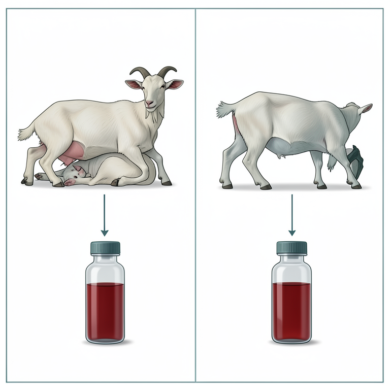
Study Comparison Framework
AI-generated conceptual illustration (female goats). It explains the idea and does not show study data.
View source evidence ↗Figures & tables
From the original paper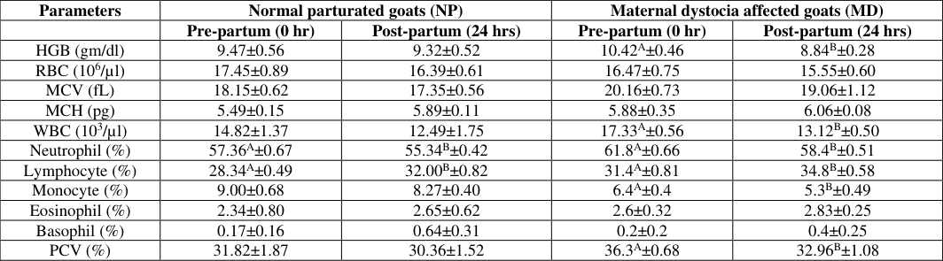 Table 1
Introduction
Dystocia, or difficult birth, is described as prolonged and difficult parturition that lasts more than one hour after rupture of the fetal membranes with frequent assistance required (Ahmed et al., 2017). Dystocia is reported to be more prevalent in goats than in ewes (Sharma et al., 1999; Mehta et al., 2002) [17,11] . Dystocia can be classified into maternal causes and fetal causes (Noakes et al., 2001) [12] . The maternal causes of dystocia includes abdominal expulsive forces failure, birth canal obstruction, uterine rupture, uterine torsion and pelvic fracture. In goats, the incidence of dystocia due to maternal factors was reported to be 31.4 percent (Purohit et al., 2006) [14] , 47.1 percent (Majeed and Taha, 1989) [9] and 57.8 percent (Sharma et al., 1999) [17] . Maternal dystocia is most often caused by a failure of cervical dilatation, followed by uterine inertia (Purohit, 2006) [15] . Parturition is a complex process which involves a sequence of endocrine events in the maternal, foetal and placental tissues that results in haemato-biochemical changes in the dam (Hussein and Abd Ellah, 2008; Vannucchi et al., 2015) [7,19] . Difficult births or dystocia is a complicated and stressful process, which makes the procedure more complicated and painful. The alteration in haemato-biochemical characteristics in dystocia and retained placenta have been extensively studied in goats (Patel et al., 2020) [13] . Variations in haematological parameters may exist between dystocia-affected and normal parturient animals. Therefore, the present research study was conducted to evaluate and compare the hematological parameters in normal kidding and assisted kidding due to maternal causes of dystocia in goats.
Materials and Methods
The present study was conducted on the goats presented to the department of Veterinary Gynaecology and Obstetrics, CVAS, Bikaner (RAJUVAS), and goats rearing at Bikaner and nearby goat farms in Bikaner city (for normal parturition). A total number of 10 goats of different parities were included in the study. The goats were divided into two groups viz. normal parturated goats (NP) and maternal dystocia affected goats (MD). Blood samples were collected from maternal dystocia affected (n=5) and normal parturated goats (n=5) just before handling of dystocia or parturition and 24 hours after manual delivery or normal parturition for the estimation of various haematological parameters. Haematological analysis was performed shortly after blood collection. Whole blood samples from EDTA vials were used to measure complete blood count including Hb, RBC, WBC, PCV, MCV, MCH, Lymphocyte, Monocyte, Eosinophils, Basophils and Neutrophil count. These parameters were analysed as per the methods described by Jain (1986) [22] . The collected data were complied, tabulated and analysed statistically by using analysis of Variance test (ANOVA) with the help of statistical software as per Snedecor and Cochran (1994) [18] . Means showing significant differences were compared by Duncan's new multiple range test (Duncan, 1955) [4] .
Results and Discussion
The haematological observations are given in Table 1.
| Parameters | Normal parturated goats (NP) | — | Maternal dystocia affected goats (MD) | — |
|---|---|---|---|---|
| Pre-partum (0 hr) | Post-partum (24 hrs) | Pre-partum (0 hr) | Post-partum (24 hrs) | |
| HGB (gm/dl) | 9.47±0.56 | 9.32±0.52 | 10.42A±0.46 | 8.84B±0.28 |
| RBC (106/µl) | 17.45±0.89 | 16.39±0.61 | 16.47±0.75 | 15.55±0.60 |
| MCV (fL) | 18.15±0.62 | 17.35±0.56 | 20.16±0.73 | 19.06±1.12 |
| MCH (pg) | 5.49±0.15 | 5.89±0.11 | 5.88±0.35 | 6.06±0.08 |
| WBC (103/µl) | 14.82±1.37 | 12.49±1.75 | 17.33A±0.56 | 13.12B±0.50 |
| Neutrophil (%) | 57.36A±0.67 | 55.34B±0.42 | 61.8A±0.66 | 58.4B±0.51 |
| Lymphocyte (%) | 28.34A±0.49 | 32.00B±0.82 | 31.4A±0.81 | 34.8B±0.58 |
| Monocyte (%) | 9.00±0.68 | 8.27±0.40 | 6.4A±0.4 | 5.3B±0.49 |
| Eosinophil (%) | 2.34±0.80 | 2.65±0.62 | 2.6±0.32 | 2.83±0.25 |
| Basophil (%) | 0.17±0.16 | 0.64±0.31 | 0.2±0.2 | 0.4±0.25 |
| PCV (%) | 31.82±1.87 | 30.36±1.52 | 36.3A±0.68 | 32.96B±1.08 |
Table text extracted from the original PDF; superscript letters are shown inline. The original table image is below.
View original table image
In the present study, when compared to the pre-partum period, haemoglobin was decreased non-significantly at 24 hours after kidding in normal parturated goats. The findings of present study are in agreement with previous research of Sangami (2017) [16] who found that haemoglobin concentration was decreased non-significantly at postpartum (24hr) period as compared to pre-partum period in normal parturated goats. In goats with maternal dystocia, haemoglobin value was found significantly (p≤0.05) decreased at 24 hours after assisted kidding as compared to pre-partum period in present study. This reduction in haemoglobin levels may be as a result of bleeding during assisted kidding in certain animals. The present findings are in agreement with previous research of Sangami (2017) [16] who found that Haemoglobin concentration was decreased significantly at post-partum (24hr) period as compared to pre-partum in dystocia affected goats. Total erythrocyte count decreased non-significantly in normal parturated as well as goats suffering from maternal dystocia at 24 hours after kidding as compared to prepartum period. The present findings are in accordance with the observations of Manat et al. (2016) [10] who found nonsignificant decrease in total RBC on the day of kidding in dystocia affected goats. No significant difference was found in the values of Mean Corpuscular Volume and Mean Corpuscular haemoglobin in normal parturated goats and goats with maternal dystocia at pre-partum period and post-partum (24 hr) period. The results of present study are in agreement with the observations of Azab and Abdel-Maksoud (1999) [2] who observed no significant difference in MCV values at 0 day and 7 days after parturition in goats. There were no significant differences in the values of WBC in normal parturated goats at pre-partum as compared to post-partum (24 hr) period. The results of present study are in agreement with the observations of Azab and Abdel-Maksoud (1999) [2] who found no significant difference in WBC count at 0 day and 7 days after parturition in goats. However, WBC count decreased significantly (p≤0.05) at post-partum (24 hr) period in goats with maternal dystocia. The results of present findings are in agreement with Sangami (2017) [16] who also found that in dystocia affected goats, total leucocyte was significantly higher as compared to normal parturated goats at pre partum period (0hr) and decreased significantly in post-partum period. The neutrophil count in goats with maternal dystocia and normal parturated goats was found significantly (p≤0.05) lower at post-partum (24hr) period as compared to perpartum period. The results of present study are in agreement with observations of Azab and Abdel-Maksoud (1999) [2] who found that neutrophils count was significantly decreased in post-partum period in goats. The values of lymphocyte were significantly higher in postpartum (24hr) period as compared to pre-partum period in maternal dystocia and normal parturated goats. These findings are in agreement with previous studies in cows, Yuksel et al. (2011) [21] who reported the haematological profile before and after parturition in cows suffering from dystocia and reported the mean lymphocytes count (%) was increased significantly following relieving dystocia in cows. The mean values of monocyte decreased significantly (p≤0.05) in goats suffering from maternal dystocia at postpartum (24 hrs) period compared to pre-partum period. Similarly, Yuksel et al. (2011) [21] also observed that mean lymphocytes count (%) decreased significantly after relieving dystocia in cows. Non-significant difference was observed in the mean values of eosinophils in normal parturated goats and goats with maternal dystocia at pre-partum and post-partum period (24 hrs). The result of present study is similar with findings of Yildiz et al. (2011) [20] who reported no significant difference in eosinophils count in dystocia affected and normal parturated cattle. Also, no significant difference was found in the values of basophils in normal parturated goats and goats with maternal dystocia at pre-partum and post-partum period (24 hrs). The result of present study are in agreement with Kaur et al. (1993) [8] also reported no significant differences in basophil count between buffaloes with uterine torsion, genital prolapse, dystocia and normal calving buffaloes. The PCV values found significantly lower (p≤0.05) in goats with maternal dystocia at post-partum (24hr) period as compared to pre-partum period. The findings of the current study are in agreement with previous findings of Yuksel et al. (2011) [21] who found significant decline in packed cell volume during the post-partum period as compared to prepartum period in goats and cows, respectively. It was concluded that Hematological parameters such as haemoglobin, WBC, monocytes, PCV decreased significantly in goats with maternal dystocia at post-partum period as compared to pre-partum and neutrophils decreased significantly in goats with maternal dystocia as well as normal parturated goats at post-partum period as compared to pre-partum period. Whereas lymphocyte increased significantly in goats with maternal dystocia as well as normal parturated goats at post-partum period as compared to pre-partum period.
Conclusion
It was concluded that Hematological parameters such as haemoglobin, WBC, monocytes, PCV decreased significantly (p≤0.05) in goats with maternal dystocia at post-partum period as compared to pre-partum and neutrophils decreased significantly (p≤0.05)in goats with maternal dystocia as well as normal parturated goats at postpartum period as compared to pre-partum period. Whereas lymphocyte increased significantly (p≤0.05) in goats with maternal dystocia as well as normal parturated goats at the post-partum period as compared to the pre-partum period. So the findings on haemato-biochemical changes in dystocia affected goats may aid in managing obstetrical problems.
References
19 in the original paper- Ahmed A, Balarabe AT, Jibril A, Sidi S, Jimoh AA, Gobe RM. Incidence and causes of dystocia in small ruminants in Sokoto Metropolis, Northwestern, Nigeria. Scholars Journal of Agriculture and Veterinary Science. 2017;4(3):114-118.
- Azab ME, Abdel-Maksoud HA. Changes in some hematological and biochemical parameters during prepartum and postpartum periods in female Baladi goats. Small Ruminant Research. 1999;34(1):77-85.
- Bihaqi SJ, Allaie IM, Banday MAA, Wani ZA, Shahardar RA. Prevalence of caprine GI helminths in temperate areas of Jammu and Kashmir. Journal of parasitic diseases. 2017;41(3):843-849.
- Duncan DB. Multiple range and multiple F tests. Biometrics. 1955;11:1-42.
- Ghuman SPS. Uterine torsion in bovines: a review. Indian Journal of Animal Science. 2010;80:289-305.
- Hegde NG. Goat development: An opportunity to strengthen rural economy in Asia and Africa. Asian Journal of Research in Animal and Veterinary Sciences. 2020:30-47.
- Hussein H, AbdEllah MR. Effects of dystocia, fetotomy and caesarian sections on the liver enzymes activities and concentrations of some serum biochemical parameters in dairy cattle. Animal reproduction science. 2008;105(3-4):384-391.
- Kaur T, Singh B, Kaur T. Reproductive disorders in buffaloes: haemato-biochemical studies. Indian Journal of Veterinary Pathology. 1993;17:118-1121.
- Majeed AF, Taha MB. Dystocia in local goats in Iraq. Small Ruminant Research. 1989;2(4):375-381.
- Manat TD, Chaudhary SS, Singh VK, Patel SB, Puri G. Hematobiochemical profile in Surti goats during the post-partum period. Veterinary world. 2016;9(1):19-24.
- Mehta V, Nagar D, Yadav RC, Garg N, Purohit GN. Obstetrics problems in goats. V National Seminar. Indian Society for sheep and goat production. 2002 Dec.30-31, Jaipur. p. 151.
- Noakes DE, Parkinson DJ, England GCW. Maternal Dystocia. In: Veterinary reproduction and obstetrics, 8thEdn. Saunders Harcourt, India. 2001:230-235.
- Patel GA, Suthar BN, Nakhashi HC, Chaudhari CF, Sutaria TV, Chaudhari RK. Haemato-biochemical profile in goats with dystocia and retained foetal membranes. Indian Journal of Small Ruminants (The). 2020;26(1):71-74.
- Purohit GN, Gupta AK, Gaur M, Sharma A, Bihani D. Periparturient disorders in goats. A retrospective analysis of 324 cases. Dairy Goat J. 2006;84(2):24-33.
- Purohit GN. Dystocia in the sheep and goat-a review. The Indian Journal of Small Ruminants. 2006;12(1):112.
- Sharma VK, Suthar BN, Parsani HR, Ojha SC. A clinical study on dystocia in small ruminants. In XV Annual Convention and National Symposium on Biotechniques in Optimizing Fertility in Farm Animals. PAU, Ludhiana, Abstracts. 1999;94:94.
- Vannucchi CI, Rodrigues JA, Silva LCG, Lúcio CF, Veiga GAL, Furtado PV, Nichi M. Association between birth conditions and glucose and cortisol profiles of periparturient dairy cows and neonatal calves. Veterinary Record. 2015;176(14):358-358.
- Yıldız H, Saat N, Şimşek H. An investigation on body condition score, body weight, calf weight and hematological profile in crossbred dairy cows suffering from dystocia. Pakistan Veterinary Journal. 2011;31:125-128.
- Yuksel M, Yildiz H, Saat N, Simsek H. Hematological profile before and after parturition in cows suffering from dystocia. F. U. Sag. Bil. Vet. Derg. 2011;25(1):1115.