
Research article · 2024 · Full text
International Journal of Veterinary Sciences and Animal HusbandryTraumatic Reticulopericarditis in murrah buffalo: Case report
Volume 9(1) · Pages 21–24 · DOI: 10.22271/veterinary.2024.v9.i1a.855 ↗
Research areasAnimal health and immunologyStreptococcal Infections and TreatmentsMicrobial infections and disease research
Abstract
Author-writtenAn 8 years old Murrah buffalo carcass was handed over to Veterinary Pathology Department, COVAS, Parbhani, and Maharashtra for post mortem examination. Clinical examination revealed subnormal rectal temperature, suspended rumination, polypnea, sluggish heart beating. Laboratory investigations revealed leukocytosis with neutrophilia, decrease hematocrit and microcytic hypochromic anemia. The liver enzyme activity found to be elevated. Postmortem examination showed, subcutaneous haemorrhages, hydroperitoneum, hydrothorax with emphysematous lung lobes. There were multiple thin repealed fluid filled cysts on lung lobes. There was suppurative and fibrinous exudate noted in pericardium. Fibrin deposition on heart gave ‘‘bread and butter’’ like appearance with adhesion to surrounding tissue. Penetrating foreign body (iron binding wire) was noted in heart with well developed ‘fibrous track’ connecting diaphragm and pericardium. Considering the above record the buffalo died due cardiorespiratory failure as result of adhesive traumatic reticulopericarditis.
Summary
Source-checkedPlain-language reading of the full paper, prepared with AI and checked against the text. Each point links to the authors' own words.
Why it matters
- Traumatic pericarditis in ruminants is a sporadic condition caused by foreign objects like nails or metal wires penetrating the pericardium from the reticulum or diaphragm. View evidence ↗
- This disease is significant because it causes cardiac dysfunction, increases treatment costs, and leads to a loss of productive capacity in animals. View evidence ↗
- In India, traumatic pericarditis has been recorded as contributing to approximately 23.38% of mortality in buffaloes. View evidence ↗
The question
- The objective of this case report was to document the necropsy and histopathological findings of a specific instance of traumatic pericarditis. View evidence ↗
What they did
- Data was collected via a haemato-biochemical report and clinical history provided by the TVCC Complex. View evidence ↗
- Heart tissue samples were preserved in 10% formal saline solution for histopathological analysis. View evidence ↗
What they found
- Biochemical analysis revealed an increased albumin-globulin ratio of 86.25 and significantly elevated levels of AST, ALT, and alkaline phosphatase. View evidence ↗
- Postmortem examination of the thoracic cavity revealed approximately 10 to 15 liters of watery fluid. View evidence ↗
- The pericardial sac was diffusely thickened and contained about 1 liter of amber-colored flocculent fluid with free-floating fibrin. View evidence ↗
- Fibrin deposits on the epicardial surface of the heart created a characteristic 'bread-and-butter' appearance. View evidence ↗
- An iron binding wire was found lodged in the myocardium, with fibrous tracks connecting it to the pericardium, diaphragm, and reticulum. View evidence ↗
- Histopathology showed marked inflammation with lymphocytic and neutrophilic infiltrates across the myocardium, epicardium, and pericardium. View evidence ↗
What it means
- The authors concluded that the Murrah buffalo died from cardiovascular failure resulting from traumatic reticulopericarditis. View evidence ↗
Visual summary
1 visualsIllustrations explain the paper; they do not show study data.
Path of Foreign Body Penetration
AI-generated conceptual illustration (Murrah buffalo thoracic and abdominal anatomy). It explains the idea and does not show study data.
View source evidence ↗Figures & tables
From the original paper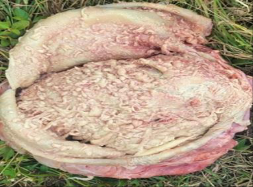 Figure 2
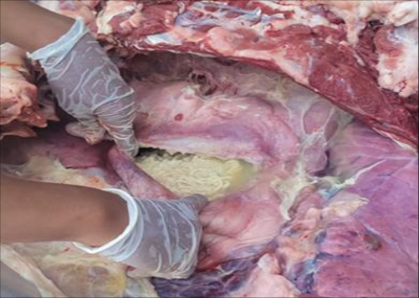 Figure 1
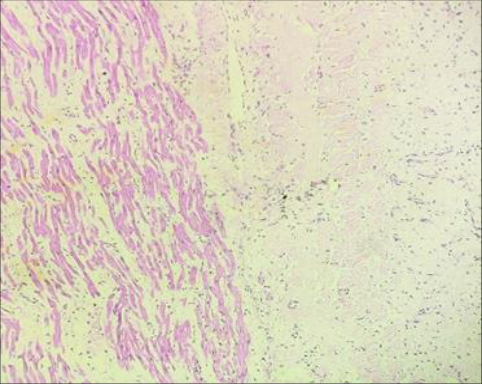 Figure 4
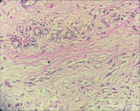 Figure 3
Introduction
In ruminants, traumatic pericarditis is a sporadic disease that is triggered by penetrating any foreign bodies such as (needles, metal wire and nails) on pericardium surface originating from the reticulum, diaphragm, and pericardial sac, resulting in traumatic pericarditis (Ibrahim and Gomaa, 2016) [13] , which lead to gathering of serous/fibrin containing inflammatory exudates results in follow of aggregating disturbances in heart function almost leads to unexpected death in animals (Braun et al., 2007 and Braun, 2009) [8,7] . Pericarditis results from many etiological factors which, spreads through the hematogenous routes by various bacterial, viral infections and sometime anaerobic infection was much less common (Grunder, 2002 and Braun, 2009) [12,7] . Increased in heart rate and sounds like friction or splashing sounds, engorgement of jugular vein and brisket oedema was the key clinical signs noted in animals. However, not all animals exhibit these symptoms, making a diagnosis difficult in some cases (Grunder, 2002) [12] . The traumatic pericarditis leading to loss of productive capacity (Aref and Abdel-hakiem, 2013) [3] , treatment cost higher (Nugusu et al., 2013) [20] and health disturbances due to cardiac function (Attia, 2016) [6] . Incidence of mortality contribute around (23.38%) due to traumatic pericarditis in bauffalo was recorded by Ramprabhu et al. (2003) [22] in India. Similar incidences were also recorded in the foreign countries due to animal mismanagement (Misk et al., 1984) [18] . It was likewise seen that as of recently calved and feeble bison have a higher pace of rate than dry and lactating buffalo (Ramprabhu et al., 2003) [22] . Frequency rate was higher in dairy cattle when contrasted with beef cattle since they were bound to be taken care of a cleaved feed, like silage or roughage (Kahn, 2005) [14] . Besides, a high frequency was recorded during the dry spell and collect seasons (Sharma and Kumar, 2006) [23] . It more commonly affects females, especially in the third trimester of gestation (Braun et al., 2007; Mohamed, 2010; Attia, 2016) [8,18,6] . Because the weight and size of the pregnant uterus makes it possible for the organ to work as a pendulum when the animal moves, applying physical pressure to the rumen and reticulum, collaborating for penetration by an existing object.
(Fubini and Divers, 2008) [8] . Because of its high significance in health and animal production, traumatic pericarditis is as yet a question of overall concern. Thus, the objective of the present case report was to highlight the necropsy along with histopathological findings observed in a case of traumatic pericarditis.
Materials and Methods
Case history and clinical findings: An eight-year-old Murrah buffalo weighed 450 kg carcass was present to the Department of Vet. Pathology for postmortem examination as referred by the TVCC Complex. The TVCC gave general information, including history, herd size, management methods, clinical symptoms shown by animals during the course, as well as a haemato-biochemical report, which include a recent parturition in the past, anorexia in the past 8 days, suspended rumination, subnormal rectal temperature, mouth breathing, sluggish heart beating, heart rhythm could not be assessed, and no feces in the rectum on per rectal examination. Leukocytosis with neutrophilia, Traumatic Reticulopericarditis Buffalo: A decrease in hematocrit, and microcytic hypochromic anemia were noted through hematological investigations. Significant increase in the levels of liver enzyme i.e., ALT, AST and alkaline phosphatase, in addition to an increased albumin-globulin ratio (A:G=86.25), was among the biochemical results.
Results and Discussion
Gross observations: At necropsy, nothing remarkable alterations were seen during external examination. The internal examinations revealed were subcutaneous hemorrhages and brisket edema. Notable amount of clear fluid were recorded in the peritoneum. On opening of the thoracic cavity, approx. around 10-15 liters of watery fluid were seen. The lungs were emphysematous; lobes were adhered with the pleura, ribs and the diaphragm. There were multiple thin, fluid-filled cysts on the lung lobes. The pericardium sac was thickened diffusely and approx. contained 1 liter of AMBE colored flocculent fluid with freefloating fibrin. There was suppurative and fibrinous exudates were noted in the pericardium surface whereas, fibrin also deposited on the epicardial surface of heart giving its appearance as bread-and-butter. The fibrin deposited on the heart and lung lobes were found to be adhered to the pleural surface. The penetrating iron binding wire piece was recovered from the heart, which were completely lodged in the myocardium layer of the heart which correlated with the well-developed fibrous tracks connecting the reticulum, diaphragm, and pericardium.
Microscopic observations:
Histopathology was performed on collected tissues of the heart samples and preserved in 10% formal saline solution. Marked depositions of fibrin on both the pericardial Traumatic Reticulopericarditis Buffalo: Epicardium surface of heart were noted. There was marked inflammation with neutrophilic and lymphocytic infiltrates in the pericardium, epicardium, and myocardium surface of the heart observed.
Discussion
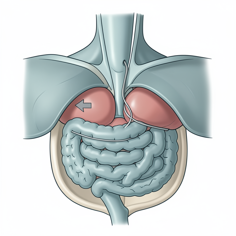
As a part of the stomach which has very high pressure on the surface of reticulum, as feed material accidentally found foreign penetrating body i.e., needle, during digestive processing due to the sharpening of the metal object perforates through the path of reticulum to diaphragm to pericardial layer to heart leads to cause traumatic pericarditis. Due to their anatomical configuration of the reticulum is of honeycomb shape, penetrating foreign bodies can easily attach to the mucosa of the reticulum without causing injury. During advanced pregnancy, physiological alteration such as uterine muscular contractions during delivery phase encourage foreign body to perforate in the reticular wall during reticular contraction (Ghanem, 2010; Anteneh and Ramswamy, 2015; Ibrahim and Gomaa, 2016) [11,2,13] . In some animals, due to foregut contractions, potential foreign bodies such as nails, needle and binding wires pierce the reticulo-diaphragm and lead to further complications. Average wall has plentiful of low sift hold strain receptors, so any injury to average reticular wall muscular build lead to brokenness of these receptors which result in lessened and hypomotility of rumeno-reticulum with variable level of tympany. Due to penetration of foreign body, leakage of reticular fluid which leads to peritonitis commences 24 hrs. Results in increase in temperature, pulse rate and rigor specially in abdomen muscles. At the site if inflammation, affected tissues release cytokines, including interleukin (IL)-1, IL-6, and tumor necrosis factor (TNF). Such proinflammatory substances leakage leads to tempt limited and general reactions, typically acute phase protein, protein depletion in muscle, pyrexia, leukocytosis and hormonal alteration. [1,15] . When that foreign body penetrate pericardial layer of heart causing an accumulation of inflammatory fluid in the pericardial sac (Kumar et al., 2012; Yildiz et al., 2019) [16,24] . Exudate further classified into three general forms, depending on the nature of contents such as effusive, fibrinous and constrictive, and their combinations can occur (Athar et al., 2012) [5] .
Conflict of interest:
Authors have no conflict of interest in this study.
Conclusion
Considering the case history, clinical observations, haematobiochemical and necropsy findings, of the Murrah buffalo might have been died due to cardiovascular failure caused by traumatic reticulopericarditis.
Acknowledgement
AcknowledgementAuthor's expresses special thanks to Associate Dean, College of Veterinary and Animal Sciences, Parabhani, MAFSU, Nagpur for extending supports to carry out the present research work.
Figures and tables from the paper
References
19 in the original paper- Abdelaal AM, Floeck M, El-Maghawry S, Baumgartner W, et al. Clinical and ultrasonographic differences between cattle and buffaloes with various sequelae of traumatic reticuloperitonitis. Veterinarian Medicinal. 2009;54(9):399-406.
- Anteneh M, Ramswamy V. Hardware disease in bovine. Acad. J Anim. Dis. 2015;4(3):146-159.
- Aref NEM, Hakiem AMA. Clinical and diagnostic methods for evaluation of sharp foreign body syndrome in buffaloes. Veterinary World. 2013;6(9):586.
- Ashfaq M, Razzaq A, Hassan S. Factors affecting the economic losses due to livestock diseases: A case study of district Faisalabad. Pakistan Journal of Agricultural Sciences, 2015, 52(2).
- Athar H, Parrah JD, Moulvi BA, Singh M, Dedmari FH, et al. Pericarditis in bovines: A Review. International Journal of Advanced Veterinary Science and Technology. 2012;1(1):19-27.
- Attia NE. Cardiac biomarkers and ultrasonography as tools in prediction and diagnosis of traumatic pericarditis in Egyptian buffaloes. Veterinary World. 2016;9(9):976.
- Braun U. Traumatic pericarditis in cattle: clinical, radiographic and ultrasonographic findings. The Veterinary Journal. 2009;182(2):176-186.
- Braun U, Lejeune B, Schweizer G, Puorger M, Ehrensperger F. Clinical findings in 28 cattle with traumatic pericarditis. Veterinary Record. 2007;161(16): 558-563.
- Ghanem MM. A comparative study on traumatic reticuloperitonitis and traumatic pericarditis in Egyptian cattle. Turkish Journal of Veterinary & Animal Sciences. 2010;34(2):143-153.
- Ibrahim HMM, Gomaa NA. Traumatic pericarditis in cattle: Risk factors, clinical features and ultrasonographic findings. Journal of Veterinary Science and Medical Diagnosis. 2016;5(3):7-15.
- Khalphallah A, Elmeligy E, Elsayed HK, Abedellaah BEA, Salman D, Al-Lethie ALA, et al. Ultrasonography as a diagnostic tool in Egyptian buffaloes (Bubalus bubalis) with traumatic pericarditis. International Journal of Veterinary Science and Medicine. 2017;5(2):159-167.
- Kumar A, Saini NS, Mohindroo J, Sood NK, et al. Ultrasonographic features of normal heart and liver in relation to diagnose pericarditis in bovine. Indian Journal of Animal Sciences. 2012;82(12):1489.
- Misk NA, Semieka MA. The radiographic appearance of reticular diaphragmatic herniation and traumatic pericarditis in buffaloes and cattle. Vet Radiol Ultrasound. 2001;42(5):426-30. DOI: 10.1111/J1740-8261.2001.tb00964.x.
- Mohamed T. Clinicopathological and ultrasonographic findings in 40 water buffaloes (Bubalus bubalis) with traumatic pericarditis. Veterinary Record. 2010;167(21): 819-824.
- Nugusu S, Velappagounder R, Unakal C, Nagappan R. Studies on foreign body ingestion and their related complications in ruminants associated with inappropriate solid waste disposal in Gondar Town, North West Ethiopia. International Journal of Animal and Veterinary Advances. 2013;5(2):67-74.
- Perkins SL, Magdesian KG, Thomas WP, Spier SJ Pericarditis And Pleuritis Caused By Corinebacterium Pseudotuberculosis In A Horse. J Am. Vet. Med. Assoc. 2004;224(7):1133-1138.
- Ramprabhu R, Dhanapalan P, Prathaban S. Comparative efficacy of diagnostic tests in the diagnosis of TRP and allied syndrome in cattle. Israel J Vet Med. 2003;58(23):68-72.
- Sharma MC, Kumar P. Foreign body syndrome in buffaloes (Bubalus bubalis): An emerging threat. Asian J Anim Vet Adv. 2006;1(1):89-98.
- Yildiz RM, Ok M, Ider U, Aydogdu, Ertürk A. Hearttype fatty acid-binding protein (H-Fabp), pentraxin-3 (PTX-3) and thrombomodulin in bovine traumatic pericarditis. Acta Vet. Hung. 2019;67(4):505-516. DOI: 10.1556/004.2019.050.