
Research article · 2023 · Full text
International Journal of Veterinary Sciences and Animal HusbandryEffect of paclitaxel chemotherapy in mammary tumors: Affected dogs
Volume 8(6) · Pages 215–222 · DOI: 10.22271/veterinary.2023.v8.i6d.840 ↗
Research areasVeterinary Oncology ResearchCancer Cells and MetastasisCancer Research and Treatments
Abstract
Author-writtenThe objective of the present study was to evaluate the therapeutic efficacy of Inj. Paclitaxel in the treatment of mammary tumors. Dogs that were presented to the veterinary clinical complex for a period of two years were screened for the presence of mammary tumors. They were subjected to various diagnostic tests to confirm mammary tumors. The dogs that were confirmed for Mammary Tumors were subjected to treatment with Inj. Paclitaxel. The clinical recovery, hematological and biochemical parameters and the therapeutic efficacy of treatment were enumerated.
Summary
Source-checkedPlain-language reading of the full paper, prepared with AI and checked against the text. Each point links to the authors' own words.
Why it matters
- Mammary gland tumors are the second most common type of tumor found in female dogs. View evidence ↗
- These tumors typically appear in dogs older than 5 to 10 years, though they can occur in dogs over 10 years old. View evidence ↗
The question
- The study aimed to evaluate how effective Paclitaxel injections are for treating mammary tumors in dogs. View evidence ↗
What they did
- Dogs were screened for mammary tumors over a two-year period at a veterinary clinical complex. View evidence ↗
- From a group of 102 dogs diagnosed with mammary tumors, 20 were selected for the therapeutic trial. View evidence ↗
- Treatment consisted of intravenous Paclitaxel at 130 mg/m2 every three weeks, for up to five doses. View evidence ↗
What they found
- Among the 20 treated dogs, 14 showed a mild reduction in tumor size by day 63 and a partial response by day 105. View evidence ↗
- Six dogs showed no change in tumor size, maintaining stable disease through the end of treatment and for three months following. View evidence ↗
- While lymph node enlargement and ulceration improved by day 105, symptoms like lethargy, anorexia, and weight loss persisted or worsened. View evidence ↗
- Treatment led to a significant decline (p<0.01) in mean hemoglobin (Hb), packed cell volume (PCV), and total erythrocyte count (TEC) from the start to day 105. View evidence ↗
- Post-therapy levels of BUN, creatinine, ALT, AST, and ALP significantly increased (p<0.01) compared to pre-therapy levels. View evidence ↗
- Side effects including alopecia, anorexia, diarrhea, and vomiting appeared in 9 dogs after the first dose and increased to 14 dogs by day 63. View evidence ↗
- Tumor growth recurred in three dogs: one in the fourth month and two in the fifth month after chemotherapy. View evidence ↗
What it means
- The authors conclude that Paclitaxel injections successfully treated dogs with mammary tumors, with side effects managed via supportive therapy. View evidence ↗
- For dogs that did not achieve complete recovery through chemotherapy, the authors utilized surgical excision. View evidence ↗
Visual summary
3 visualsIllustrations explain the paper; charts are replotted from the paper's own table values.
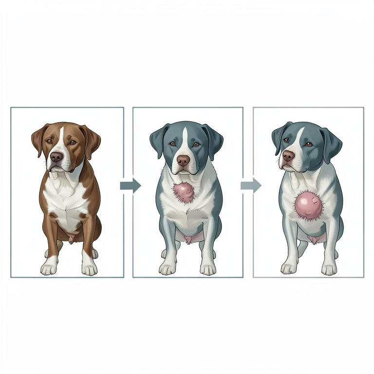
Paclitaxel Treatment Cycle and Effects
AI-generated conceptual illustration (canine mammary tumors). It explains the idea and does not show study data.
View source evidence ↗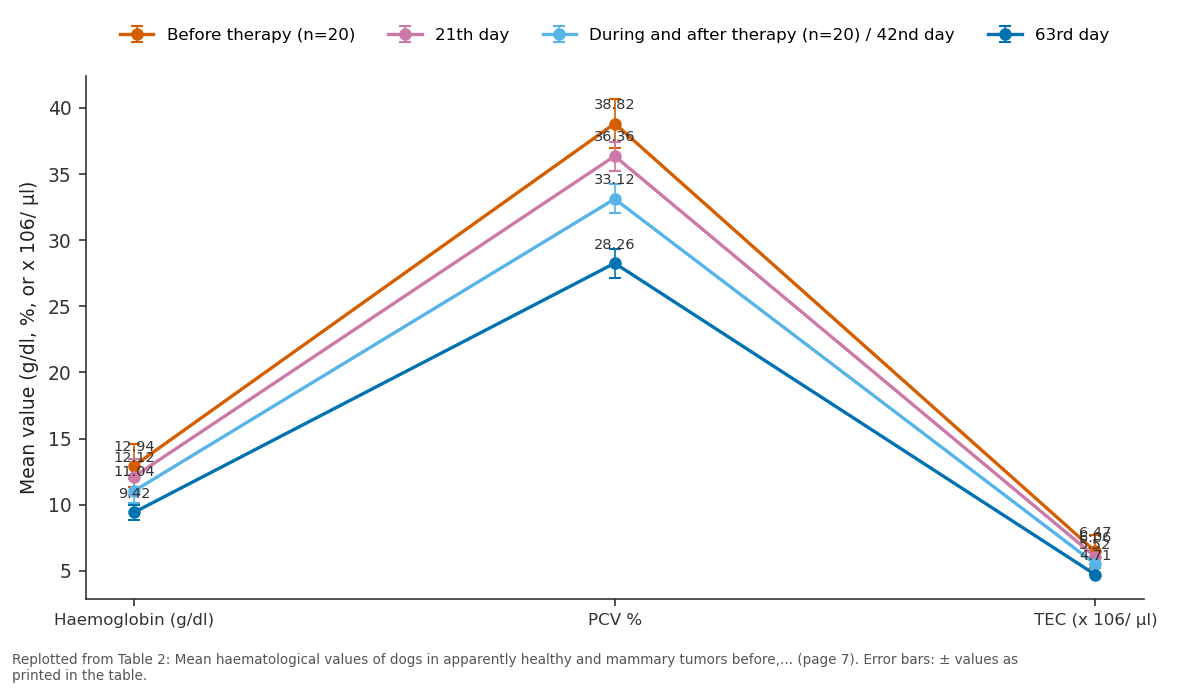
Before therapy (n=20) and 21th day and more: Haemoglobin (g/dl) vs PCV % vs TEC (x 106/ µl)
Replotted from Table 2 (page 7) of the original paper. Every bar is the paper's own table value.
View source evidence ↗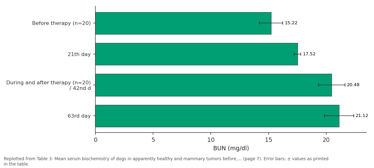
BUN (mg/dl): Before therapy (n=20) vs 21th day vs During and after... vs 63rd day
Replotted from Table 3 (page 7) of the original paper. Every bar is the paper's own table value.
View source evidence ↗Figures & tables
From the original paper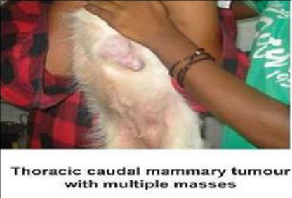 Figure 1
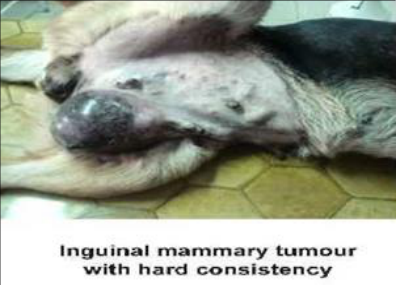 Figure 1
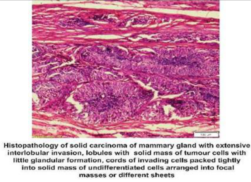 Figure 1
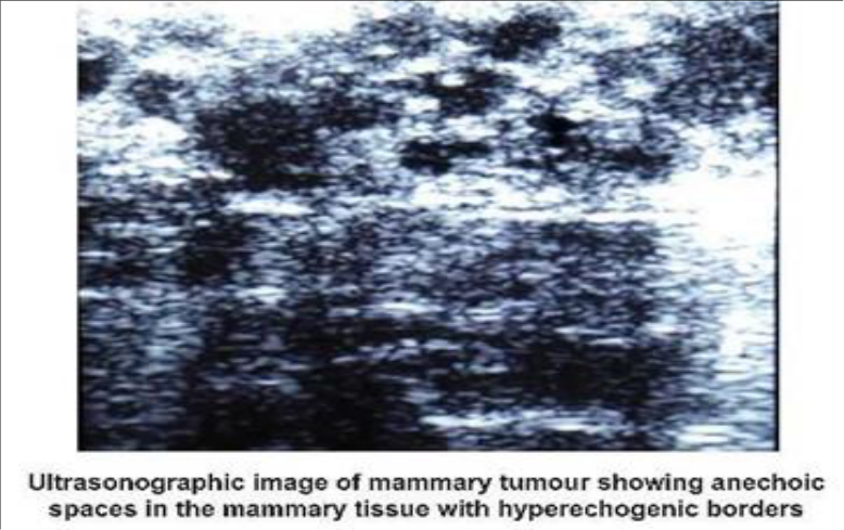 Figure 2
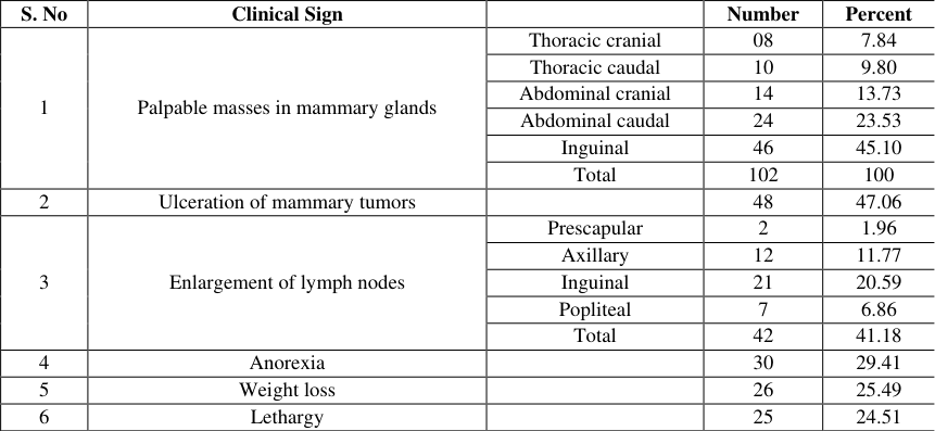 Table 1
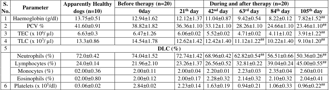 Table 2
 Table 3
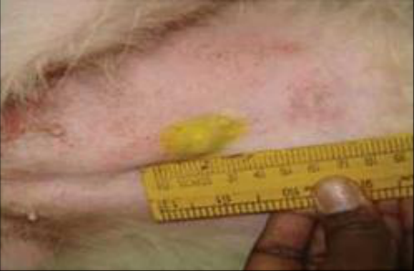 Figure 3
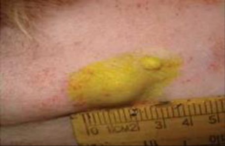 Figure 3
Introduction
Mammary gland tumours represent the second most frequently recognized tumours in female dogs. The average age of onset is > 5-10 years, but cases in dogs more than 10 years of age were also reported (Todorova et al., 2005) [13] . They also occur in male dogs, but the prevalence is only 1.96%. The inguinal mammary glands are more often affected than cranial and caudal glands. Pure bred dogs are more significantly affected with mammary cancer.
Materials and Methods
The dogs presented to the clinic and those referred from various hospitals in and around Hyderabad to the Veterinary Clinical Complex, Bhoiguda over a period of 2 years formed the basis for the present study. Whole blood and serum samples of all the identified dogs formed the clinical material for laboratory examination. Blood and serum were also collected from apparently healthy dogs to establish normal values. Two ml of whole blood was withdrawn from the cephalic or saphenous vein in a vacutainer with K-EDTA for the estimation of hematological parameters. Serum was separated immediately after clotting by centrifugation at 3000 rpm for 5 minutes and collected in Eppendorf tubes. All the haematological and biochemical parameters were estimated on the same day of collection. Hematological parameters such as Hb, PCV, TEC, TLC and DLC were estimated as per the standard procedures. Biochemical parameters such as BUN, Creatinine, ALT, AST, ALT and TP were estimated by a semi-auto analyzer. Dogs selected for therapy had a BSA of 0.6 to 0.8 m 2
Results and Discussion
Results and Discussion
Diagnosis
Diagnosis of malignant mammary tumours was made based on symptomatology, FNAC and histopathology. Among the 458 dogs diagnosed with neoplasia, dogs affected with malignant mammary tumours were the highest with 22.27% incidence (102 dogs). Out of these diagnosed dogs, twenty dogs were included in the present study.
Clinical signs
Among the 102 malignant mammary tumour cases, the most common clinical sign observed was palpable masses in mammary glands (100 per cent) of which inguinal mammary glands were affected the most i.e., in 46 dogs followed by abdominal caudal glands in 24 dogs, abdominal cranial in 14 dogs, thoracic caudal in 10 dogs and least in thoracic cranial i.e., 8 dogs.
The present findings were also in agreement with the findings of Radmehr et al. (2013) [10] and Pankaj et al. (2014) [8] who explained that most often mammary tumours occurred in inguinal mammary (60%) and abdominal (27%) glands. Affected mammary glands had solitary nodules in 59 dogs and multiple masses in 43 dogs. In 48 dogs one gland was affected, in 28 dogs 2 glands, in 18 dogs 3 to 4 glands and in 8 dogs more than 4 glands were affected ( [5] who reported that out of 55 cases of mammary tumours, 38 cases had solitary growths, 17 cases had multiple growths. In the present study the higher percentage of solitary nodules when compared to multiple masses might be due to better care taken by the owners in detecting the tumours at an earlier stage. The other signs such as enlargement of lymph nodes was seen in 44 dogs. Among them pre scapular, axillary, inguinal and popliteal lymph node enlargement was seen in 2, 12, 23 and 7 dogs depicting 1.96%, 11.77%, 22.55% and 6.86% respectively. Bilateral lymphadenopathy was observed in 6 dogs while ipsilateral lymphadenopathy was observed in 38 dogs affected with mammary tumors. Ulceration of the skin over the mammary glands was seen in 48 dogs showing 47.06% of occurrence, anorexia was seen in 30 dogs, weight loss in 26 dogs and lethargy in 25 dogs. (Table 1 andFig 1). Among 20 dogs taken up for study palpable masses in mammary glands were seen in all the dogs and other clinical signs like ulceration, enlargement of lymph nodes, anorexia, weight loss and lethargy were observed in 11, 12, 8, 11 and 8 dogs respectively.
| S. No | Clinical Sign | — | Number | Percent |
|---|---|---|---|---|
| 1 | Palpable masses in mammary glands | Thoracic cranial | 08 | 7.84 |
| Thoracic caudal | 10 | 9.80 | ||
| Abdominal cranial | 14 | 13.73 | ||
| Abdominal caudal | 24 | 23.53 | ||
| Inguinal | 46 | 45.10 | ||
| Total | 102 | 100 | ||
| 2 | Ulceration of mammary tumors | 48 | 47.06 | |
| 3 | Enlargement of lymph nodes | Prescapular | 2 | 1.96 |
| Axillary | 12 | 11.77 | ||
| Inguinal | 21 | 20.59 | ||
| Popliteal | 7 | 6.86 | ||
| Total | 42 | 41.18 | ||
| 4 | Anorexia | 30 | 29.41 | |
| 5 | Weight loss | 26 | 25.49 | |
| 6 | Lethargy | 25 | 24.51 |
Table text extracted from the original PDF; superscript letters are shown inline. The original table image is below.
View original table image
TNM Staging of Mammary Tumours
In the present study, a total of 102 mammary tumors were classified based on the WHO TNM Classification. The mammary tumors were divided into four stages based on the tumor size, lymph node involvement and distant metastasis. Among these, 20 dogs were selected from Stage I and Stage II. It was noticed that the tumor size was less than 3 cm in 19 dogs i.e., (T 1 ) and 1 dog had tumor of 3-5 cm (T 2 ) size. Among these 20 dogs of the study, 15 dogs did not show any regional lymph node involvement (N 0 ) whereas 5 dogs showed unilateral lymph node involvement (N 1 ). Out of the 20 dogs, distant metastasis was not seen in any of the dogs (M 0 ). Sorenmo et al. (2009) [12] stated that in their study, only 3% of malignant tumours were smaller than 1 cm and the risk of being malignant increased gradually as the tumour diameter increased; 70% of the malignant tumours were larger than 3cm.
Fine needle aspiration Cytology (FNAC)
Examination of stained smears collected by FNA in dogs affected with mammary tumours revealed that neoplastic cells of varied sizes were normally round exhibiting anisocytosis and anisokaryosis, one or more basophilic nucleoli and coarse chromatin while cytoplasmic vacuoles were seen in some cells. These findings were in agreement with the findings of Yogita et al. (2015) [14] who stated that cytological evaluation is a quick inexpensive and promising diagnostic technique to differentiate benign and malignant tumours and Mohapatra et al. (2016) [7] stated that FNAC samples showed anisocytosis, anisokaryosis, pleomorphism, hyperchromatic nuclei and increased nucleus to cytoplasmic ratio, binucleated cells in malignancies.
Histopathology
Histopathological examination of biopsy samples stained with H&E of dogs affected with mammary tumours revealed epithelioid cells of round to spindle shape, large nucleus, one or two nucleoli and multiple vacuoles. Out of the 102 mammary tumours diagnosed 79 tumours were diagnosed to be simple malignant tumours, among them adenocarcinomas were seen in 29 dogs, tubulo-papillary adenocarcinomas were seen in 22 dogs, ductular adeno carcinoma were seen in 19 dogs and solid carcinomas were 9. Mixed mammary carcinomas were diagnosed in 28 dogs out of which Myxochondroid carcinoma and fibrosarcoma were 7, Myxolipoadeno carcinoma were 5 and liposarcoma were 4. Pankaj et al. (2014) [8] enumerated that out of 35 cases, histopathology revealed that 33 were neoplastic (32 malignant and1benign) and two were non-neoplastic and Yogita et al. (2015) [14] who enumerated the various mammary tumours. Malignant adenocarcinoma of the mammary gland was composed of malignant epithelial cells. Proliferation of acinar cells showed increased pleomorphism, hyperchromatic nuclei, numerous mitotic figures, stromal invasion and epithelial stratifications in papillary fashion. The lumens were completely or partially occluded with epithelial cells (Fig 2).
Electron microscopy
Ultra-structural examination of mammary tumours revealed numerous cells with epithelium like cell morphology. The nucleus was large and had heterochromatin covered by irregular nuclear membrane. The cytoplasm of the cells had protruding microvilli on the cell surface. In addition, the cytoplasm contained numerous vacuolar structures, free ribosomes, endoplasmic reticulum, mitochondria and intermediate filaments with some lipid accumulation. Similar findings were observed by Kusewitt et al. (1992) [4] in spindle cell carcinoma of mammary glands.
Radiographic study
Radiographic examination of the thorax for pulmonary metastasis in 102 dogs revealed the presence of metastasis in 6 dogs out of 17 dogs with stage III and 8 dogs out of 20 dogs with stage IV. Pulmonary radiographic examination revealed the presence of well-differentiated nodules in 2 dogs, pulmonary masses in 3 dogs, single/solitary nodules in 5 dogs, multiple small nodules in 2 dogs and diffuse interstitial pattern in 2 dogs. The abdominal radiographs did not reveal any abnormality or metastasis to the organs. Jain and Raghunath (2007) [3] reported distant metastasis in 54.05 per cent of canine mammary tumours which appeared as nodular interstitial and diffused patterns radiographically. They also stated that the lung was the most common site for distant metastasis in dogs with malignant mammary tumors (Fig 2).
Ultrasonographic examination
Abdominal sonographic examination in dogs with canine mammary tumours did not reveal any abnormal echo pattern of abdominal organs suggesting metastasis. Ultrasonographic examination of the canine mammary tumour mass revealed the presence of anechoic fluid-filled cystic collections, hyperechoic areas with diffuse margins and anechoic areas with hyperechoic margins. Jain and Raghunath (2007) [3] evaluated the nature and the extent of neoplastic nodules in canine mammary tumours by ultrasonography and recorded that 54.05% of tumours had extensive growth patterns, 21.60% had an infiltrative type and 23.35% of tumours had both types of growth patterns. While Ayhanbastan et al. (2009) [1] evaluated the efficacy of ultrasonographic imaging of malignant tumours in dogs and reported that these were irregular in shape (78%), margins were multilobulated (57%), echo texture was heterogenous (84%) the internal echogenicity was varied (84%), enhancement of acoustic transmission (52%), presence of pseudo capsule (90%) and also documented that the size and mean length of tumours was 6.2 cm (Fig 2).
Haematology
In the present study, in dogs affected with mammary tumours an insignificant decrease of mean haemoglobin (13.01±1.20 g/dl), PCV (38.96±0.72 percent) and TEC (6.46±0.12x10 6 /μl) was noticed when compared to that of apparently healthy dogs (13.75±0.51g/dl, 41.60±0.91 percent and 6.63±0.3 x10 6 /μl) respectively. The mean leukogram of mammary carcinoma cases revealed an insignificant elevation of total WBC count (14.75±0.24x10 3 /μl) and neutrophils (73.61±0.52 percent); decrease in lymphocytes (21.86±0.18 percent) and normal monocytes as well as eosinophils (2.25±0.09 percent and 2.30±0.12 percent), when compared to that of apparently healthy dogs (13.30±0.86x10 3 /μl, 72.0±0.32 percent, 24±0.14 percent, 2.00±0.36 percent and 2.00±0.80 percent), respectively. Similarly, an insignificant decrease was noticed with respect to platelet levels (2.92±0.36x10 5 /dl) when compared to that of apparently healthy ones (3.06±0.24 x10 5 /dl) (Table No. 2). As explained similarly by Todarova et al. (2005) [13] and Mohaopatra et al. (2016) [7] the leukogram of mammary carcinoma cases revealed insignificant elevation of total WBC count and neutrophils, which indicates inflammatory reaction and possible bacterial infection in the tumour affected dogs. The haematological findings were in concurrence with the findings of Todarova et al. (2005) [13] and Mohapatra et al. (2016) [7] who stated that haematological parameters in mammary tumour cases did not show any deviations from the reference range.
Serum Biochemistry
Serum biochemical estimations in dogs with mammary tumours revealed an insignificant decrease in BUN values while an insignificant increase in serum creatinine, ALT, AST, ALP and total protein was observed as depicted in Table No. 3 Similar biochemical observations were also represented by Todarova et al. (2005) [13] who stated that serum biochemical parameters in mammary tumour cases did not show any deviations from the reference range, Pankaj et al. (2014) [8] who stated that the mean values of serum AST and ALT were within normal physiological range before treatment and Mohapatra et al. (2016) [7] who opined that no significant difference was observed between the healthy control group and mammary tumour affected dogs with respect to serum biochemical parameters
Therapy
Out of the 102 dogs diagnosed with mammary tumours, 20 dogs were taken up for the therapeutic trail. The dogs were treated with Paclitaxel (Inj. Paclitax) @ 130 mg/m 2 mixed with Normal saline and administered IV once in 3 weeks for a maximum of 5 doses. Dogs selected for therapy in both groups had a BSA of 0.6 to 0.8 m 2 .
Clinical improvement
In 20 dogs taken up for therapy with Inj. Paclitaxel, 14 dogs showed mild reduction in tumour size by day 63 (i.e., 21 days after 3 rd dose) and partial response was seen by day 105 (i.e.21 day after the 5 th dose) while no response of tumour size was noticed in 6 which showed a stable disease even after 5th dose of Inj. Paclitaxel administration until 3 months after completion of chemotherapy. The other clinical signs like ulceration, enlargement of lymph nodes showed marked alleviation by day 105 whereas weight loss, anorexia and lethargy were persistent throughout the treatment period which further aggravated in treatment dogs. Poirer et al. (2004) [9] studied the efficacy of Paclitaxel in treatment of canine malignant tumours and stated that Paclitaxel was given at an initial dose of 165 mg/m2 IV with subsequent dose reduction if toxicity was observed. Treatment was repeated every 21 days for at least 2 treatments or until progression of disease was noted. 30 to 60 minutes before the Paclitaxel infusion dogs were premedicated with dexamethasone sodium phosphate@ 2 mg/kg IV. Inj. Paclitaxel (6mg/ml) was diluted in 10 times its volume of Normal Saline. Infusion was administered @ 30 ml / hr for 10 mins. If no allergic reaction was noticed the rate of infusion was increased to 60 ml/10 mins. If dog had allergic reactions the infusion was stopped and restarted 15 mins later @ 30 ml/hr. The response rate (20%) to Inj. Paclitaxel treatment was observed as two of 3 dogs with metastatic mammary carcinoma experienced partial remission (Fig 3).
Haematological findings
Haematological examination before during and after therapy revealed that the mean Hb, PCV, TEC had significantly declined (p<0.01) from day 0 to day 105. There was insignificant increase in leucocytes on day 0 i.e., before treatment when compared to apparently healthy dogs. But the mean leucocyte count showed a significant decline by day 63 during therapy (p<0.01) and continued until day 105. There was insignificant neutrophilia on day 0. But the mean neutrophil count significantly declined (p<0.01) by day 63 during therapy and continued until day 105. The mean lymphocyte count significantly increased (p<0.01) during the course of therapy while monocytes and eosinophils did not show any significant alterations. During therapy platelet count showed a significant decline continuously till day 105 (p<0.01). These findings were similar to the findings of Poirer et al. (2004) [9] who reported that anemia, leucopenia, neutropenia and thrombocytopenia were the important changes which occur during treatment of mammary tumours with Inj. Paclitaxel. Serum biochemical parameters showed a significant increase (p<0.01) in the mean post-therapeutic levels of BUN, creatinine, ALT, AST and ALP when compared to pretherapeutic means. However, a mild significant decline (p<0.05) in total protein level was noticed posttherapeutically when compared to pre-therapeutic mean. Cizmeci et al. (2012) [2] explained that BUN increase may be linked to decreased glomerular filtration rate or increased protein catabolism caused by necrosis of tumour or metabolic side effects of neoplasia. The increase in Serum creatinine was attributed to the increase in catabolic activity. However, a significant decline (p<0.05) in total protein level was noticed post-therapeutically when compared to the pre-therapeutic mean.
Side effects
The side effects associated with Inj. Paclitaxel included vomiting, diarrhoea, anorexia and alopecia. These side effects started in 9 dogs from day 21 (after the first dose) slowly and aggravated in 14 dogs by day 63. These side effects were also observed by Todorova et al. (2005) [13] who studied the effect and toxicity of chemotherapy which effectively suppressed the development of new neoplasm and metastases, but was accompanied by general adverse reactions as lethargy, anorexia, vomiting, hair loss, fever, hypochromic anaemia and strong immunosuppresion.
6. Recurrence: Three dogs showed a recurrence of tumour growth in the fourth month (1 dog) and fifth month (2 dogs) after chemotherapy. These dogs were then treated by surgical excision of which one died due to metastasis of lungs and regional lymph nodes on day 35 after surgical excision. Similar recurrences of mammary tumours after chemotherapy were also reported by Sharma et al. (2010) [11] . **: Significant at p<0.01 when compared to apparently healthy dogs, *: Significant at p<0.05 when compared to apparently healthy dogs ##: Significant at p<0.01 when compared to before therapy, #: Significant at p<0.05 when compared to before therapy 6.70±0.14 7.12±0.68 7.02±0.01 6.98±0.14 6.80±0.01 6.97±0.72 6.64±0.01 # **: Significant at p<0.01 when compared to apparently healthy dogs, *: Significant at p<0.05 when compared to apparently healthy dogs ##: Significant at p<0.01 when compared to before therapy, #: Significant at p<0.05 when compared to before therapy Mammary tumor of the thoracic caudal gland Before treatment After treatment
Conclusion
In the present study dogs affected with mammary tumours were successfully treated with Inj. Paclitaxel and the dogs showed side effects were managed with supportive therapy. The dogs that failed to recover completely were subjected to surgical excision.
Figures and tables from the paper
| S. No. | Parameter | Apparently Healthy dogs (n=10) | Before therapy (n=20) | — | During and after therapy (n=20) | — | — | — |
|---|---|---|---|---|---|---|---|---|
| 0day | 21th day | 42nd day | 63rd day | 84th day | 105th day | |||
| 1 | Haemoglobin (g/dl) | 13.75±0.51 | 12.94±1.62 | 12.12±1.37 | 11.04±0.87 | 9.42±0.54 | 8.22±0.12 | 7.82±1.52## |
| 2 | PCV % | 41.60±0.91 | 38.82±1.82 | 36.36±1.10 | 33.12±1.10 | 28.26±1.10 | 24.66±1.10 | 23.46±1.10## |
| 3 | TEC (x 106/ µl) | 6.63±0.3 | 6.47±1.26 | 6.06±0.02 | 5.52±0.02 | 4.71±0.02 | 4.11±1.02 | 3.91±1.22## |
| 4 | TLC (x 103/ µl) | 13.3±0.86 | 14.54±1.78 | 12.62±1.42 | 12.42±1.40 | 11.12±1.22## | 10.22±1.40 | 9.10±1.20## |
| 5 | DLC (%) | |||||||
| Neutrophils (%) | 72.0±0.42 | 74.04±1.52 | 72.74±1.42 | 68.96±0.42 | 62.82±0.54## | 56.51±0.66 | 50.36±0.26## | |
| Lymphocytes (%) | 24.0±0.14 | 21.96±2.10 | 23.26±1.37 | 26.56±0.52 | 32.81±0.22 | 39.04±0.24 | 45.00±0.55## | |
| Monocytes (%) | 02.00±0.36 | 2.00±0.11 | 2.00±0.04 | 2.20±0.01 | 2.23±0.03 | 2.35±0.04 | 2.60±0.01 | |
| Eosinophils (%) | 02.00±0.80 | 2.00±0.12 | 2.00±0.17 | 2.28±0.32 | 2.14±0.32 | 2.10±0.32 | 2.04±0.41 | |
| 6 | Platelets (x 105/dl) | 03.06±0.02 | 2.84±0.02 | 2.23±0.14 | 1.63±0.19 | 0.94±0.21 | 1.06±0.33 | 0.96±0.22## |
Table text extracted from the original PDF; superscript letters are shown inline. The original table image is below.
View original table image
| S. No. | Parameter | Apparently Healthy dogs (n=10) | Before therapy (n=20) | — | During and after therapy (n=20) | — | — | — |
|---|---|---|---|---|---|---|---|---|
| 0day | 21th day | 42nd day | 63rd day | 84th day | 105th day | |||
| 1 | BUN (mg/dl) | 16.29±0.44 | 15.22±1.02 | 17.52±0.28 | 20.48±1.14 | 21.12±1.26 | 25.38±1.70 | 29.72±2.01## |
| 2 | Creatinine (mg/dl) | 0.95±0.62 | 1.16±0.62 | 1.46±0.04 | 1.43±0.01 | 1.46±0.02 | 1.65±0.02 | 2.10±1.02## |
| 3 | ALT (IU/L) | 30.47±0.98 | 36.74±1.36 | 41.60±1.25 | 44.50±0.72 | 45.08±0.02 | 49.56±0.01 | 56.74±1.28## |
| 4 | AST (IU/L) | 41.52±0.36 | 41.26±0.48 | 43.26±1.20 | 44.64±0.32 | 49.02±0.70 | 50.13±0.10 | 55.10±1.50## |
| 5 | ALP (IU/L) | 42.90±0.80 | 45.12±1.24 | 56.66±0.72 | 60.24±1.22 | 69.42±1.36 | 74.68±1.10 | 89.14±2.28## |
| 6 | Total Protein (g/dl) | 6.70±0.14 | 7.12±0.68 | 7.02±0.01 | 6.98±0.14 | 6.80±0.01 | 6.97±0.72 | 6.64±0.01# |
Table text extracted from the original PDF; superscript letters are shown inline. The original table image is below.
View original table image
References
14 in the original paper- Ayhanbastan, Ozenc E, Yagci IP, Acar DB. Ultrasonographic Evaluation of Mammary Tumours in Bitches. Kafkas University Vet Fak Derg. 2009;15(1): 81-86.
- Cizmeci SU, Kose AM, Aydin I, Dinc DA, Maden M, Kose SI. Clinical efficiency of Doxorubicin and Cisplatin in treatment of transmissible venereal tumour of bitches. Revue Med. Vet. 2012;163(11):516-521.
- Jain V, Raghunath M. Evaluation of combination of doxorubicin and cyclophosphamide for management of canine mammary neoplasms. Indian Journal of Veterinary Surgery. 2007;28(2):94-97.
- Kusewitt DF, Hahna FF, Muggenburg BA. Ultrastructure of a Spindle Cell Carcinoma in the Mammary Gland of a Dog Veterinary Pathology. 1992;29:179-181.
- Maiti SK, Manikandan N, Shivakumar MU, Kumar N, Saikumar G, Gupta OP. Therapeutic evaluation of methotrexate with or without COX-2 inhibitor in the management of canine mammary tumours. Indian Journal of Canine Practice. 2011;3(2):117-126.
- Marconato L, Lorenzo RM, Abramo F, Ratto A, Zini E. Adjuvant gemcitabine after surgical removal of aggressive malignant mammary tumours in dogs. Veterinary and Comparative Oncology. 2008;6(2):90101.
- Mohapatra AK, Das D, Panda SK, Jena B, Singh J. Spontaneous Canine Mammary Neoplasia: A Clinico Pathological Study. Journal of Cell and tissue Research. 2016;16(2):5661-5666.
- Pankaj G, Raghunath M, Gupta AK, Ankur S, Kawardeep K. Clinical study for diagnosis and treatment of canine mammary Neoplasms (CMNs) using different modalities Indian Journal of Animal Research. 2014;48(1):45-49.
- Poirer VJ, Hershey AE, Burges KE, Phillips B, Turek MM, Forest LJ, et al. Efficacy and toxicity of Paclitaxel (Taxol) for the treatment of Canine Malignant Tumours. Journal of Veterinary Internal Medicine. 2004;18:219222.
- Shafiee R, Javad J, Nahid A, Pegah K, Danial K, Alimohammad B, et al. Diagnosis classification and grading of canine mammary tumours as a model to study human breast cancer a clinico cytohistopathological study with environmental factors influencing public health and medicine. Diagnostic Pathology. 2013;8:136.
- Sharma A, Dhakate MS, Upadhye SV. Chemotherapeutic Management of Canine Mammary Tumours, 2010, 5(1).
- Sorenmo KU, Kristiansen VM, Cofone MA, Shofer FS, Breen AM, Langeland M, et al. Canine mammary tumours; a histological continuum from benign to malignant; clinical and histopathological evidence. Veterinary and Comparative Oncology. 2009;7(3):162172.
- Todorova G, Simeonova R, Simeonov, Dinev D. Efficacy and toxicity of doxorubicin and cyclophosphamide chemotherapy in dogs with spontaneous mammary tumours. Trakia Journal of. Sciences. 2005;3:51-58.
- Yogitapawar, Dattatray K, Gajendra K, Renuka N. Gross and cytological Evaluation of Canine Spontaneous Mammary Neoplasms and Its Correlation with Histopathology and Morphometric Analysis. International Journal of Veterinary Science. 2015;4(3):104-101.
Cited by 1
From OpenAlex, OpenCitations and reference lists read by Scinovex
- Incidence of canine mammary carcinoma
Pathi Suman, S. Ayodhya, VVV Amruth Kumar, B. Anil Kumar, Swathi Bora · International Journal of Advanced Biochemistry Research · 2025
The present investigation aimed to determine the incidence of canine mammary carcinoma in dogs presented to veterinary hospitals. Out of a total of 42, 505 dogs examined, 422 cases were diagnosed with malignant neoplasms, indicating an overall incidence rate of 0.99%. Canine mammary carcinoma was most frequently observed in dogs aged between 6 and 10 years (66.67%), while the lowest incidence was recorded in the 0 to 5-year age group (7.41%). Among the affected breeds, Labrador Retrievers exhibited the highest breed-specific incidence at 38.28%, with a marked prevalence in intact female dogs (