
Research article · 2023 · Full text
International Journal of Veterinary Sciences and Animal HusbandrySchistosomus reflexus syndrome-induced dystocia in sheep: Unveiling an exceptionally rare hereditary anomaly and its obstetrical implications
Volume 8(6) · Pages 21–23 · DOI: 10.22271/veterinary.2023.v8.i6a.798 ↗
Research areasCongenital Anomalies and Fetal SurgeryAssisted Reproductive Technology and Twin Pregnancy
Abstract
Author-writtenSchistosomus reflexus is a condition that is most commonly observed in cattle, but it has also been reported in some cases in other animals, including sheep, goats, and swine. A two-and-a- half-year-old non-descript pluriparous sheep was brought to the District Veterinary Polyclinic in Nellore with a history of labor pain lasting for eight hours, but there was no progress in parturition. Upon clinical examination, it was observed that the visceral organs of the fetus were protruding from the vulva of the ewe. Further examination through vaginal examination revealed that the fetus in the pelvic cavity was deformed, leaving insufficient space for fetal manipulation. A tentative diagnosis of dystocia due to fetal deformity was made based on these findings. To relieve the dystocia, a caesarean section was performed. Upon examination of the defective fetus, it was confirmed to be a case of true schistosomus reflexus in a sheep. The characteristics of the deformity included spinal inversion, ankylosis (fusion) of the hind limbs, and the protrusion of organs such as the liver, intestine, stomach, and kidney from the abdominal opening of the fetus. This case report highlights the occurrence of schistosomus reflexus in a sheep, which, as mentioned earlier, is a condition more commonly associated with cattle but can occasionally affect other livestock as well.
Summary
Source-checkedPlain-language reading of the full paper, prepared with AI and checked against the text. Each point links to the authors' own words.
Why it matters
- Schistosomus reflexus is a congenital fetal disorder that occurs most frequently in cattle and rarely in small ruminants. View evidence ↗
- The condition is characterized by a severe failure of the abdominal cavity to close and often includes skeletal issues, which typically creates challenges during birth. View evidence ↗
- The cause of the syndrome is primarily linked to the transfer of autosomal recessive genes with incomplete penetrance during embryonic development. View evidence ↗
The question
- The authors aim to document and present a rare case of Schistosomus reflexus in a sheep to contribute to the limited literature on this condition in small ruminants. View evidence ↗
What they did
- The subject was a two-and-a-half-year-old pluriparous sheep at full term who presented with eight hours of straining. View evidence ↗
- Clinical parameters were measured, showing a rectal temperature of 103.5 °F, heart rate of 90 bpm, and respiratory rate of 50 breaths per minute, all of which were normal. View evidence ↗
- Because manual traction failed, an emergency caesarean section was performed following standard procedures to save the animal. View evidence ↗
- The surgical procedure utilized 2% Lignocaine as local anesthesia administered via a leaner infiltration method. View evidence ↗
What they found
- Initial physical examination revealed that fetal legs and abdominal organs were protruding through the vulva. View evidence ↗
- Vaginal examination showed a completely dilated cervix, but the birth canal was obstructed by the fetus's limbs and organs. View evidence ↗
- Post-extraction examination confirmed true schistosomus reflexus, characterized by spinal inversion, fused hind limbs, and the protrusion of the liver, stomach, kidney, and intestine. View evidence ↗
- The ewe recovered without complications and was able to stand following the surgery. View evidence ↗
What it means
- The authors conclude that veterinarians must be prepared for rare fetal presentations and should consider fetal size and condition when choosing the delivery method. View evidence ↗
- The authors recommend caesarean sections as the safest and most practical approach for delivering abnormal fetuses with joint ankylosis, unless the fetus is very small. View evidence ↗
- The authors note that congenital defects in sheep are likely more prevalent than reported because sporadic cases are often underreported. View evidence ↗
Visual summary
1 visualsIllustrations explain the paper; they do not show study data.
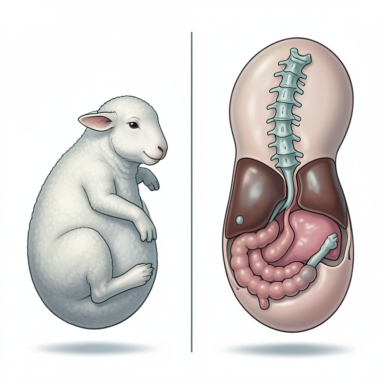
Anatomy of Schistosomus Reflexus
AI-generated conceptual illustration (sheep fetus). It explains the idea and does not show study data.
View source evidence ↗Figures & tables
From the original paper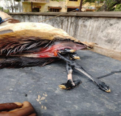 Figure 1
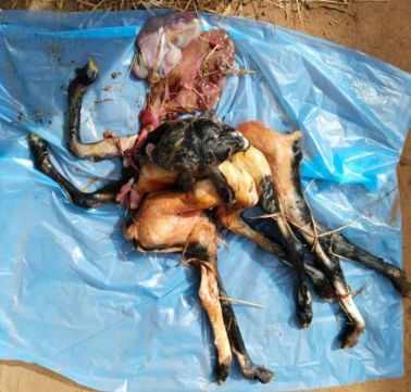 Figure 2
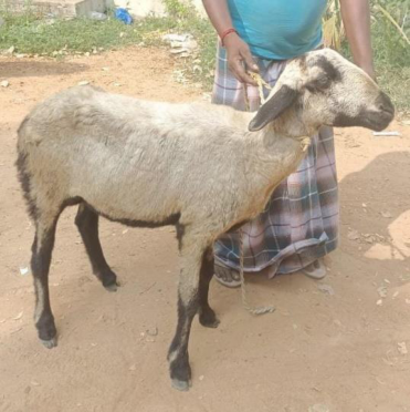 Figure 3
Introduction
Schistosomus reflexus, a congenital fetal disorder, is predominantly observed in cattle, with only rare occurrences in small ruminants (Roberts, 1971) [5] . This condition represents an extreme closure defect of the abdominal cavity, often accompanied by skeletal abnormalities, and it typically presents as an obstetric challenge. While Schistosomus reflexus is most frequently reported in cattle, there have been sporadic instances of its occurrence in sheep (Smith, 1969; Dennis, 1972; Mukasa-Mugerwa and Bekele, 1989) [16,11,14] and goats (Bedford, 1967) [10] as documented in the literature. Among farm animals, it is noteworthy that Schistosomus reflexus is considerably more common in cattle and comparatively rare in sheep and goats (Selvaraju et al. 2020) [20] . The etiology of Schistosoma is primarily attributed to the transfer of autosomal recessive genes with incomplete penetrance to the developing embryo (Laughton et al., 2005 and Balamurugan et al., 2020) [2,1] . A true case of Schistosoma is classified by the presence of both exposed viscera and an inversion of the spine. This congenital syndrome is characterized by spinal inversion, resulting in a distinct ventral convex curvature, exposure of abdominal viscera due to a ventral abdominal wall fissure, limb ankylosis, limb positioning adjacent to the skull, and lung hypoplasia (Roberts, 1986) [15] . Schistosomus reflexus is a significant congenital anomaly that arises during embryonic development. For fully developed Schistosomus reflexus cases, foetotomy or caesarean section is imperative, whereas small-sized monster fetuses may be expelled per vaginam without obstetrical assistance (Kalita et al., 2004) [17] . It's worth noting that congenital defects in sheep are likely more prevalent than reported. Cases of defective lambs are often underreported unless their numbers are substantial. Therefore, numerous sporadic cases, including various genetic defects, often go unreported (Dennis and Leipold, 1986) [12] .
Due to the rarity of documented instances of this condition in sheep, this paper presents a case of Schistosomus reflexus encountered at the District Veterinary Polyclinic in Nellore.
Case History and Observation
A pluriparous sheep, aged two and a half years, at full term and with no prior history of abnormal deliveries, was brought to the District Veterinary Polyclinic in Nellore due to straining for the past 8 hours. During the physical examination, protruding fetal legs and abdominal viscera were observed hanging through the vulvar lips. Upon per vaginal examination, it was found that the cervix was completely dilated, and the birth canal was fully obstructed by fetal limbs and visceral contents. The precise positioning of the fetus could not be determined, making manual intervention for relief uncertain. Notably, there were no visible abdominal or uterine contractions or signs of straining. The sheep's clinical parameters, including a rectal temperature of 103.5 °F, a heart rate of 90 beats per minute, and a respiratory rate of 50 breaths per minute, fell within the normal range. The mucous membranes appeared pinkish (indicative of normalcy), and the capillary refill time was less than 2 seconds. Based on the findings from the vaginal examination and the presence of fetal deformity, a tentative diagnosis of dystocia induced by schistosomus reflexus was made. Efforts to manipulate the fetus and alleviate the dystocia through traction proved unsuccessful due to the limited space within the pelvic cavity. Consequently, a decision was made to address the dystocia through a caesarean section, which was carried out using standard procedures as outlined by (Morrow 1986) [13] as an emergency intervention to save the life of animal.
Treatment
In brief, the ewe was positioned in right lateral recumbency, and the left flank area was prepared using aseptic techniques. The fetal masses protruding from the vulva were also cleaned with warm water and antiseptic. Following the aseptic preparation, local anesthesia with 2% Lignocaine was administered using a leaner infiltration method on the right side between the milk vein and the para median line. A 5-6 inch incision was made through the skin, abdominal muscle, and peritoneum. The uterine horn containing the fetus was exposed through this incision. A four-inch longitudinal incision was made on the dorsal curvature of the right uterine horn, allowing for the extraction of the fetus while an assistant pushed back the parts hanging from the vulva into the pelvic cavity. Subsequently, the fetal membranes were removed, and both the uterine and abdominal incisions were closed following standard surgical procedures. Upon examination of the fetus, it was determined that since there were no visceral organs inside the fetal abdomen upon opening, the dystocia was indeed due to the schistosomus reflexus condition of the fetus. After the surgery, the wound was dressed with antiseptic Himax ointment. Wound dressing was performed daily for four consecutive days. The animal received a course of injections, including ceftriaxone 300 mg (Intacef) IM, meloxicam 5mg IM, and Nurocare IM, following established protocols. Following the caesarean operation, the animal was able to stand on her legs and exhibited a smooth and uncomplicated recovery.
Discussion: This report highlights a noteworthy case of schistosomus reflexus in a sheep, a condition that is more commonly associated with cattle and buffaloes (Padma Rao et al. in 1993 and Srivastava et al. in 1998) [18,19] . Schistosomus reflexus is characterized by the abnormal herniation of abdominal contents, making it a rare but intriguing fetal monstrosity. In cases where schistosoma reflexus presents with visceral herniation, it's interesting to note that natural births are relatively common (Noakes et al., 2009) [3] . This suggests that this condition, while challenging, can sometimes resolve spontaneously during the birthing process. This natural birthing phenomenon has been further supported by reports from (Suthar et al., 2011, Singh et al., 2017) [7,6] , who all described successful per vaginal deliveries in cases of visceral schistosomus reflexus in ewes and does. However, the situation becomes more complicated when schistosoma reflexus presents with extremities and joint ankylosis (fusion of joints), as this can make manipulative delivery exceedingly difficult. In such instances, a cesarean operation is often recommended (Noakes et al., 2009) [3] . This surgical intervention is considered the most practical and safe approach to ensure both the safety of the ewe and the successful delivery of the abnormal fetus, unless the fetus is exceptionally small. This case emphasizes the need for veterinarians and livestock practitioners to be prepared for unusual presentations during childbirth, as well as the importance of considering the size and condition of the fetus when determining the appropriate course of action. Schistosomus reflexus, while rare, serves as a valuable case study in veterinary obstetrics.
Acknowledgement
AcknowledgementThe authors express their gratitude to Dr. Krishnamurthy for his skillful execution of the surgery and for offering invaluable guidance that contributed to the completion of this article.
Figures and tables from the paper
References
20 in the original paper- Balamurugan B, Reddy YP, Jyothi K. Successful pervaginal delivery of Schistosoma reflex foetal monster in a nondescript buffalo: A case report. The Pharma Innovation Journal. 2020;9(4):57-58.
- Laughton KW, Fisher KR, Halina Partlow WG. Schistosomus reflexus syndrome: A heritable defect in ruminants. Anatomia, histologia, embryologia. 2005;34(5):312-8.
- Pramod K, Sasi G, Banu P, Rajendra M, Tipu S. Dystocia due to Schistosomus reflexus in a Marwari ewe. Haryana Veterinarian. 2020;59(2):285-286.
- Roberts SJ. Veterinary Obstetrics and Genital Diseases, 2 nd edn. SJ Roberts, Ithaca, NY. 1971;605:70-73.
- Singh D, Kumar P, Kumar A, Kumar A, Ruhel S. Dystocia due to schistosomus reflexus (co-twin) in a Marwari goat-a case report. Life Sciences Leaflets. 2017;94:10-12.
- Suthar DN, Sharma VK, Dabas VS, Bhoi DB. Pervaginal handling of Schistosomus reflexus as a cause of dystocia in a Goat. Veterinary World. 2011;4(7):330-331.
- Dennis SM, Mayer EP. Schistosomus reflexus in a sheep, Vet. Rec. 1965;77:1386.
- Wani NA, Wani GM, Bhat AS. Schistosomus reflex us in a Corriedale ewes. Small Ruminant Research. 1994;14:95-97.
- Indian Bedford PGC. Schistosoma reflexus in a goat: A case report. Veterinary Record. 1967;80:326.
- Dennis SM. Schistosomus reflexus in conjoined twin lambs. The Veterinary Record. 1972;90:509-510.
- Dennis SM, Leipold HW. Congenital and inherited defects in sheep. Current Therapy in Theriogenology. Morrow, D.A. (Eds), W. B. Saunders, Philadelphia Toronto, USA. 1986, 864-866.
- Morrow DA. Current Therapy in Theriogenology. 2 nd Edition. Philadelphia Toronto, USA. W. B. Saunders, 1986.
- Mukasa-Mugerwa E, Bekele T. Schistosomus reflexus in indigenous Ethiopian sheep. Bulletin of Animal Health Production in Africa. 1989;37:399.
- Roberts SJ. Veterinary Obstetrics and Genital diseases. 3 rd Edition. Woodstock. 1986.
- Smith ID. Schistosomus reflexus in conjoined twin lambs. Veterinary Record. 1969;85:138-139.
- Kalita D, Bhuyan D, Mukit A, Islam S. Dystocia due to Schistomus reflexus in a goat. Indian J Anim. Reprod. 2004;25(1):76-77.
- Padma Rao V, Ramchandraiah K, Mohan Reddy AR, Venkata Subbiah D, Chandrasekhar B. Schistosomus reflexus in a monocephalus tripus dibrachius calf. Indian Vet. J. 1993;70:1083.
- Srivastava KK, Sharma AK, Ahlawat SPS, Maithy SK. Schistosomus reflexus with perosomus elumbis in Holstein Friesian cow. Indian J Anim Reprod. 1998;19(1):75.
- Selvaraju M, Prakash S, Varudharajan V, Ravikumar K, Palanisamy M, Gopikrishnan D, et al. Obstetrical disorders in farm animals: A review. The Pharma Innovation J. 2020;SP-9(9):65-74.
- Cavalieri J, Farin PW. Birth of a Holstein freemartin calf co-twinned to a schistosomus reflexus fetus. Theriogenology. 1999;52:815-826.