
Research article · 2023 · Full text
International Journal of Veterinary Sciences and Animal HusbandrySurgical management of evisceration in female dog after ovariohysterectomy: A case report
Volume 8(5) · Pages 421–422 · DOI: 10.22271/veterinary.2023.v8.i5g.779 ↗
Research areasUrological Disorders and TreatmentsHuman-Animal Interaction StudiesVeterinary Medicine and Surgery
Abstract
Author-writtenA 6 months old desi female dog was presented to Veterinary clinic with history of ovariohysterectomy. On clinical examination it was found that there was wound dehiscence and evisceration. Emergency surgical intervention was performed by restraining the animal under proper anaesthetic protocol. The dog had a complete recovery without any complication.
Summary
Source-checkedPlain-language reading of the full paper, prepared with AI and checked against the text. Each point links to the authors' own words.
Why it matters
- Ovariohysterectomy is a very common surgical procedure performed on companion dogs. View evidence ↗
- Inexperienced veterinary practitioners may cause postoperative complications such as evisceration or acute incisional hernia during these surgeries. View evidence ↗
- Evisceration occurs when internal abdominal contents move outside the abdominal cavity through a surgical wound that has opened (dehisced). View evidence ↗
The question
- The study aimed to provide efficient techniques to prevent postoperative complications, which can be adapted as veterinarians gain more experience. View evidence ↗
What they did
- The subject of the case report was a 6-month-old female desi dog with a history of ovariohysterectomy. View evidence ↗
- The dog was anesthetized using an intramuscular combination of Xylazine (1 mg/kg) and Ketamine (15 mg/kg). View evidence ↗
- During the emergency surgery, the contaminated edges of the wound, including the rectus fascia and peritoneum, were removed. View evidence ↗
- The abdominal wall was repaired using chromic catgut No.1 in a simple continuous pattern with 5-mm fascia bites and specific knot throws (5 caudal, 6 cranial), while the skin was closed with nylon using a simple interrupted pattern. View evidence ↗
- Post-operative care for 10 days included intravenous ceftriaxone and tazobactam (20 mg/kg), oral Meloxicam (0.2 mg/kg), and antiseptic dressings. View evidence ↗
What they found
- Initial examination revealed a reddish, painful eviscerated mass with echymotic skin, accompanied by tachypnoea and restlessness, though temperature remained normal. View evidence ↗
- The skin sutures were successfully removed on the 8th day following the corrective surgery. View evidence ↗
- The dog achieved a complete recovery within 10 days through the use of regular dressings and combination antibiotic therapy. View evidence ↗
What it means
- The authors conclude that evisceration can be prevented by avoiding predisposing factors, which are categorized as either suture-related or tissue-related. View evidence ↗
- The authors successfully describe the surgical management and recovery of a 6-month-old female dog suffering from evisceration after ovariohysterectomy. View evidence ↗
Visual summary
1 visualsIllustrations explain the paper; they do not show study data.
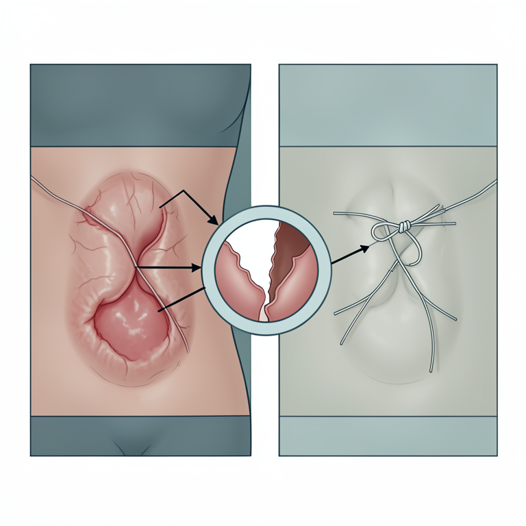
Causes of Evisceration
AI-generated conceptual illustration (female dog abdominal wall and surgical sutures). It explains the idea and does not show study data.
View source evidence ↗Figures & tables
From the original paper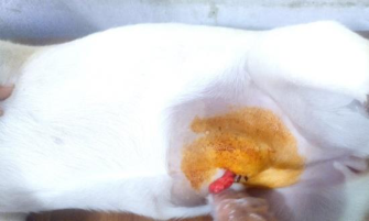 Figure 1
Introduction
Most of the beginner veterinary practitioners are inexperience in performing ovariohysterectomy in dog and cat, which can result in postoperative complication like acute incisional hernia and evisceration. Evisceration is the uncontrolled exteriorization of intraabdominal contents through the dehisced surgical wound outside of the abdominal cavity (Carlson, 1997) [2]. Canine ovariohysterectomy is one of the most frequently used surgical procedures in the case of companion animals (Howe, 2006) [4]. Ventral midline celiotomy is common surgical approaches for abdominal surgery in small animals.
Failure of the suture line used, to close the celiotomy result in postoperative complication and have serious consequences (Bellenger, 2007) [1]. The aim was to contribute the efficient techniques for prevention of post-operative complication which can be modified as veterinarian gain experience. History and Clinical findings A 6 months old desi female dog was brought to Veterinary Clinic with the history of ovariohysterectomy and complaint of wound dehiscence and evisceration twice repeatedly after suturing. On clinical examination, echymotic skin, painful and reddish around the eviscerated mass were noticed (Fig. 1).
The dog showed restlessness and tachypnoea with normal temperature. Emergency surgical procedure was done to save the life of the animal. VET 2023; 8(5): 421-422 Daniel Kakki Farm Manager, Department of Dairy Unit, Central Agricultural University, Pasighat, Arunachal Pradesh, India Daniel Kakki Farm manager, Department of Dairy Unit, Central Agricultural University, Pasighat, Arunachal Pradesh, India Fig 1: Evicerated mass Treatment and Discussion The animal was anaesthetized by using combination of Xylazine 1 mg/kg and Ketamine 15 mg/kg through intramuscular route respectively (Subhan et al., 2017) [6] along with fluid https://www.veterinarypaper.com therapy and was placed on dorsal recumbency. The affected area and eviscerated mass was cleaned with normal saline.
The mass was examined thoroughly and the sutures were removed to reduce the eviscerated mass into the abdominal cavity. The contaminated edges of the wound including a combination of the peritoneum and rectus fascia are excised. After correction, sutures were placed, incorporating at least 5- mm bites of fascia to carefully repair the abdominal wall defect using chromic catgut No.1. A simple continuous pattern was used with square knots of 5 throws at the beginning (caudal knot) and 6 throws at the end (cranial knot) and the skin was opposed with simple interrupted suturing pattern using nylon.
Post-operatively, animal was given ceftriaxone and tazobactam combination injection (Intacef Tazo Peta, 20 mg/kg i/v) and Meloxicam (Melonex oral suspensionb, 0.2 mg/kg) along with antiseptic dressing with bectodinec solution, D-magd spray and kiskine ointment for 10 days. The wound was inspected for inflammation, swelling, drainage, seroma and any other complications. After 8th day of post surgical the skin sutures were removed. The patient had complete recovery over a period of 10 days on a combination therapy of antibiotic and regular dressing.
Evisceration can be prevented by avoiding the predisposing factors that lead to its occurrence (Gower et al., 2009) [3]. The causes of evisceration can be grouped into tissue and suture related factors (Smeak, 2012) [5]. Tissue related factors are extreme tightening of the sutures, incorporating excessive amounts of rectus abdominis muscle and failure to incorporate large secure bites of external rectus fascia in the abdominal wall repair. Suture related factors are inappropriate handling of the suture, poor knot security, inappropriate suture material choice and poor choice of suture size.
In rare cases, evisceration can develop from self trauma to the wound after surgery, straining to defecate or urinate, unrestricted exercise and excessive abdominal pressure from coughing. (A, B, D, E)-Brand of Intas Animal Health, Ahmedabad (c)- Brand of Glide Chem Pvt. Ltd., Himachal Pradesh.
Conclusion
Successful treatment of a case of evisceration after ovariohysterectomy in 6 months old female dog is described.
References
6 in the original paper- Bellenger CR. Parede abdominal (3rd Edn.). Rio de Janeiro, Manole; c2007, p. 405-413.
- Carlson MA. Acute wound failure. Surg Clin N Am. 1997;77:607-636.
- Gower SB, Weisse CW, Brown DC. Major abdominal evisceration injuries in dogs and cats: 12 cases. JAVMA. 2009;234(12):1566-1572.
- Howe LM. Surgical methods of contraception and sterilization. Theriogenology. 2006;66(3):500-509.
- Smeak DD. Veterinary Surgery Small Animal (1st Edn.), Elsevier Saunders, United States; c2012, p.1353-1379.
- Subhan U, Majid A, Muhammad S, Sardar H, Zaheer A, Nasar K, et al. Effect of xylazine and ketamine on pulse rate, respiratory rate and body temperature in dog. Int J Avian & Wildlife Biol. 2017;2(4):137-139.