
Research article · 2023 · Full text
International Journal of Advanced Biochemistry ResearchRole of β-hydroxybutyrate as regulator of cell death pathway in experimental cancer model
Volume 7(2) · Pages 39–42 · DOI: 10.33545/26174693.2023.v7.i2a.219 ↗
Research areasDiet and metabolism studiesCancer, Hypoxia, and MetabolismDiet, Metabolism, and Disease
Abstract
Author-writtenCancer is a disease of unlimited cellular growth and division which lead to the disturbance of cellular homeostasis, destruction to the immune system, and different impairment that can be catastrophic to the cells. Chronic inflammation is widely recognized as a major risk factor for cancer formation, but the underlying mechanisms are poorly understood. Recently, it was shown that Gasdermin D (GSDMD) protein drives pyroptotic cell death in macrophages on cleavage by inflammatory caspases Allow massive release of pro-inflammatory mediators. In this study, we examined the role of B.hydroxy butyrate as a regulator of cell death pathway in Ehrlich’s Ascites Carcinoma (EAC).50 female albino mice randomly divided into five equal groups:Group N:negative control, Group E (Ehrlich ascetic bearing mice), Group E+K (EAC- bearing group co-treated with ketone bodies, Group K+E+K (ketone bodies pretreated group), GroupK:ketone bodies control group.Ascetic cells and mammary gland tissue were homogenized and used for the assay of the gasdermin D level by ELISA. Keto diet significantly increase trypan blue positive dead cells. This was associated with decreased gasdermin D in all treated groups when compared to the untreated group.Conclusion: The current study obscure that inflammatory cell death pathways pyroptosis which is one of key element of tumor microenvironment was significantly reduced through keto diet administration.
Summary
Source-checkedPlain-language reading of the full paper, prepared with AI and checked against the text. Each point links to the authors' own words.
Why it matters
- Cancer is characterized by uncontrolled cell growth and division, which disrupts cellular homeostasis and damages the immune system. View evidence ↗
- Chronic inflammation is considered a significant risk factor for the development of cancer, although the exact mechanisms are not fully understood. View evidence ↗
- The protein Gasdermin D (GSDMD) is a key driver of pyroptosis, a form of programmed cell death that releases pro-inflammatory mediators. View evidence ↗
The question
- The study aimed to investigate how β-hydroxybutyrate regulates cell death pathways in a model of Ehrlich’s Ascites Carcinoma (EAC). View evidence ↗
What they did
- The experiment used 50 female albino mice, aged 8 to 10 weeks and weighing between 20 and 25 g. View evidence ↗
- Gasdermin D levels were measured in mammary gland and ascitic cell homogenates using ELISA. View evidence ↗
- The trypan blue exclusion method was used to count viable and non-viable EAC cells. View evidence ↗
What they found
- Final body weight increased in the EAC-bearing group (Group E: 32.650±1.944 g) compared to the negative control (Group N: 28.848±1.478 g), while all ketone-treated groups showed a decrease in final weight. View evidence ↗
- The ketogenic diet increased the number of dead cells (trypan blue positive) in the E+K and K+E+K groups compared to the untreated EAC group. View evidence ↗
- Gasdermin D levels were significantly higher in the EAC-bearing group (1653±705 ng/mg protein) than in any other group, including the ketone-treated groups. View evidence ↗
- There was no significant difference in Gasdermin D levels between the ketone-treated groups (E+K, K+E+K) and the ketone control group (K). View evidence ↗
What it means
- The authors conclude that the ketogenic diet significantly reduces pyroptosis, an inflammatory cell death pathway that is a key element of tumor development. View evidence ↗
- The findings suggest that pyroptosis could be a potential therapeutic target for treating cancer. View evidence ↗
- The authors state that the primary limitation of the study was its short duration. View evidence ↗
Visual summary
1 visualsIllustrations explain the paper; they do not show study data.
Mechanism of Ketogenic Diet on Pyroptosis
AI-generated conceptual illustration (cancer cells in a laboratory model). It explains the idea and does not show study data.
View source evidence ↗Figures & tables
From the original paper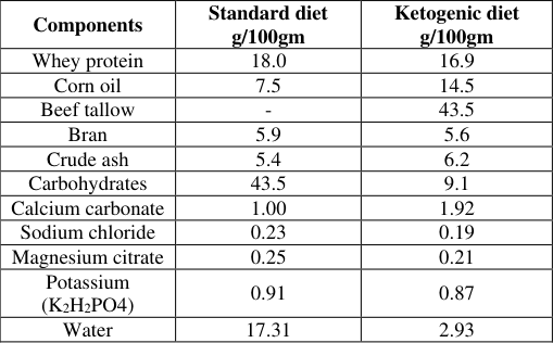 Table 1
 Table 2
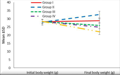 Figure 1
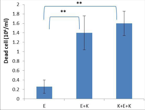 Figure 2
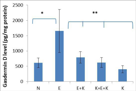 Figure 3
Introduction
Cancer is a process results from multiple factors such as genome mutations, infections, environmental agents. Genetic mutations involving genes of the DNA repair, cell cycle or cell death pathways were associated with malignancy (Gu and Lee, 2022) [4] . Otto Warburg found that the cancer cells used glycolytic pathway for production of ATP not oxidative phosphorylation. Cancer cell is able to increase its anabolism, maintain cellular hemostasis and reprogram gene expression to support their proliferation (Wang, Patti, 2023) [14] . Using keto diet in different varieties of cancer which provide diverse biological effects, protecting normal cells while increase response of tumor cells to antitumor therapy (Barrea et al. 2022) [2] . Metabolic incapacity of cancer cells to metabolize ketone bodies due to the reduced ketolytic enzymes and compromised mitochondria. So ketogenic diet result in cancer cells starvation and apoptosis (Wang et al. 2018) [15] . Gasdermin D (GSDMD), a member of the gasdermin family, has been identified as a key factor in the genesis of pyroptosis and secretion of several inflammatory mediators, such as IL-18 and IL-1β. The inflammatory factors released during the process of pyroptosis will lead to the development of inflammation. Over the past few years, the effects of molecules, inflammasomes, gasdermins, and inflammatory products during pyroptosis on tumorigenesis have been well investigated, but the conclusions are controversial (Wei et al. 2022) [16] .
Animal experiment procedure
The current study was performed on 50 female albino mice aged 8 to 10 weeks and weighted 20 to 25 g. The mice were fed a standard chow diet and had free access to food and water.
Cancer cells inoculation
A mouse bearing Ehrlich ascites carcinoma (EAC) was purchased from the National Cancer Institute, Cairo University, Egypt. The Ehrlich carcinoma cells were then harvested from the parent line by an intraperitoneal puncture using sterile insulin syringe. The cells were counted using hematocytometer. The ascitic fluid containing the malignant cells was diluted in 0.9% saline solution so that each 1 ml of the ascitic fluid contain one million EAC cells. Mice were randomly divided into five equal groups of 10 mice each as follows: Group N (negative control group) that received intraperitoneal injection of saline at a dose of 20 ml /kg.Group E (EAC-bearing group) which was injected intraperitoneally by Ehrlich's ascites carcinoma (EAC) cells (1X 10 6 ) cells/mouse. The day zero was designated as the day of the first injection (Kasumi and Sato, 2019) [6] . Group E+K (EAC-bearing group co-treated with ketone bodies) and these mice were received a ketogenic diet at the day of tumour inoculation for two weeks (Kasumi and Sato, 2019) [6] . Group K+E+K (ketone bodies pretreated group): Mice bearing EACs received a ketogenic diet for two weeks before and for 2 weeks after tumour inoculation. Group K (ketone bodies control group): that received a ketogenic diet daily for 2 weeks. Composition of standard and ketogenic diet in grams/100 gram diet is shown in (Table 1) (Morscher et al., 2015) [10] . Components of each diet were mixed, pelleted and stored at -20 ºC. Tumor growth was monitored by weighting the mice every day.
| Components | Standard diet g/100gm | Ketogenic diet g/100gm |
|---|---|---|
| Whey protein | 18.0 | 16.9 |
| Corn oil | 7.5 | 14.5 |
| Beef tallow | - | 43.5 |
| Bran | 5.9 | 5.6 |
| Crude ash | 5.4 | 6.2 |
| Carbohydrates | 43.5 | 9.1 |
| Calcium carbonate | 1.00 | 1.92 |
| Sodium chloride | 0.23 | 0.19 |
| Magnesium citrate | 0.25 | 0.21 |
| Potassium (K2H2PO4) | 0.91 | 0.87 |
| Water | 17.31 | 2.93 |
Table text extracted from the original PDF; superscript letters are shown inline. The original table image is below.
View original table image
Methods
Methods
Specimens collection and cell counting
The ascetic fluid was aspirated from the peritoneal cavity by a syringe. The tumor cells were counted using Neubauer hemocytometer and viable and non-viable EAC cells were counted using trypan blue exclusion method (El-Magd et al., 2017) [3] .
Preparation of Ehrlich ascitic carcinoma cells (EAC) homogenate
The ascetic fluid was centrifuged using Labofuge 200 (Labofuge, Germany) at 3000 rpm for 15 minutes to obtain a) clear ascetic fluid was separated and stored at -80 °C and b) to get the cell deposit which was homogenized in phosphate buffer saline (pH = 7.4) to obtain 5% homogenate using a Potter-Elvenhjem tissue homogenizer and was centrifuged at 4 °C at 4000 rpm for 15 minutes using Eppendorf 5804R centrifuge (Eppendorf, Germany). The supernatant was separated and stored at -80 °C.
Biochemical assay
Gasdermin D level in the ascitic cells homogenate and mammary gland homogenate was determined by an enzyme-linked immune-sorbent assay (ELISA) using a commercially available kit (Catalogue number: 201-02-5841) supplied by Sun Red Biotechnology Company, Shanghai, China. Total protein was estimated using Bradford method
Statistical Analysis
Statistical analysis was performed as mean±standard deviation (SD) using SPSS version 23. One-way analysis of variance (ANOVA) was used for the multiple comparisons to evaluate the statistical significance between the different studied groups followed by Tukey's post-hoc test.The correlation study was calculated using Pearson's correlation. p value < 0.05 was considered significant.
Results
Results
Effect of ketogenic diet on mice weight
Concerning weight, at the end of the experiment, there was statistically significant differences in the final body weight values among all studied groups (F value = 24.950, p<0.001*). There was a significant decrease of final body weight in all treated groups as compared with the weight at the beginning of the experiment. While Final body weight was significantly increased in groups E when compared to initial body weight in the same group (Table 2; fig1).
The effect of ketogenic diet on cancer cell viability in
Group E, Group E+K and Group K+E+K For the cell viability, keto diet treated groups (E+K) and (K+E+K) had a significant increase of trypan blue positive dead cells count, relative to the untreated EC group (Table 2; fig 2).
The effect of ketogenic diet on Gasdermin D level of cancer cells of Ehrlich ascitic carcinoma cells (EAC)
For Gasdermin D, There were statistically significant differences among all studied groups (F value = 19.79, p< 0.001*). There was statistically significant increase of Gasdermin D level in Group E as compared to other studied groups. However there was insignificant difference between group (E+K, K + E + K and K) (Table 2; Fig. 3).
Discussion
Cancer is considered as one of the diseases causing high mortality and morbidity and it is characterized by genomic instability, cellular energetics disturbance, tissue invasion and metastasis, sustained angiogenesis and tumor-promoting inflammation (Hanahan and Weinberg, 2011) [5] . To have these abilities, the cancer cells developed ability to create a tumor microenvironment that supports the high growth rate and the ability to metastasize. Since its evolution in 1972, the ascetic model of Ehrlich carcinoma had become a wellknown model for the study of the behavior of cancer cells.
That is mainly because it had a huge resemblance to human tumor cells, in its higher un-differentiation potential, and rapid growth rate (Tayel, 2021) [13] . Unlike healthy cells, most cancer cells cannot utilize ketone bodies as energy source due to their genetic mutations, and must depend on glucose as main fuel (Maurer et al., 2011) [9] . The present study resulted in a significant decrease in final body weight in keto diet treated groups (E+K, K+ E+ K and K) as compared to initial body weight in the same groups. The current work also came in line with (Noh et al.
2004) [11] who reported that during the dietary treatment, body weights of the keto diet fed rats exhibited markedly reduced weight gain compared to those maintained on normal diet fed rats (Noh et al., 2004) [11] . Also similar results obtained by Kasumi and Sato. 2019 [6] revealed that the body weight of ketogenic group was significantly lower than regular diet group in a mouse model of peritoneal dissemination of colorectal cancer Kasumi and Sato. 2019 [6] . Human metabolism is characterized by great flexibility, as it is able to utilize different metabolic substrates depending of their availability to obtain energy. Ketogenesis is a metabolic process leading to the production of ketone bodies -acetoacetate, beta-hydroxybutyrate, acetone -an alternative fat-derived metabolic fuel for vital organs in states of nutrient deprivation, such as fasting, glucose deprivation, and prolonged physical exercise (Youm et al.
Inflammation is one of the hallmarks of cancer.
Inflammasomes are the most critical components of the response to cancer promoting inflammation. Once activated by diverse danger signals of pathogenic or non-pathogenic origin, inflammasomes can trigger the maturation and secretion of pro-inflammatory cytokines, such as interleukin-1β (IL-1β) and IL-18. Strong interplay between dysregulation of inflammasomes and malignant diseases highpoint the importance of this pathway in cancer management (Lin and Zhang, 2017) [7] . Pyroptosis is a gasdermins mediated programmed cell death. GSDMD acts as the final and direct executor of pyroptotic cell death.
Owing to the selective targeting of the inner leaflet of the plasma membrane with the pore-forming that determines pyroptotic cell death, GSDMD could be a potential target to control cell death.
To our knowledge, there were no direct studies assessing the effect of ketogenic diet on Gasdermin D. In the present study there was statistically significant increase of Gasdermin D level in Group E as compared to other studied groups including group E+K, K+E+ K and K respectively. The anti-pyroptotic effect of BHB was supported by (Tajima et al., 2019) [12] who evaluated the impact of BHB on renal ischemia/reperfusion injury (IRI).Mice were treated with a continuous infusion of BHB. Tajima T et al. showed that BHB through its epigenetic effect on FOXO3 expression. It upregulates the expression of apoptosis repressor with caspase recruitment domain (ARC), which leads to downregulation of the expression of caspase-1 and downstream proinflammatory factors for pyroptosis (Tajima et al., 2019) [12] .
Limitations
The main limitations of the present study were short period of the study.
Conclusion
The current study implied that inflammatory cell death pathways pyroptosis which is a crucial element of tumerogenesis was significantly reduced through keto diet administration. This highlighted pyroptosis as a potential therapeutic target for cancer.
Recommendations
In view of the assessed data, role of Gasdermin D as a protein of pyroptosis in cancer development and its underlying mechanisms deserve further studies.
References
18 in the original paper- Abdelwahab MG, Fenton KE, Preul MC, Rho JM, Lynch A, Stafford P, et al. The ketogenic diet is an effective adjuvant to radiation therapy for the treatment of malignant glioma. PLoS One. 2012;7(5):e36197.
- Barrea L, Caprio M, Tuccinardi D, Moriconi E, Di Renzo L, Muscogiuri G, et al. Obesity Programs of nutrition, Education, Research and Assessment (OPERA) group. Could ketogenic diet "starve" cancer? Emerging evidence. Crit Rev Food Sci. Nutr. 2022;62(7):1800-1821.
- El-Magd MA, Khamis A, Nasr Eldeen SK, Ibrahim WM, Salama AF. Trehalose enhances the antitumor potential of methotrexate against mice bearing Ehrlich ascites carcinoma. Biomed Pharmacother. 2017;92:870878.
- Gu CY, Lee TKW. Preclinical mouse models of hepatocellular carcinoma: An overview and update. Exp Cell Res. 2022;412(2):113042.
- Hanahan D, Weinberg RA. Hallmarks of cancer: the next generation. Cell. 2011;144:646-74.
- Kasumi E, Sato N. A ketogenic diet improves the prognosis in a mouse model of peritoneal dissemination without tumor regression. J Clin Biochem Nutr. 2019;64(3):201-208.
- Lin C, Zhang J. Inflammasomes in InflammationInduced Cancer. Front Immunol. 2017;8:271.
- Martínez-Reyes I, Chandel NS. Cancer metabolism: looking forward. Nat Rev Cancer. 2021;21(10):669680.
- Maurer GD, Brucker DP, Bähr O, Harter PN, Hattingen E, Walenta S, et al. Differential utilization of ketone bodies by neurons and glioma cell lines: A rationale for ketogenic diet as experimental glioma therapy. BMC Cancer. 2011;11:315.
- Morscher RJ, Aminzadeh-Gohari S, Feichtinger RG, Mayr JA, Lang R, Neureiter D, et al. Inhibition of Neuroblastoma Tumor Growth by Ketogenic Diet and/or Calorie Restriction in a CD1-Nu Mouse Model. PLoS One. 2015;10(6):e0129802.
- Noh HS, Lee HP, Kim DW, Kang SS, Cho GJ, Rho JM, et al. A cDNA microarray analysis of gene expression profiles in rat hippocampus following a ketogenic diet. Brain Res Mol Brain Res. 2004;129(1-2):80-7.
- Tajima T, Yoshifuji A, Matsui A, Itoh T, Uchiyama K, Kanda T, et al. β-hydroxybutyrate attenuates renal ischemia-reperfusion injury through its anti-pyroptotic effects. Kidney Int. 2019;95(5):1120-1137.
- Tayel F, Mahfouz ME, Salama AF, Mansour MA. Ethoxyquin Inhibits the Progression of Murine Ehrlich Ascites Carcinoma through the Inhibition of Autophagy and LDH. Biomedicines. 2021;9(11):1526.
- Wang Y, Patti GJ. The Warburg effect: A signature of mitochondrial overload. Trends Cell Biol. 2023:S09628924(23)00070-3.
- Wang Y, Xia Y, Lu Z. Metabolic features of cancer cells. Cancer Commun (Lond). 2018;38(1):65.
- Wei X, Xie F, Zhou X, Wu Y, Yan H, Liu T, et al. Role of pyroptosis in inflammation and cancer. Cell Mol Immunol. 2022;19(9):971-992.
- Youm YH, Nguyen KY, Grant RW, Goldberg EL, Bodogai M, Kim D, et al. The ketone metabolite βhydroxybutyrate blocks NLRP3 inflammasomemediated inflammatory disease. Nat Med. 2015;21(3):263-9.
- Fine EJ, Segal-Isaacson CJ, Feinman RD, Herszkopf S, Romano MC, Tomuta N, et al. Targeting insulin inhibition as a metabolic therapy in advanced cancer: A pilot safety and feasibility dietary trial in 10 patients. Nutrition. 2012;28(10):1028-35.