
Research article · 2023 · Full text
International Journal of Veterinary Sciences and Animal HusbandryClinical outcome by effective physiotherapy after excision arthroplasty in a Labrador
Volume 8(4S) · Pages 77–79 · DOI: 10.22271/veterinary.2023.v8.i4sb.641 ↗
Research areasVeterinary Orthopedics and NeurologyHip disorders and treatmentsVeterinary Equine Medical Research
Abstract
Author-writtenThe study was conducted in a Labrador, 10 months of age, and weighted 24 kg to evaluate effectiveness of physiotherapy after femoral head and neck excision surgery. Femoral head and neck excision was performed with osteotome from base of trochanter minor to medial edge of greater trochanter. Effective physiotherapy by physical massage with cold/warm pack application, passive exercise, laser therapy, and swimming. Animal showed complete weight bearing by 4th post-operative day. Post-operative monitoring was done for 3 weeks. On serial follow up examinations dog showed good range of motion, with no or non-significant lameness and pain relief with excellent clinical outcome.
Summary
Source-checkedPlain-language reading of the full paper, prepared with AI and checked against the text. Each point links to the authors' own words.
Why it matters
- Canine hip dysplasia is a hereditary condition that leads to degenerative joint disease in young dogs. View evidence ↗
- Femoral Head and Neck Excision (FHNE) is used as a salvage procedure to improve a pet's quality of life by removing pain. View evidence ↗
- Compared to Total Hip Replacement (THR), FHNE cases often show more prominent muscle atrophy, limb shortening, movement restrictions, and pain. View evidence ↗
The question
- The study aims to describe how analgesic therapy and physiotherapy can achieve satisfactory gait and early pain-free mobility in a Labrador following FHNE surgery. View evidence ↗
What they did
- The subject of the study was a 10-month-old Labrador weighing 24 kg. View evidence ↗
- The surgical procedure involved an ostectomy performed from the base of the trochanter minor to the trochanter major after incising the acetabulum and the femoral head and neck capsule. View evidence ↗
- Physiotherapy included cold or warm packs for the first 3 days, passive hip and femur exercises three times daily, and daily laser therapy (6 Joules for 5 minutes) for 7 days. View evidence ↗
- Swimming sessions of 30 minutes daily for 2 weeks were recommended if poor muscle mass was observed during the second post-operative week. View evidence ↗
What they found
- The dog achieved complete weight bearing by the 4th day after the operation. View evidence ↗
- Follow-up examinations indicated the dog was in good condition with either no lameness or non-significant lameness. View evidence ↗
- Serial radiographic evaluations showed that the patient maintained good muscle mass and developed a good false joint. View evidence ↗
- A three-week post-operative regimen of laser therapy, massage, passive exercise, and swimming resulted in an excellent clinical outcome, including pain relief and good range of motion. View evidence ↗
What it means
- The authors recommend physiotherapy programs to minimize the long-term use of NSAIDs, which can cause renal failure and gastric ulcers. View evidence ↗
- The authors noted that limb shortening occurred in younger patients, potentially due to the excision or damage of the physis. View evidence ↗
Visual summary
1 visualsIllustrations explain the paper; they do not show study data.
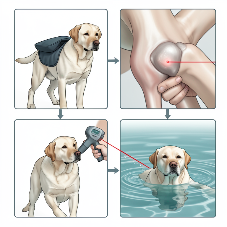
Physiotherapy Regimen
AI-generated conceptual illustration (Labrador Retriever). It explains the idea and does not show study data.
View source evidence ↗Figures & tables
From the original paper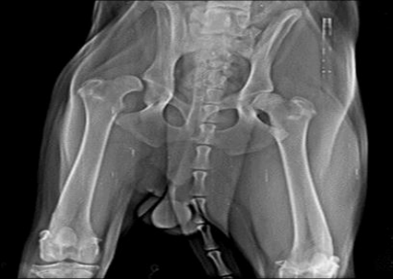 Figure 6
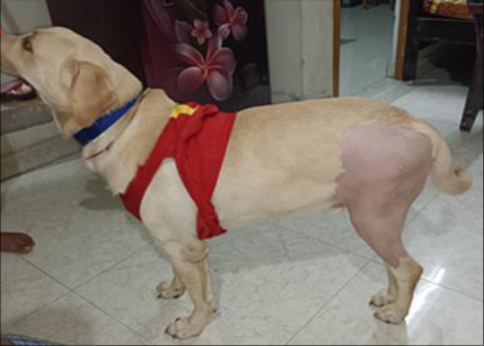 Figure 5
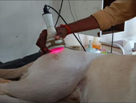 Figure 4
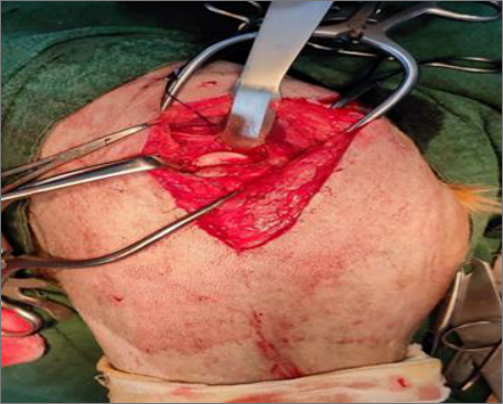 Figure 3
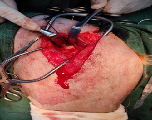 Figure 2
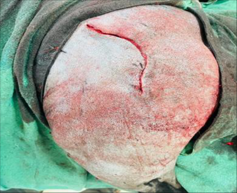 Figure 1
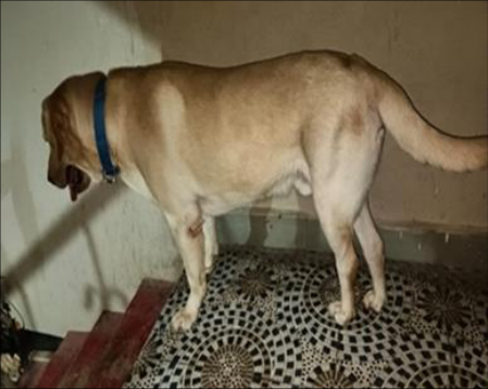 Figure 8
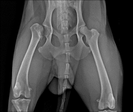 Figure 7
Introduction
Canine hip dysplasia is a hereditary tendency to cause degenerative joint disease in younger dogs. Todhuhter and Lust (2003) reported that hip dysplasia was a developmental problem, mainly affecting medium to large-breed dogs. Commonly encountered coxo-femoral joint affections are hip dysplasia (54%), osteoarthritis (28%) and hip luxation (18%) are most common which require newer treatment protocols to satisfy the ethical and welfare concerns (Arunprasad et al., 2012) [8] . Physiotherapists have therapeutic treatments mainly focusing on correcting muscle dysfunctions, relieving pain, joint mobilizations, and advisement on lifestyle modifications could be equally beneficial to the canine patient. Dogs of irrespective sizes, suffering hip disease and pain, underwent Femoral Head and neck excision (FHNE) instead of Total Hip Replacement (THR) failed to restoring all hip mobility and comfort. Presence of pain, movement restrictions, limb shortness and muscle atrophy were more prominent in FHNE cases than THR cases (Jahandideh A and Tabary PZ, 2020) [3] . Femoral Head and Neck Excision (FHNE) is considered as a salvage procedure for improving the quality of life of many pets by eliminating of pain (Ganesh et al., 2017) [2] . The conventional radiographic method (CRM) is used as a definitive diagnosis, and it is based on evidence of joint laxity, degenerative joint disease (DJD) or both (Kapatkin et al., 2002) [9] . The present study describes analgesic therapy and physiotherapy procedures to achieve early pain-free mobility and satisfactory gait in a Labrador after Femoral head and neck excision surgery.
Case History and Observation
The study was conducted on a Labrador, aged 10 months, and weighting 24kg, presented with history of pain, lameness, exercise intolerance, unable to climb stairs and difficulty in jumping. Physical examination revealed joint enlargement, muscle atrophy, stiffness of joint, reduced range of motion and dysplastic femoral head from acetabulum. Standard hip extended ventrodorsal radiographs revealed hip dysplasia. Radiographic observations included hip joint congruity, flattening of acetabulum and marked subluxation.
Treatment and Discussion
Animal premedicated with xylazine @ 1 mg/kg and ketamine @ 10 mg/kg intramuscularly.
Induction with Butorphanol 0.2 mg/kg and diazepam 0.3 mg/kg slow IV then maintained with 2% Isoflurane. Animals were stabilized with intravenous fluid therapy, use of ceftriaxone sodium has effectively prevented post-operative infection. The craniolateral approach to the hip joint was used, incision curves cranial to greater trochanter and straightened to one-third the length of the femur distally (Fig 1). Then fascia lata incised and vastus lateralis retracted cranially and biceps femoris caudally. Superficial and middle gluteal muscles retracted dorsally (Fig 2) and partial tenotomy of deep gluteal muscles tendon provided best visualization of hip joint (Fig 3). Capsule around head and neck of femur and acetabulum incised and ostectomy was performed from base of trochanter minor to trochanter major. Any rough prominences were removed with rongeurs and joint capsule sutured (DeCamp et al., 2016) [1] . Pre-emptive analgesia with meloxicam 0.2 mg/kg IV followed by carprofen 2.2 mg/kg twice a day post-operatively until pain relief. The skin sutures were removed on 14 th post-operative day. Application of cold or warm pack application for initial 3 days. Passive exercise like flexion of femur and extension of hip joint in lateral recumbency or standing 3 times a day. Laser therapy 6 Jouls @ 5 minutes daily once for 7 days (Fig 4). If poor muscle mass was reported in 2 nd post-operative week suggested for swimming sessions 30 minutes daily for 2 weeks. Dog showed complete weight bearing by 4 th post-operative day (Fig 5). On serial follow-up examination, the dog was in good condition with no or non-significant lameness. FHNE indicated in degenerative joint disease resulting from hip dysplasia, Legg-Calve-Perthes disease or aseptic/ avascular necrosis of the femoral head and neck, femoral neck fractures, complex acetabular fractures, and irreducible or chronic cox-femoral luxations. Kasstrom (1975) [5] stated that the higher incidence in young animals below one year might be due to very rapid growth, improper skeletal and muscular growth, overloading of articular surfaces and rupture of round ligaments leads to dysplasia. In these cases Total Hip Replacement was not possible technically due to lack of availability of implants (owner's affordability and/or availability). Serial follow-ups with physical and radiographic examinations were best used for patient evaluation concurs with study of Sabiza S et al., 2019 [6] (Fig 5)
Figures and tables from the paper
References
8 in the original paper- Ganesh TN, Bridglal Singh S, Legall C. Femoral head and neck excision arthroplasty in 38 dogs: A two-year study with special reference to surgical indication and outcome. Indian Journal of Veterinary Surgery. 2017;38(2):77-80.
- Jahandideh A, Tabary PZ. Comparison of Results of Total Hip Replacement (THR) with Femoral Head Ostectomy (FHO) in Dogs: Review of Literatures. Pakistan Journal of Health. 2020 Sep;14(3):1722.
- Kapatkin AS, Fordyce HH, Mayhew PD, Smith. Canine hip dysplasia: The disease and its diagnosis. Compendium on Continuing Education for the Practising Veterinarian. 2007;24:526-537.
- Kasstrom, H. Nutrition, Weight Gain, and Development of Hip Dysplasia: An experimental investigation in growing dogs with special reference to the effect of feeding intensity. Acta Radiol Suppl. 1975;344:135-179.
- Soroush Sabiza, Ali Ronagh, Ahmad Khajeh. Effective Medical Management and Physiotherapy Program of Femoral Head and Neck Ostectomy in 24 Dogs and Cats; Clinical Report. Iranian Journal of Veterinary Surgery. 2019;14(1):Serial No 30.
- Todhunter RJ, Lust G. Hip dysplasia: Pathogenesis In: Textbook of small animal surgery, Slatter DH, (Ed), 3 rd Ed, WB Saunders company, Philadelphia. 2003;2:200201.
- Mohanty SK, ArunPrasad P. Managing the Performance of Highly Mobile Skilled Individuals in Hi-Tech Firms. In Comparing High Technology Firms in Developed and Developing Countries: Cluster Growth Initiatives 2012 (pp. 167-184). IGI Global.
- Davis GJ, Kapatkin AS, Craig LE, Heins GS, Wortman JA. Comparison of radiography, computed tomography, and magnetic resonance imaging for evaluation of appendicular osteosarcoma in dogs. Journal of the American Veterinary Medical Association. 2002 Apr 15;220(8):1171-6.