
Research article · 2023 · Full text
International Journal of Veterinary Sciences and Animal HusbandryDual demodicosis in a dog: Diagnosis and micrometry analysis
Volume 8(3) · Pages 90–92 · DOI: 10.22271/veterinary.2023.v8.i3b.528 ↗
Research areasDermatological diseases and infestationsInsect and Pesticide ResearchEntomological Studies and Ecology
Abstract
Author-writtenA nondescript dog aged six months was presented with a history of alopecia, severe itching, thickening of skin on ears, around eyes, forehead and lateral aspects of abdomen. Clinical examination revealed dermatitis, irregular skin lesions, crust formation, erythema, pruritis and alopecia on the affected area. Upon microscopic analysis of the skin scrapings, it was found that there were abundant short tailed Demodex cornei mites and long tailed Demodex canis mites. Micrometry was carried out for the two different species of Demodex mites and statistically analysed. The dog was treated with oral ivermectin @ dose of 400 μg/kg body weight for 21 days along with topical application of amitraz. A marked improvement was observed as the dog achieved complete clinical recovery, accompanied by the absence of mites in skin scrapings during two consecutive weekly examinations.
Summary
Source-checkedPlain-language reading of the full paper, prepared with AI and checked against the text. Each point links to the authors' own words.
Why it matters
- Canine demodicosis is a common parasitic skin disease caused by the overgrowth of Demodex mites in a dog's sebaceous glands and hair follicles. View evidence ↗
- Three different species of Demodex mites are known to infect dogs: D. canis, D. injai, and D. cornei. View evidence ↗
- Different Demodex species cause different symptoms: D. canis leads to folliculitis or furunculosis, D. injai causes oily skin/hair, and D. cornei causes itchy (pruritic) skin disease. View evidence ↗
The question
- The study aims to describe a case of a dog concurrently infested with both D. cornei and D. canis, analyze the measurements of these mites, and detail the treatment used. View evidence ↗
What they did
- The subject was a six-month-old female nondescript dog presenting with a three-week history of itching, hair loss, and skin thickening on the abdomen, forehead, ears, and around the eyes. View evidence ↗
- Diagnosis was performed by collecting deep and superficial skin scrapings from affected areas after applying paraffin oil. View evidence ↗
- Morphometric analysis was conducted using ten adult mites (both male and female) of each species. View evidence ↗
- The researchers used SPSS software and Student's t-test for the statistical analysis of the mite measurements. View evidence ↗
What they found
- Microscopic analysis revealed that D. cornei was abundant in superficial scrapings, while D. canis was found in large numbers in deep scrapings. View evidence ↗
- D. cornei had a significantly shorter total body length (149.46 ± 11.48 μm) compared to D. canis (217.29 ± 5.24 μm), representing about 55-72% of the latter's length. View evidence ↗
- The average opisthosoma length of D. cornei (67.19 ± 8.27 μm) was roughly half that of D. canis (132.86 ± 6.05 μm). View evidence ↗
- Statistically significant differences (P < 0.01) existed for total body and opisthosoma lengths, but no significant differences (P > 0.05) were found for podosoma or gnathosoma lengths. View evidence ↗
- The treatment regimen consisted of oral ivermectin (400 μg/kg) for 21 days and topical 12.5% amitraz (5 mL/L) applied twice weekly for 4 weeks. View evidence ↗
- Clinical improvement and new hair growth began after 7 days, with complete recovery and negative skin scrapings achieved after 21 days of treatment. View evidence ↗
What it means
- The authors conclude that the combination of oral ivermectin and supportive therapy successfully eradicated the mixed mite infestation and led to full recovery. View evidence ↗
Figures & tables
From the original paper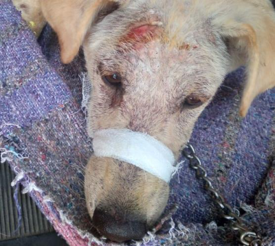 Figure 1
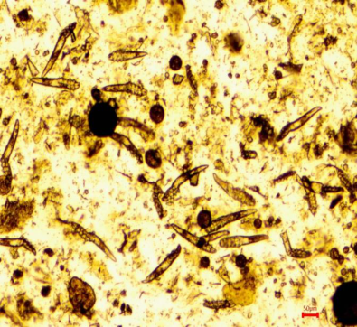 Figure 2
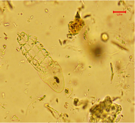 Figure 3
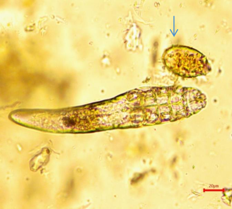 Figure 4
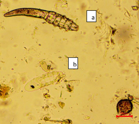 Figure 5
Introduction
Canine demodicosis is a prevalent parasitic skin disease frequently encountered in veterinary practice, resulting from the proliferation of Demodex mites in the hair follicles and sebaceous glands of affected dogs (Kelly et al., 2022) [2] . Scientific evidence has conclusively demonstrated that the Demodex mite is a natural part of the healthy canine skin microflora (Mueller et al., 2020) [5] . Furthermore, it has been observed that puppies acquire these mites from their mother during the initial days of their life (O'Neill et al., 2020) [6] . There are three distinct species of Demodex mites known to affect dogs, which include Demodex canis (D. canis), Demodex injai (D. injai), and Demodex cornei (D. cornei) (JN Izdebska and Rolbiecki, L, 2018) [1] . D. canis, primarily residing in hair follicles, is commonly found in the majority of clinical cases (Plant et al., 2011) [7] . Additionally, the long-bodied D. injai, which inhabits the sebaceous glands, has also been implicated. Male D. injai mites were observed to be more than twice the length of male D. canis mites (Moskvina et al., 2017) [3] . A short-bodied D. cornei mite is found in the superficial layer of the epidermis, measuring approximately half the length of a female D. canis mite. D. canis causes demodectic folliculitis and/or furunculosis in dogs, while D. Injai is associated with the development of oily skin and hair coat on the trunk of dogs. On the other hand, D. cornei is responsible for pruritic skin disease in canines (Sakulploy and Sangvaranond, 2010) [8] . The definitive diagnosis of canine demodicosis is achieved by detecting a large number of adult mites and/or immature forms in deep skin scrapings, hair plucking, or acetate tape impressions. In certain cases, otic swabs and histopathology may be utilized based on the type and location of the lesion (Mueller et al., 2017) [4] . This paper describes occurrence of concurrent infestation by D. cornei and D. canis in a dog. The study includes a detailed analysis of mite measurements and outlines the therapeutic approach utilized for effective management of the infestation.
Materials and Methods
A six months old female nondescript dog was presented to the Veterinary Clinical Complex, Veterinary College and Research Institute, Udumalpet with a history of hair loss, pruritis, itching and thickening of the skin on ears, around eyes, forehead and lateral aspect of abdomen since 3 weeks.
Clinical examination revealed alopecia, papules, pustules, erythema, crust formation, thickening of the skin and cellulitis. The lesions were distributed over the ears, forehead, around eyes and lateral aspects of abdomen (Fig 1). Superficial and deep skin scrapings were collected from the affected areas following the application of paraffin oil to moisten the skin. The dog was treated for 21 days with oral ivermectin at a dosage of 400 μg/kg body weight. Additionally, topical application of 12.5% amitraz at a concentration of 5 mL per litre of water was administered twice weekly for a duration of 4 weeks (Saravanan and Palanivel, 2018) [11] . Clinical signs resolved and new hair growth on affected skin started after 7 th day of treatment and complete uneventful recovery observed after 21 days of treatment. Two consecutive skin scrapings at weekly interval were found negative. Ten adult mites, both male and female of D. canis and D. cornei were used for morphological studies and morphometric analysis. Demodex spp. exhibits a characteristic overall structure, comprising the head or gnathosoma containing the mouthparts, as well as the idiosoma and extremities. The idiosoma consists of the podosoma, where the legs are attached, and the opisthosoma, located distally to the legs. Notably, Demodex species are characterized by an elongated, cigar-like idiosoma, ring-like segmentation of the opisthosoma, and very short legs with claw-like hooks at the tips. D. cornei mites exhibited a long body structure with short and stumpy legs on the podosoma region, notably possessing a shorter opisthosoma compared to D. canis. Measurements of the gnathosoma, podosoma, opisthosoma, and total body length were recorded for both mite species (Table 1). Statistical analysis was conducted using Student's ttest in the SPSS software.
Results and Discussion
Based on the measurements of the mites, it was observed that D. cornei exhibited a shorter body length compared to D. canis. Specifically, the total body length of D. cornei (149.46 ± 11.48 μm) accounted for approximately 55-72% of the total body length of D. canis (217.29 ± 5.24 μm). Moreover, the average opisthosoma length of D. cornei (67.19 ± 8.27 μm) was approximately 50% of that observed in D. canis (132.86 ± 6.05 μm). These findings are consistent with the results reported in previous studies conducted by Sakulploy and Sangvaranond (2010) [8] as well as Sivajothi et al., 2015 [9] . Significant differences (P < 0.01) were observed in the lengths of the total body and opisthosoma between the two types of mites. However, there were no significant differences (P > 0.05) in the lengths of the podosoma and gnathosoma. In this study, D. cornei infestation manifested as scaly and pruritic skin diseases, which is consistent with previous reports (Tater and Patterson, 2008; Sivajothi et al., 2015) [10,9] . Furthermore, the authors observed that the intensity of pruritus in D. cornei infestations was correlated with the mite burden, suggesting a relationship between the number of mites and the degree of pruritus. In contrast, canine demodicosis resulting from D. canis infestation commonly presents as alopecic skin diseases rather than pruritic skin diseases, unless there is a concurrent secondary bacterial infection, in which case the intensity of pruritus may be influenced by the severity of the secondary infection (Muller et al., 2020). The technique used for mite collection can provide valuable diagnostic information. Since D. cornei resides in the stratum corneum of the epidermis, superficial skin scraping or tape preparation techniques are suitable for its collection. On the other hand, D. canis inhabits hair follicles, sebaceous glands, and deeper layers of the dermis, necessitating deeper skin scrapings or hair plucking techniques for effective diagnosis (Sakulploy and Sangvaranond, 2010) [8] .
Conclusion
This study presents a case of canine demodicosis caused by a mixed infestation of Demodex canis and Demodex cornei mites. Species identification of the mites was determined by considering clinical signs, mite habitats, morphology, and morphometric analysis. The treatment approach involved administering oral ivermectin at a dosage of 400 μg/kg body weight for duration of 21 days, accompanied by appropriate supportive therapy. The treatment administered in this study achieved a successful eradication of the mite infestation in the dog, leading to a complete recovery from the condition.
Figures and tables from the paper
References
5 in the original paper- O'Neill DG, Turgoose E, Church DB, Brodbelt DC, Hendricks A. Juvenile-onset and adult-onset demodicosis in dogs in the UK: prevalence and breed associations. Journal of Small Animal Practice. 2020;61:32-41.
- Plant JD, Lund EM, Yang M. A case-control study of the risk factors for canine juvenile-onset generalized demodicosis in the USA. Veterinary Dermatology. 2011;22:95-99.
- Sakulploy R, Sangvaranond A. Canine Demodicosis caused by Demodex canis and short opisthosomal Demodex cornei in Shi Tzu dog from Bangkok Metropolitan Thailand. Kasetsart Veterinarians. 2010;20:1.
- Sivajothi S, Reddy BS. Demodicosis caused by Demodex canis and Demodex cornei in dogs. Journal of Parasitic Diseases. 2015;39(4):673-676.
- Ramesh RR, Muralidharan V, Palanivel S. Preparation and application of unhairing enzyme using solid wastes from the leather industry: An attempt toward internalization of solid wastes within the leather industry. Environmental Science and Pollution Research. 2018;25:2121-2136.