
Research article · 2023 · Full text
International Journal of Veterinary Sciences and Animal HusbandryHistomorphological and histochemical study of small intestine of adult age of local pigeon Columba livia
Volume 8(1) · Pages 116–122 · DOI: 10.22271/veterinary.2023.v8.i1b.478 ↗
Research areasAnimal Nutrition and PhysiologyComparative Animal Anatomy Studies
Abstract
Author-writtenOur study was aimed to morphological and histological results, so the study was included some special stains for histochemical study of adult age of local pigeon Columba livia. Six pigeons were sacrificed. The morphological study had three parts of the small intestine duodenum, jejunum and ileum. The duodenum was U-like to connect between gizzard and jejunum. Ascending and descending duodenum includes the pancreas. The jejunum was characterized coil tube and anastomosed at anteriorly with duodenum and caudally with ileum. Ileum was the last part of small intestine. Generally, the histological sections were included entire the small intestine. Light microscope showed histological structures to mucosa, submucosa, musculais and serosa. Mucosa was included long finger project is called villi. These finger structures were different in length during duodenum, jejunum and ileum. Where lengths of the duodenum were taller. So it's lined by simple columnar epithelia with basal nuclei. Either lamina properia was located under epithelia which it's characterized loose connective tissue and included intestinal glands with crypts of Lieberkuhn is lined by simple columnar cells. Muscularis layer was last tunica in mucosa, it's composed from longitudinal smooth muscles. Others tunica, submucosa was thin layer of dense connective tissue almost no obvious during histological sections. So tunica musculais was consisted of two orientations smooth bundles, first longitudinal orientation is outer while inner layer had been circular smooth fibers. Final tunica was serosa. The present study showed histochemical reaction between goblet cells, where it's appeared the periodic acid Schiff the mucin secretion of their cells is neutral positive reaction while alcian blue stain showed acidic positive reaction of the secreted goblet cells.
Summary
Source-checkedPlain-language reading of the full paper, prepared with AI and checked against the text. Each point links to the authors' own words.
Why it matters
- The digestive system is a sensitive part of a bird's life and can be affected by diet or chemical treatments. View evidence ↗
- Birds have high metabolic rates and high food consumption, with variations occurring between species based on environmental changes. View evidence ↗
The question
- The study aimed to describe the morphological and histochemical characteristics of the entire small intestine in adult local pigeons. View evidence ↗
What they did
- The experimental design involved the use of six adult local pigeons. View evidence ↗
- Samples from the duodenum, jejunum, and ileum were preserved in 10% formalin for 72 hours. View evidence ↗
- General structures were stained with hematoxylin and eosin, collagen fibers with Masson's trichrome, and histochemical analysis was performed using Periodic Acid Schiff (PAS) and Alcian blue (AB). View evidence ↗
What they found
- The duodenum was found to be U-shaped, consisting of descending and ascending segments that form a tube containing the pancreas. View evidence ↗
- The jejunum was characterized by cone-like spiral coils that form centripetal coils. View evidence ↗
- The ileum was observed to be a longer portion than the other two segments, extending as a straight part located between the caeca. View evidence ↗
- Histological analysis showed that the villi in the duodenum were longer than those in the jejunum and ileum. View evidence ↗
- The submucosa of the duodenum in local pigeons was found to lack Brunner's glands. View evidence ↗
- The submucosa was less developed in local pigeons, appearing as a thin layer or being absent in some histological sections. View evidence ↗
- Histochemical staining showed that PAS reacted with neutral mucin secretions, while Alcian blue reacted with acidic mucin from goblet cells. View evidence ↗
What it means
- The authors concluded that the small intestine of adult local pigeons is similar to other bird species, though variations exist in the shape and size of villi across the intestine. View evidence ↗
Visual summary
1 visualsIllustrations explain the paper; they do not show study data.
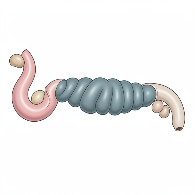
Morphology of the Pigeon Small Intestine
AI-generated conceptual illustration (adult local pigeon (Columba livia) small intestine). It explains the idea and does not show study data.
View source evidence ↗Figures & tables
From the original paper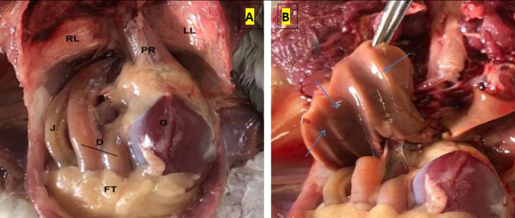 Figure 1
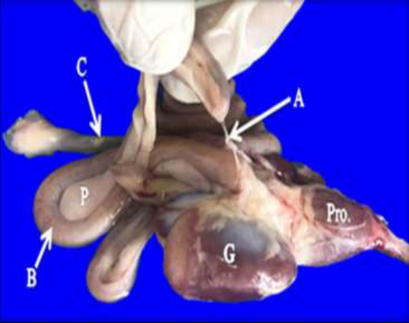 Figure 2
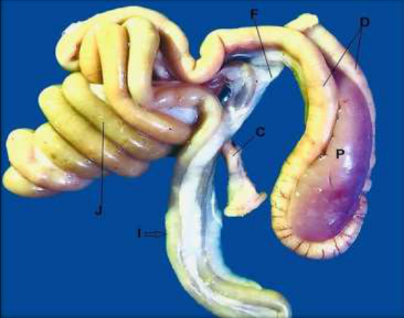 Figure 3
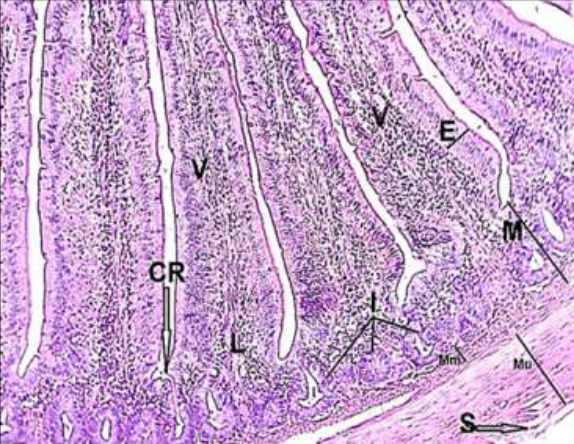 Figure 4
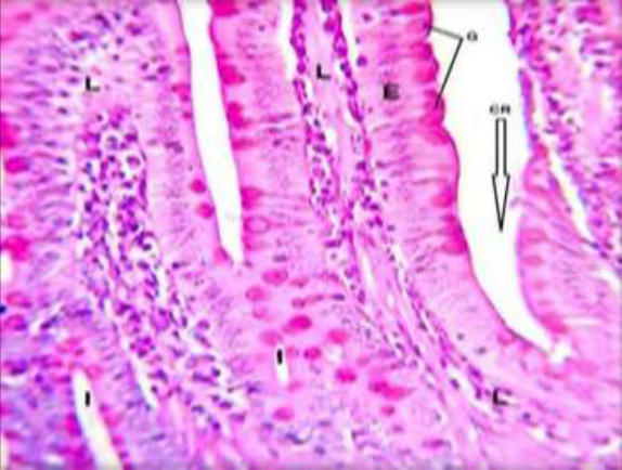 Figure 5
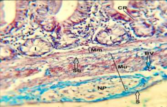 Figure 6
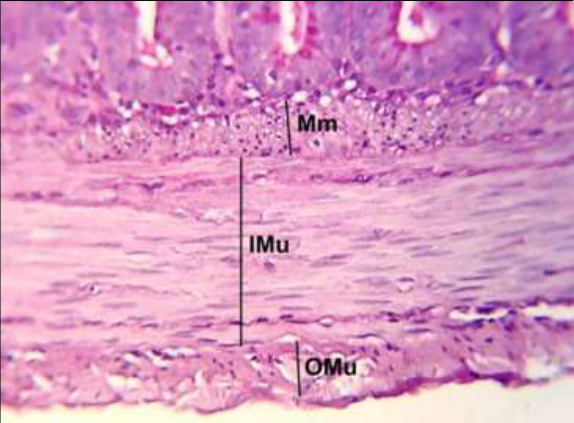 Figure 7
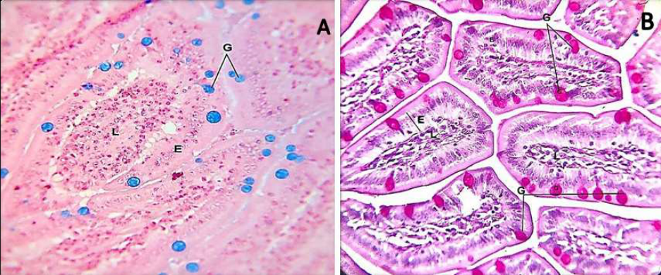 Figure 8
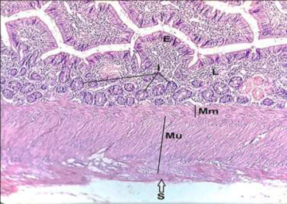 Figure 9
Introduction
The pigeon Columba livia are wide pervasion around the world. There are more than 8000 species [1] . The digestive and others systems are considered a sensitive parts to play role in birds life, where it's affected by have diet, if was treated by some chemical materials [2] . The birds have high metabolism therefor consume a lot of diet. These variations are occurred between the birds species according to the environmental changes [3] . The small intestine is composed from three portions, duodenum, jejunum and ileum. The duodenum is first part which it's connected anterior with gizzard and posterior extend to jejunum [4] . The small intestine is different between other birds species of the formation such as black-winged kite haveonly duodenum and ileum [5] . The parts of the small intestine are separated by diverticulum called vitellin (Meckel's) which spilt jejunum and ileum, the longest part of small intestine [6] . The duodenum has U-shaped loop in indigenous duck, and divided into descending and ascending limb [7] . The jejunum is the second part after the duodenum, it has a spiral coils structure (cone) in African pied crow. The cone is appeared centripetal and centrifugal parts from apex of cone [8] . Ileum is last part, it's started from end diverticulum toward the cecum. Histological features are included four tunics, mucosa, submucosa, muscularis and serosa. Light microscope showed that layers, so they are included epithelium, lamina properia and muscularis within mucosa layer. However, the mucosa of intestine glands are characterized extensive project know villi. These villi processes are varied during three segments of small intestine (duodenum, jejunum and ileum), [9] . The intestinal glands are situate in lamina properia with crypts.
It's called Lieberkuhn's crypt which covered by simple columnar epithelium [10] . The submucosa is located under the mucosa layer, included dense connective tissue [7] . Third layer is called muscularis which has circular and longitudinal muscle fibers. Either serosa layer is last part of tunics represented by loose connective tissue with cover mesothelium. The study is aimed to describe the morphological and histochemical sections of entire the small intestine of adult age of local pigeons.
Materials and methods
Experimental design: Six adult local birds were used. These birds were bought from central market in Baghdad. Then the birds were put on dissected board for the sacrificed after dislocated neck region. Later the pigeons were done -midline incision in ventral region. Three parts of small intestine (duodenum, jejunum and ileum) were put inside container fill 10% formalin during 72 hour. The samples were cleaned carefully by syringe carry the buffer solution to remove of material residues. The histological techniques were included dehydration across processing of specimens to alcohol through 2 hour (70, 80, 90, 100%), these specimens were passing to clearing and later embedded inside paraffin wax for their cutting by microtome machine. The slides were stained by hematoxine and eosin for general structure, masson's trichrom for describe collagen fiber. For histochemical technique were used periodic acid shift PAS and Alcian blue AB [11] .
Results
Results
Morphological study
The small intestine of adult age of local pigeon Columba livia were composed from three segments, duodenum, jejunum and ileum. Generally the study showed that the duodenum Ushape, formed from descending (ventral) and ascending (dorsal) segments lead to duodenal tube, where the pancreas located inside upper and lower parts of duodenum (Figure 1, 2, 3). The duodenum was anastomosed to the jejunum at ascending limb (Figure 3). The jejunum located at ending part of ascending duodenum. The jejunum was characteristics features cone-like of spiral coils, this forming centripetal coils. The jejunum is occupied almost the caudal part of coelomic cavity (Figure 1, 3). The dorsal part of jejunum was beneath right lobe of liver with impression on it (figure 1b). The last part of small intestine was called ileum, the present study were observed that ileum a long portion of other two segments. Which it's extended to straight part, located between the caeca (figure 1,3).
Histological study
Histological study appeared the three segments, duodenum, jejunum and ileum were formed from mucosa, submucosa, muscularis and serosa. Histologically, the light microscope showed the duodenum mucosa had epithelium, lamina properia and muscularis. The epithelia is simple columnar with basal nuclei. In addition to villi is projected finger extensive processes which extended from base of lamina properia to free part of epithelia. The histological structures showed villi differences of lengths between entire the small intestine. Where the villi length of duodenum was longer than jejunum and ileum villi (figure 4,9). The present study showed crypts of Lieberkuhn, it's located between villi and lining by simple columnar epithelium (figure 5). Submucosa of duodenum has no Brunner's glands in local pigeon (figure 6). The intestine glands were distributed along lamina properia during duodenum, jejunum and ileum (figure 6,7,9). Tunica muscularis were well developed during the entire three segments, represented by longitudinal smooth muscle fibers (figure 7,9). Submucosa were less developed in local pigeon, seem thin layer or lost in some histological sections (figure 6). Generally, third tunica is called musculais, this layer had longitudinal outer layer, while inner layer was circular (figure 7). Serosa is final layer which included loose connective tissue with simple squamous epithelium (figure 9).
Histochemical study
Third target of this study represented in the histochemical study, where it's showed the stains by periodic acid schiff (PAS) and alcian blue (AB) stains, where appeared that stains were positive reaction with secretion of mucous (goblet cell) mucin. Hence the PAS stain appeared neutral activity with mucin secretion (figure 5, 8b), while Alcian blue showed acidic reaction with mucin of the goblet cell (figure 8a).
Discussion
The small intestine of adult age of local pigeon C. Livia were included duodenum, jejunum and ileum in order. Morphology study showed the duodenum U-like, anastomosing with lower part of gizzard, formed from lower (descending) and upper (ascending) parts, these parts were taken shape duodenal tube. The pancreas is situated between two parts of duodenum. These results of gross findings were accepted with some previously studies of avian species such as adult kestrel Falco tinnunculus [12] and in starling and pigeons [13] whom stated the duodenum was formed from two segments (upper and lower parts), then the upper parts of duodenum anastomosed with jejunum. The jejunum was began at ending part of ascending duodenum. The jejunum was characterized conelike of spiral coils, and which it's consisted of centripetal coils. The jejunum is occupied almost the caudal part of coelomic cavity, so the study appeared liver impressions. These results no corresponding with [14] in human and domestic rabbit whom said the dorsal part of jejunum was beneath right lobe of liver which it's appeared impression on it. The last part of small intestine was called ileum, the present study were observed that ileum a long portion of duodenum and ileum. Which it's extended to straight part, located between the caeca. These observations disagreement with [15] Coturnix coturnix japonica and [16] whom showed the ileum was continuous with the colorectum and no caeca in yellow and blue macaw. Histological study was proved the small intestine it's formed from three segments, duodenum, jejunum and ileum. The histological structure was included mucosa, submucosa, muscularis and serosa. The current results parallel with observations [5] . In black winged kite and accepted with [17] in rock dove. Also in adult rock dove. The light microscope showed the duodenum mucosa, it's consisted of simple columnar cells called epithelia, eitherlamina properia and muscularis were located underlying the epithelia. These results accepted with said [18] as well as villi is projected finger extensive processes which extended from base of lamina properia, it's longer in duodenum from rest other two segments. Either Submucosa of C. livia was thin layer may see or absent, and has no Brunner's glands. This result conflict with said [18] in Guinea pig. Either [19] in 20 day age in pigeon. The present study showed crypts of Lieberkuhn, it's located between villi and lining by simple columnar epithelium. The intestine glands were distributed along lamina properia during duodenum, jejunum and ileum. These findings were corresponding with said [20] in chicken. And [16] , in yellow and blue macaws, and accepted with [21] in Uttar fowl. Tunica muscularis were well developed during the entire three segments, represented by longitudinal smooth muscle fibers. These results accepted with said [7,22] in mallard, in and owl and kadaknath fowl respectively. The histochemical sections were stained by periodic acid schiff (PAS) and alcian blue (AB) stains, where appeared that stains were positive reaction with secretion of mucous (goblet cell) mucin secretion. Hence the PAS stain appeared neutral activity with mucin secretion, while Alcian blue showed acidic reaction with mucin of the goblet cell. These results accepted with [23] . Turkey and Cockatiel whom proved that goblet mucin were positive reaction with some special stains. Muscularis tunica was third in order and had longitudinal outer layer, while inner layer was circular. Serosa is final layer which included loose connective tissue with simple squamous epithelium. These results accepted with [24,25] in pigeon and esophagus of squirrels [26] .
Conclusions
small intestine of adult age of local pigeons were appeared a similar with other birds species. There are some varies during arrangement of the shape and size villi in entire the small intestine. The histochemical techniques showed positive reaction of mucin secretion during periodic acid Schiff and alcian blue stain.
Acknowledgement
AcknowledgmentsThe current study was supported by department of anatomy and histology and College of Veterinary Medicine, University of Baghdad.
Figures and tables from the paper
References
22 in the original paper- King AS, Mclelland J. Outline of avian anatomy 1st ed. Bailliere, Tindall, London; c1975. p. 91-98. https://doi.outlines-of-avian-anatomy/oclc/1975.3070357
- Maty HN, Al-Maatheedi MS, Ahmed SM. Effect of Oregostem® and imbalance diet on body performance and reproductive efficiency in male quails. Iraqi Journal of Veterinary Sciences. 2022;36(1):29-37. DOI: 10.33899/ijvs.2021.128810.1602.
- Hamdi H, El-Ghareeb A, Zaher M, AbuAmod F. Anatomical, Histological and Histochemical Adaptations of the Avian Alimentary Canal to Their Food Habits: IIElanus caeruleus. International Journal of Scientific Engineering and Research. 2013;4(10):1355-1364. https://doi.ijser.2013.4.10.
- Zaher M, El-Ghareeb A, Hamdi H, AbuAmod F. Anatomical, histological and histochemical adaptations of the avian alimentary canal to their food habits: I-Coturnix coturnix. Life Sciences. 2012;9(3):253-75. http://doi.org/10.7537/marslsj090312.13
- Halse SA. Diet and size of the digestive organs of Spurwinged Geese. Wild Fowl. 1985;36(36):129-134. https://doi.1985.36/711/71
- Bezuidenhout AJ, Vanswegen GA. light microscopic and immune cytochemical study of the gastrointestinal tract of the Ostrich (Struthio camelus). Onderstepoort Journal of Veterinary Research. 1990;57(1):37-48. https://doi.OnderstepoortJVeter.Rese.1990.57.1.2338997
- Luna LG. Manual of histologic staining method of armed forces. Institute of pathology. 3 ed . New York, USA; c1968. p. 123. https://doi.US. 1968.123.201300459165
- Al-Haaik AG. A gross anatomical and histological study of pancreas in adult Kestrel (Falco tinnunculus). Iraqi Journal of Veterinary Sciences. 2019;33(2):175-180.
- AL-Taai SAH. Morphological comparison of proventriculus and gizzard in starling birds Sturnus vulgaris and pigeon Columba livia. International Journal of Veterinary Sciences and Animal Husbandry. 2022;7(1):15-18. DOI: http://dx.doi.org/10.22271/2022.7.1.4
- Al-Hamdany MZ. Comparative anatomical, histological, and histochemical study of liver in human and domestic rabbit. Iraqi Journal of Veterinary Sciences. 2019;33(2)437-446. DOI: IJVS.2019.33.2.20437-446.
- Ahmad JM, Mamde CS, Patil VS, Khandare SM. Gross Anatomical Study of Small Intestine in Japanese Quail (Coturnix japonica). Indian Journal of Veterinary Anatomy. 2012;24(2):82-83. http:// doi.IJVA.2012.27091
- Aizawa J, Tivane C, Rodrigues MN, Wagner PG, Campos DB, Guerra RR, et al. Gross anatomical features of the gastrointestinal tract of Blue-and-Yellow Macaws (Ara ararauna) -Oesophagus to Cloaca. Histology and Anatomy. Embry; c2012. https://doi.A.H.E.10.1111.2012.2341.4512
- Al-Bideri AW, Jawad AN. Comparative Anatomical and Histological Study of Duodenum between Laughing Dove Streptopelia Senegalensis and White breasted Kingfisher, Halcyon Smyrnensis. Zoology Classification QL. c2015. p. 801-950. https://DOI.JOPS.2015.703.699
- Al-Saffar FJ, Nasif RH. Histoarchitecture and Histochemical Study of the Duodenum in Adult Guinea Pigs (Cavia porcellus). Indian Journal of Natural Sciences. 2019;10(57):17815-24. https://doi:12096/8827/2019.10.27.87.
- AL-Muhammdi HH, Al-Taai SAH. Histological study of small intestine during twenty day age in Iraqi pigeon (Columba livia). Plan Arch. 2020;20(2):5323-5327. https://doi.org/10.51470/Plantarchives.2020.v20.no2.343.
- Nasrin M, Siddiqi MNH, Masum MA, Wares MA. Gross and histological studies of digestive tract of broilers during postnatal growth and development. Journal of the Bangladesh Agricultural University. 2012;10(1):69-77. https://doi.JBAU.2012.10.1.12096.8827
- Rana J, Dhote BS, Ambwani TK, Kumar S. Histochemical studies on small intestine of Uttara Fowl. International Journal of Science, Environment and Technology. 2016;5(3):1181-1188. https://doi; Int. J Sci. Enviro.Techn.984.2016.5.3.543.
- Kalita PC, Singh GK, Kalita A. Gross morphological and morphometrical studies of small intestine in post hatched kadaknath fowl. Indian Journal of Veterinary Anatomy. 2012;24(2):74-75. http://doi.IJVA.2012.24.2.27083.
- Taha AM. Comparative histological and histochemical study of the ileum in two different birds. Iraqi Journal of Veterinary Sciences. 2021;35(3):479-487. DOI: 10.33899/ijvs.2020.127046.1447
- Al-Saffar FJ, Al-Samawy ERM. Histomorphological and histochemical study of stomach of domestic pigeon (Columba livia domestica). Iraqi Journal of Veterinary Medicine. 2016;40(1):89-96. https://doi.org/10.30539/iraqijvm.v40i1.144
- AL-Muhammadi HH, AL-Taai SAH. Histological study of small intestine during twenty day age in Iraqi pigeon (Columba livia), Plant Archives. 2020;20(2):5323-5327.
- Suhaib AH, AL-Taai, Nsaif RH, Masarat S. Almayahi: Histomorphological study of esophagus in squirrel (Sciurus anomalus). Biochemical and Cellular Archives. 2021;21(1):1391-1394. DocID: https://connectjournals.com/ 03896.2021.21.1391
Cited by 9
From OpenAlex, OpenCitations and reference lists read by Scinovex
- A new anatomical insight into the architecture of biliary duct system, gall bladder, and Duodenal Papilla with the extrahepatic bile ducts of white pekin duck (Anas platyrhynchos) ↗
Zeinab K. Aboghanima, Ashraf El Sharaby, Asmaa Aboelnour, Mohamed M. A. Abumandour, Ahmed G. Nomir · Tissue and Cell · 2025
❝ 7 - Integrated Morphological Analysis Of The Small Intestine In The Pekin Duck's (Anas platyrhynchos) Using Gross Anatomical, Histological, Histochemical, and Scanning Electron Microscopic Techniques ↗
Youssef Elsabbgh, Ashraf El Sharaby, Asmaa Aboelnour, Mohamed M. A. Abumandour, Ahmed G. Nomir · Tissue and Cell · 2026
❝ 1 - Comparative histological and histochemical study of the duodenum in two birds with different feeding habits: the carnivorous slender-billed gull (Chroicocephalus genei) and the grainivorous cockatiel (Nymphicus hollandicus) ↗
Ameer M. Taha, Dalal Sulaiman Alshaya, Eman Fayad, Diaa Massoud, Mohamed M. A. Abumandour · Zoomorphology · 2025
❝ 1 - Detection of virulence factors of Pseudomonas aeruginosa from dog’s wound infections by using biochemical and molecular methods
Sara Abdalkareem Raheem, Dalia Abdalkareem Abdalshheed · International Journal of Veterinary Sciences and Animal Husbandry · 2023
This investigation's goal was to find Pseudomonas aeruginosa virulence factors in canine wound infections. 100 samples were gathered and tested for eight virulence variables utilizing biochemical and molecular techniques for this purpose. According to the findings, women (72%) had the highest isolation ratio. The virulence factors were found in various ratios, with capsule being found in (16.6%) of them, amylase enzyme detected in (3.3%), hemolysin enzyme detected in (70%), protease detected in (10%), phospholipase enzyme detected in (83.3%), urease enzyme detected in (100%), DNAase enzyme det
- Histological and Histochemical Characterization of the Small Intestine of German Nun Pigeons ↗
Shaima Khazaal Waad, Iman K. M. ALABADI, Shadan Qais Ahmed · European Journal of Theoretical and Applied Sciences · 2026
The present study aimed to describe the histological and histochemical characteristics of the small intestine in adult German Nun pigeons (Columba livia domestica). Samples from the duodenum, jejunum, and ileum were collected from six adult birds of both sexes and processed using routine hematoxylin and eosin (H&E) staining, as well as periodic acid–Schiff (PAS). The three distinct segments of the…
❝ 0PDF - Anatomical and histological insights into gastrointestinal adaptations of adult Egyptian quail ↗
Eman El- Nahass, Mona Elwan · Tissue and Cell · 2026
❝ 0 - Exploring the pathobiology of colibacillosis in broiler chickens reared in Ganderbal district of North Kashmir
Majid Shafi, Shabia Shabir, Omer Khalil Baba, Masood Saleem Mir, Shayaib Ahmad Kamil, Andleeb Rafiq · International Journal of Biology Sciences · 2025
The aim of the study was to examine the Pathomorphological changes in the naturally occurring Colibacillosis in broiler chickens reared in commercial broiler farms at Ganderbal district of Kashmir. During this study, a total of 200 cases of Colibacillosis were identified at 10 different broiler farms and the dead birds were sent to the Division of Veterinary Pathology for a thorough postmortem investigation. Colibacillosis is a complex disease mainly occurring in the age groupof 3-4 week age of age and is characterized by air sacculitis, pericarditis, peritonitis, salpingitis, synovitis, osteo
- Histopathological changes in the small intestine of domestic pigeons induced by cestodes infections ↗
Nidhal Yousif Mohammed, Ibrahem A. Abdulzahra · International Journal of Advanced Research in Medicine · 2025
The present investigation was conducted to assess and document the microscopic tissue alterations occurring in the small intestines of pigeons as a consequence of Cestoda parasite infections. Sixty pigeons of different ages and sexes were randomly collected from local markets in Muthanna Governorate, southern Iraq. Results displayed that 20 of the 60 tested were infected with cestoda, with a prevalence
- Coccidial occurrence among domesticated pigeons (Columba Livia Domestica) In Babylon Province, Iraq. ↗
Mohammed KA Altamimi, Muntadher M Flayyih, Haider Alwan · Acta Entomology and Zoology · 2025
The parasite Eimeria spp., an Apicomplixian protozoa, responsible for significant disease known as Coccidiosis of health concern in birds globally. Our study making a survey for Eimeria infecting domestic pigeons (Columba Livia Domestica) across several cities within Babylon province, Iraq. Two handred fecal samples gathered during the period between January to October 2023 and the diagnosis depend on the direct