
Research article · 2023 · Full text
International Journal of Veterinary Sciences and Animal HusbandrySuccessful cosmetic outcome of eyelid lacerations in a young male Marwari horse
Volume 8(3) · Pages 222–224 · DOI: 10.22271/veterinary.2023.v8.i3d.547 ↗
Research areasSurgical Sutures and AdhesivesLeech Biology and ApplicationsEntomological Studies and Ecology
Abstract
Author-writtenA 2-year-old Marwari horse presented with right-side eyelid laceration along with lateral canthus injury after 2 hours of accident. Because of animals’ temperament, laceration was repaired surgically under general anaesthesia. Gross appearance i.e. near to normal was reported after 20 days of surgery.
Summary
Source-checkedPlain-language reading of the full paper, prepared with AI and checked against the text. Each point links to the authors' own words.
Why it matters
- Traumatic injuries, particularly eyelid lacerations and other ocular pathologies, are common in horses because of their agile nature. View evidence ↗
- The upper eyelid is critical for protecting the eyeball and spreading the tear film; if it is non-functional, it can lead to corneal diseases. View evidence ↗
- Immediate treatment of eyelid lacerations is necessary to avoid chronic eye irritation and complications that make surgical repair more difficult. View evidence ↗
The question
- The study aimed to perform a successful surgical repair of multiple eyelid lacerations in a young male horse using general anesthesia. View evidence ↗
What they did
- The subject was a 2-year-old male horse weighing 360 kg that presented with a right-eye trauma approximately two hours after the incident. View evidence ↗
- The horse was diagnosed with a laceration of the upper eyelid and an injury to the lateral canthus. View evidence ↗
- Due to the animal's hyperactive temperament and lack of cooperation with regional blocks, general anesthesia was chosen for the procedure. View evidence ↗
- The anesthetic protocol included premedication with Xylazine (1.1 mg/kg) and Butorphanol (0.02 mg/kg), induction with Ketamine (2.2 mg/kg) and Diazepam (0.02 mg/kg), and maintenance with 1-2% Isoflurane. View evidence ↗
- The laceration was repaired in two layers: the conjunctival tearing was sutured with absorbable Vicryl (3-0), and the skin was aligned with non-absorbable nylon (zero number) for a cosmetic result. View evidence ↗
What they found
- Immediately following the surgery, the repaired laceration appeared cosmetically near to normal. View evidence ↗
- An anatomically normal gross appearance was achieved 22 days after the surgical procedure. View evidence ↗
- Post-operative evaluation showed that the repaired eyelid was properly mobile, the cornea was normal, and the horse exhibited positive menace and pupillary light reflexes (PLR). View evidence ↗
- The horse experienced no postoperative complications following the repair. View evidence ↗
What it means
- The authors conclude that multiple eyelid lacerations in a young male horse can be successfully repaired using general anesthesia. View evidence ↗
Visual summary
1 visualsIllustrations explain the paper; they do not show study data.
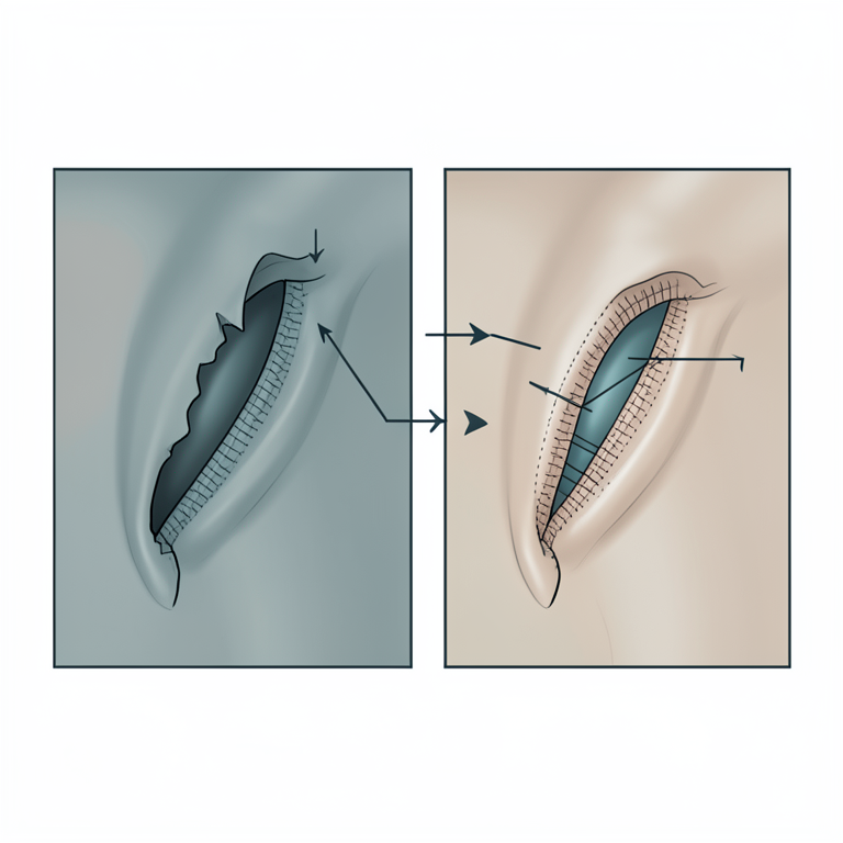
Two-Layer Suture Technique
AI-generated conceptual illustration (Marwari horse eyelid). It explains the idea and does not show study data.
View source evidence ↗Figures & tables
From the original paper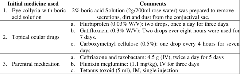 Table 1
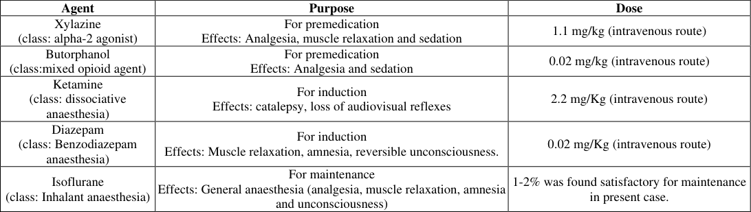 Table 2
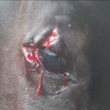 Figure 1
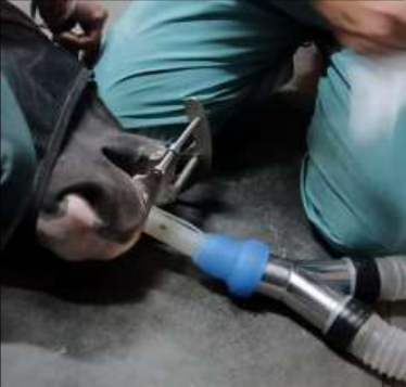 Figure 2
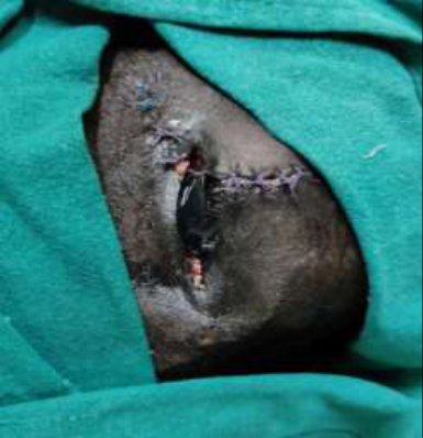 Figure 3
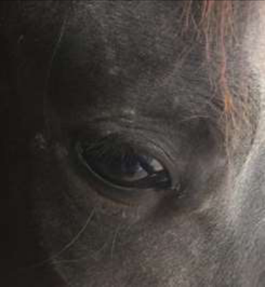 Figure 4
Introduction
Traumatic injuries are common in equine due to their agile nature (Brooke, 2006) [1] . Majority of adenexa and ocular pathology in equines are eye lid laceration, puncta blockage, corneal ulcers and recurrent uveitis therefore encountered frequently (Plumer, 2005) [4] . Eyelid (especially upper eyelid) helps in eyeball protection through its physical activity and hence it should be well functional. A non functional upper eyelid is immobile and not able to spread tear film over the cornea which further imparts in corneal diseases (Brooks and Matthews, 2007) [2] . Case if presented with complete laceration must be transferred to surgical facility. Prompt treatment is required before further ocular complications. Actually delayed cases are problematic for surgical repair and hence desired recovery cannot be achieved. Moreover irregular eyelid margins are responsible for chronic eye irritation.
Case presentation findings and initial care
A 2-year old male horse (body wight; 360 kg) having history of trauma to right eye (Fig. 1) was presented to GADVASU-clinic (in late evening), approximately two hours after diseases occurrence. Mild fresh bleeding from injured (lacerated) tissue was noticed for timing of injury. Usual feeding and walking were observed. Case has been examined and diagnosed as upper eye-lid laceration along with lateral canthus injury. Ophthalmic examination revealed that globe was functional having normal Menace and Pupilary light reflex. Initially (till surgical attempt), eye was treated with collyria, topical eye drops and parentral drug administration. 2. Topical ocular drugs a. Flurbiprofen (0.03% W/V): two drops, once a day for three days. b. Gatifloxacin (0.3% W/V): Two drops ever eight hours were used for 7 days. c. Carboxymethyl cellulose (0.5%): one drop every 4 hours for seven days.
3. Parentral medication a. Ceftriaxone and tazobactam: 4.5 g (IV), twice a day for 5 days b. Flunixin meglumine: (1.1 mg/kg), IV for three days c. Tetanus toxoid (5 ml), IM, single injection
Anaesthesia and surgical management
Animal was not cooperating for regional block and because of such hyperactive temperament; patient has been selected for general anaesthesia. Blood picture revealed healthy parameters. Animal was kept off-feed before general anaesthesia. The equine was pre-anaesthetized with a mixture of inj. Xylazine @ 1.1 mg/Kg and inj. butorphanol @ 0.02 mg/kg, IV in standing position. Once, the equine was head down upto knees, the general anaesthesia was induced with a mixture of inj. Ketamine hcl @2.2mg/Kg and inj. Diazepam @ 0.02 mg/Kg, IV. The equine lied down within 2 minutes and was intubated (ID. 20). The anesthesia was further maintained on Isoflurane in 100% oxygen. (Fig. 2). For maintenance Effects: General anaesthesia (analgesia, muscle relaxation, amnesia and unconsciousness)
1-2% was found satisfactory for maintenance in present case.
Note: Xylazine and Butorphanol were given at the same time for premedication whereas both Ketamine and Diazepam were mixed in a syringe and injected for induction. After induction ET was introduced and animal was taken on inhalant anaesthesia Surgical site was prepared aseptically and lacerated margins were freshened using B.P. blade no. eleven. Laceration was sutured in two layers (Fig. 3). Absorbable suture material (Vicryl, 3-0) was used for suturing of conjunctival tearing whereas skin was aligned to achieve cosmetic appearance using non-absorbable suture (nylon, zero number). Animal was recovered successfully from general anaesthesia without any complications.
Results and Discussion
Because of good blood supply to eyelids (Schaer, 2007) [5] , laceration was repaired successfully and cosmetically looks near to normal immediately after surgery. Topical medications (eye drops) along with parentral drugs were recommended postoperatively (Table : 1). Successful outcomes with anatomically normal gross appearance were reported after 22 days of surgery. Verma et al., (2018) [6] also repaired lower eyelid laceration with successful recovery within 10-days. However sutures were removed after 2-weeks of surgery. Proper apposition is mandatory to prevent keratitis in postoperative days (Lavach et al., 1984) [3] and similarly in present case also proper alignment has been done under general anaesthesia. Affected eyelid, cornea and retina were evaluated for mobility, ulcer and vision respectively (Fig. 4). Repaired eyelid showed proper mobile, cornea was normal and positive menace and PLR were noticed. Eyelids should be well reapposed otherwise vision impairment can occur and hence it is very important affection (Schaer, 2007) [5] similarly present case has been taken for surgery and repaired successfully. In present case there was no any postoperative complication but Brooks (2006) [1] stated that complications usually occur after surgical eyelid repair. The study reports the successful repair of multiple eyelid lacerations in a young male horse under general anaesthesia.
Figures and tables from the paper
References
4 in the original paper- Lavach D, Severen GA, Roberts SM. Lacerations of the equine eye: A review of 48 cases. Journal of the American Veterinary Medical Association. 1984;184(10):1243-1248.
- Plummer CE. Equine Eyelid Disease. Current Techniques in Equine Practice. 2005;4(1):95-105.
- Schaer B. Ophthalmic emergencies in horses. Veterinary Clinics of North America Equine Practice. 2007;23(1):49-65.
- Verma MK, Khangembam SD, Gangwar AK. Surgical Repair of Equine Eyelid Laceration. Special issue, RKVY nov-2018 seminar. Nduat Ayodhya; c2018. p. 8.