
Research article · 2022 · Full text
International Journal of Veterinary Sciences and Animal HusbandryLumpy skin disease (LSD) in cattle caused by the lumpy skin disease virus (Neethling virus): A member of the genus capripoxvirus within the family poxviridae
Volume 7(6) · Pages 25–28 · DOI: 10.22271/veterinary.2022.v7.i6a.451 ↗
Research areasPoxvirus research and outbreaksVirus-based gene therapy researchVector-Borne Animal Diseases
Abstract
Author-writtenLumpy skin disease (LSD) causes major economic losses in the livestock industry of most African countries. It is caused by Lumpy Skin Disease Virus (LSDV), a member of the Poxviridae family, with the prototype Nettling strain. LSDV belongs to the capripox virus family which includes sheep pox virus and goat pox virus. It is spread by insect vectors between pastures and watering places and cattle congregating in the same barn. LSD is an enzootic infectious, eruptive and rarely fatal disease of cattle characterized by skin nodules. Cattle and water buffalo are the only animal species affected, with high morbidity rates but low mortality, however, mortality is high in calves. LSD reduces milk and beef production, causes abortion in females and infertility in males. The origin of LSD was in Zambia in 1929. LSD is considered an endemic disease in the African continent. However, the disease has moved out of Africa since the 1980s. The disease is reported in LSD-free countries (Jordan, Syria, Lebanon, Turkey, Iran and Iraq) with potential economic losses to the livestock industry. Therefore, mass vaccination along with other appropriate control measures is the most effective way to limit the spread and economic impact of lumpy skin disease.
Summary
Source-checkedPlain-language reading of the full paper, prepared with AI and checked against the text. Each point links to the authors' own words.
Why it matters
- Lumpy skin disease (LSD) is a significant problem because it causes major economic losses within the livestock industries of most African nations. View evidence ↗
- The disease is economically damaging because it lowers beef and milk production and impairs reproduction through infertility in males and abortions in females. View evidence ↗
- While LSD is endemic to Africa, it has expanded its geographic reach beyond the continent since the 1980s. View evidence ↗
The question
- The paper aims to provide a comprehensive overview of Lumpy Skin Disease in cattle, focusing on the causative Neethling virus and its classification within the Poxviridae family. View evidence ↗
What they did
- The author reviews the disease's etiology, identifying it as an infectious condition caused by the Lumpy Skin Disease Virus (LSDV), a member of the capripox virus genus. View evidence ↗
- The review examines host susceptibility, noting that natural infections occur specifically in cattle and Asian water buffalo. View evidence ↗
- The paper details transmission methods, identifying arthropod vectors, including specific mosquitoes, biting flies, and male ticks, as the primary means of infection. View evidence ↗
What they found
- The prevalence of LSD is significantly higher in cattle (30.8%) compared to Asian water buffalo (1.6%). View evidence ↗
- Clinical signs of LSD include mild fever for 2-3 days, weakness, enlarged lymph nodes, and skin or visceral nodules measuring 2-5 cm in diameter. View evidence ↗
- The disease can result in mortality rates of up to 20% and causes economic loss through damaged coats and secondary bacterial infections. View evidence ↗
- General morbidity rates for the disease are reported between 10-20%, with mortality rates typically ranging from 1-5%. View evidence ↗
- The incubation period varies by infection type, taking 7 to 14 days in experimental cattle and 2 to 5 weeks in natural cases. View evidence ↗
- LSDV can be isolated from ulcerated nodules on mucous membranes for at least 35 days. View evidence ↗
- Recovery time for severely affected animals can be prolonged, taking up to 6 months for full recovery. View evidence ↗
What it means
- The author concludes that vaccination is the most effective control method and recommends the use of live homologous vaccines containing a Neethling-like strain. View evidence ↗
- The author recommends that timely and accurate diagnosis is essential for implementing effective control measures. View evidence ↗
- The author notes that while insect control is commonly used during outbreaks, its actual effectiveness remains unclear. View evidence ↗
Visual summary
1 visualsIllustrations explain the paper; they do not show study data.
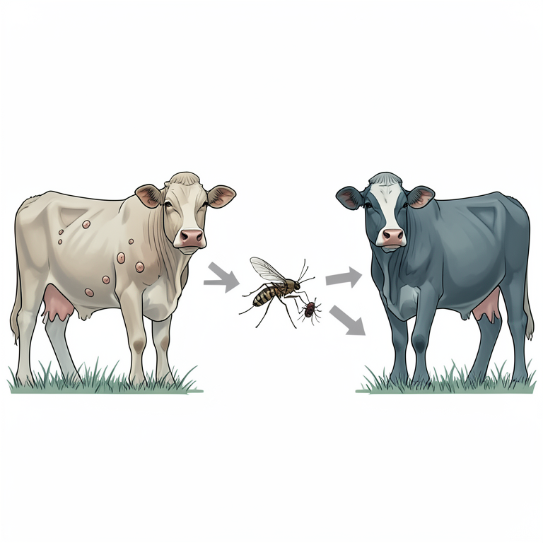
LSD Transmission Cycle
AI-generated conceptual illustration (cattle and arthropod vectors). It explains the idea and does not show study data.
View source evidence ↗Figures & tables
From the original paper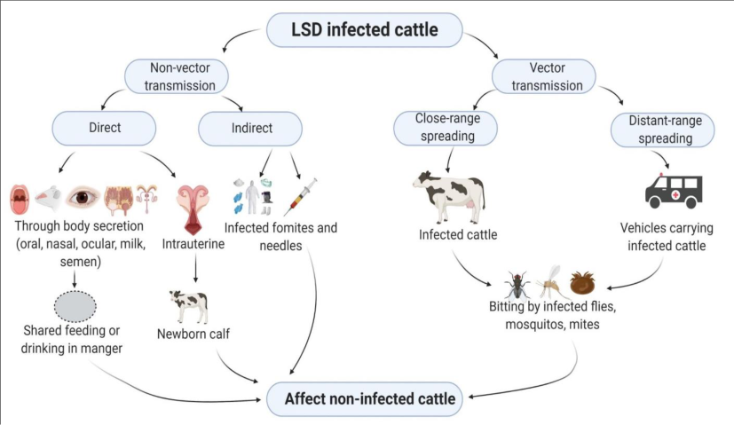 Figure 1
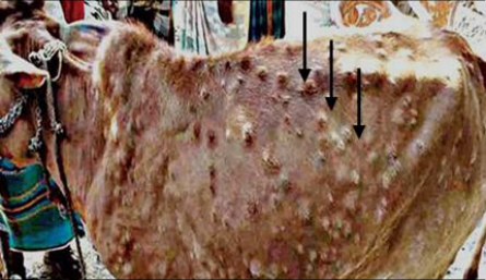 Figure 2
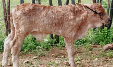 Figure 2
Introduction
Lumpy skin disease (LSD, Pseudo-urticaria, Neethling virus disease, exanthema nodularis bovis, and knopvelsiekte) is an infectious disease. It is caused by a virus (LSDV) of the Poxviridae family, capripox virus, (Alexander et al., 1957) [5] . Capripoxvirus (CaPVs) is one of eight species in the Chordopoxvirinae subfamily of Poxviridae and includes Lumpy Skin Disease Virus (LSDV), Sheep Pox Virus (SPPV), and Goat Pox Virus (GTPV). These viruses are responsible for economically important diseases of domestic ruminants in Africa and Asia, (CFSPH, 2008) [26] . LSD causes significant economic losses in affected countries, but while all stakeholders in the livestock industry experience loss of income, poor, small and marginal farmers suffer the most, (FAO, 2017) [17] . LSD is a disease of domestic cows and buffaloes. Although LSD has not been detected in goats or sheep. Sheep, goats, giraffes, impala, and Grant's gazelles housed near sick cattle develop skin lesions, (OIE, 2016) [23] . Lumpy skin disease (LSD) is a borderline high-impact cattle pox disease. Clinical symptoms include lesions in mouth, pharynx and respiratory tract, skin and visceral nodules (2-5 cm in diameter), mucous membrane and internal organs, mild fever for 2-3 days, weakness, enlarged lymph nodes, swelling of the skin and sometimes death, (OIE, 2016; Buller R.M., 2005) [23,10] . Nodules are circumscribed, firm, round, large, and involve the skin, subcutaneous tissue, and sometimes muscle. It is a vector-borne disease transmitted by various biting and biting blood-feeding arthropods such as mosquitoes, flies and ticks. Mortality rates up to 20%, economic losses due to condition deterioration, reduced milk production, abortions, infertility, secondary bacterial infections and damaged coats, (Anonymous, 1988) [32] . Although infected animals recover within 2-3 weeks, milk production decreases in lactating cattle for several weeks. The morbidity rate is about 10-20% and the mortality rate is about 1-5%, (Advisory and guidelines). The double-stranded DNA virus, LSDV, like other Poxviridae, has a large size, a property that has made this virus widely used as a vector to express foreign viral genes and to produce recombinant multivalent vaccines. Another potentially beneficial property of LSDV is its remarkable stability and ability to survive for long periods of time at ambient temperature, particularly in dried form. The virus is also very resistant to inactivation.
History and Geographic Distribution:
The disease was first identified in Northern Rhodesia (Zambia) in 1929. Lumpy skin disease (LSD) was known, a skin disease of cattle called "pseudo-urticaria" found in what was then known as Northern Rhodesia. The disease was probably a lumpy skin disease, but at the time it was thought to be caused by insect bite wounds, (Mac-DONALD, 1931) [21] . The disease appeared in the following years and plant poisoning was also the cause, (L.E. Roux, 1945) [20] . Another epidemic of this disease occurred in October 1943 in Botswana and was named "Ngamiland Cattle Disease" because it was initially observed in Ngamiland. During this period there was evidence that the disease was caused by an infectious agent, (Abdulqa H.Y., 2016) [1] . Additional cases occurred between 1943 and 1945 in Botswana, Zimbabwe, and the Republic of South Africa. An estimated 8 million cattle were affected by panzootic infection in South Africa in 1949, causing enormous economic losses. LSD spread across Africa between the 1950s and 1980s, affecting cattle in Kenya, Sudan, Tanzania, Somalia, and Cameroon. The disease was first reported in Israel in 1989, and since then sporadic outbreaks have occurred in several countries in the Middle East. In 2012, the disease appeared on Israel's northeastern border and spread to an unprecedented scale in the Middle East, (OIE, 2016) [23] . Outbreaks were reported from Lebanon, the Palestinian Autonomous Territories, Jordan, Kuwait, Saudi Arabia, Iraq, and Iran. The first entry of the disease was reported in Turkey in 2013, where LSD is currently endemic, (Bowden T.R. et al., 2008).
Hosts and susceptibility
LSD has a narrow vertebrate host range. Lumpy skin disease is host-specific, causing natural infection in cattle and Asian water buffalo (Bubulus bubalis), although the prevalence of disease in buffalo (1.6%) is significantly lower than in cattle (30.8%), (Spickler, 2008) [26] . Some LSDV strains can replicate in sheep and goats. Although mixed herds of cattle, sheep and goats are common, to date there is no epidemiological evidence on the role of small ruminants as reservoirs for LSDV, (Al-Salihi K., et al., 2014) [7] . Lumpy skin disease does not affect humans. Clinical signs of LSD have been observed following experimental infection in impala (Aepyceros melampus) and giraffe (Giraffa Camelopardalis), (El-Nahas E.M., 2011) [15] . The disease has also been reported in the Arabian oryx (Oryx leucoryx) and (Greth a,) springbok (Antidorcas marsupialis). The susceptibility of wild ruminants or their potential role in the epidemiology of LSD is not known.
Sources of the virus
Nodules found on the mucous membranes of the eyes, nose, mouth, rectum, udder, and genitalia also ulcerate and release large amounts of LSDV that can serve as a source of virus. Virus can be separated from this material for up to 35 days and likely for longer, (Tuppurainen E.S., 2012) [30] . LSDV can be isolated from blood, saliva, eye and nasal secretions, and semen. LSDV is detected in the blood (viraemia), usually at lower levels 7 to 21 days after infection than in skin lesions. Ejaculation may be prolonged; LSDV has been isolated from the semen of experimentally infected bulls 42 days after inoculation. There has been one report of placental transmission of LSD. LSD does not cause chronic disease. It does not indicate a delay and the disease does not recur. The main means of transmission is believed to be through arthropod vectors. Consequently, hosts become infected with the virus through the bites of mosquitoes (e.g., Culex mirificens and Aedes natrionus), biting flies (e.g., Stomoxys calcitrans and Biomyia fasciata), and male ticks (Riphicephalus appendiculatus and Amblyomma hebraeum). Could play a role in virus transmission. The importance of different 2 arthropod vectors may vary in different areas depending on the abundance and feeding behavior of the vector, (Tuppuraine E.S. and Alexandrov T., 2017) [28] . Infected bulls can excrete the virus in semen; however, LSD has not been transmitted through infected semen, (Annandale C.H., 2014) [8] . It is not known whether infection can be transmitted by fomites, for example by ingestion of food and water contaminated with infected saliva, (Ali H., 2012) [6] . Animals can be infected by experimental inoculation with skin nodules or blood material. Direct contact is thought to play a minor, if any, role in the transmission of the virus.
Clinical signs
The time between inoculation and the first observation of typical clinical signs in experimentally infected cattle is 7 to 14 days, regardless of route of infection, and 2 to 5 weeks in natural cases. (Carn V.M., 1995) [11] . Clinical symptoms may include nodules on the skin, mucous membranes and internal organs, fever, emaciation, enlarged lymph nodes, nasal discharge, lacrimation, abortion, skin edema, and sometimes death. In acutely infected animals the first symptoms are fever, followed by the formation of swellings or nodules on the skin that give the disease its name. These nodules are 2-7 cm in diameter and are mainly found on the head, neck, udder and perineum. Nodules, which may number in the hundreds, are painful, affect the skin, subcutaneous tissue, and sometimes the underlying muscles, and become necrotic, causing deep pitting, (Alemayehu G., 2013) [4] . Other symptoms include a significant decrease in milk yield, depression, weakness, excessive salivation, and eye and nasal discharge. Pox lesions also occur in the mouth, nose, testicles, and bladder. Superficial lymph nodes, the draining area of the affected skin are significantly enlarged and the limbs become swollen, making the animal reluctant to move. Secondary bacterial infections commonly affect the teats, tendons, and joints, and may cause fly strikes at the site of skin lesions, (EFSA. 2015) [14] . Miscarriages can occur and aborted fetuses have sometimes been reported to be included in the nodules. Infected bulls and cows may become temporarily or permanently sterile, (Constable P.D., 2017) [13] .
Clinical Diagnosis
Although the field incubation period is poorly described, in experimental infections, fever occurs 6-9 days after inoculation and the first skin lesions appear at the inoculation site 4-20 days, (Hosamani M. et al., 2004) [19] . LSD should be suspected when the characteristic skin nodules, fever and enlarged superficial lymph nodes are seen. Painful nodules of 2-7 cm in diameter develop over the entire body, particularly on the head, neck, udder and perineum between 7 and 19 days after virus inoculation, (Abera Z., 2015) [2] .
Differential Diagnosis
Although severe LSD is highly characteristic, mild forms include pseudo-lumpy skin disease/bovine herpes mammalities (bovine herpes virus 2), dermatophilosis, ringworm, insect or tick bites, besnoitiosis, hypoderma, bovis infestation, photosensitization, and bovine herpes. It can be confused with such diseases as popular stomatitis (Parapoxvirus), urticaria and skin tuberculosis. Most of these diseases can be distinguished from lumpy skin disease by the duration of the disease, as well as clinical signs, including histopathology and other laboratory tests, (Radostits O.M. et al., 2006) [25] .
Control and prevention
Control and prevention of lumpy skin disease relies on four tacticsmovement control (quarantine), vaccination, culling campaigns and management strategies. Specific national control plans vary from country to country and advice should therefore be sought from the relevant authorities and veterinarians, (EFSA, 2015) [14] . Lumpy skin disease can be introduced into new areas by infected animals, contaminated skin and other animal products, or infected insects. Early identified outbreaks have sometimes been eradicated through quarantine, population, and cleaning and disinfection of infected premises, but in some large outbreak's vaccination was an important component of eradication plans, (Murray L., 2013) [22] . Quarantine and movement control are unlikely to completely prevent transmission when LSDV is vector-borne; however, they may prevent infected animals from transmitting the virus to distant centers. Insect control is commonly used during outbreaks of lumpy skin disease, although its effectiveness is still unclear. Sometimes an insecticide treatment of the carcasses helps prevent the flies from contracting the virus. This is especially important if the carcasses are transported from non-infected areas. Vaccination is the most effective means of control, and live homologous vaccines containing a Neethling-like strain of LSDV are recommended, (Thomas L., 2002) [27] .
Treatment
There is no specific treatment for lumpy skin disease, but supportive care, including antibiotics, anti-inflammatory drugs or vitamins, is used in some cases to treat secondary bacterial infections or to improve fever or inflammation and the animal's appetite. Wound dressings have been used to reduce fly strikes and secondary infections. Complete recovery may take several months and may be prolonged if a secondary bacterial infection occurs. Treatment is directed at preventing or controlling secondary infection. Animals severely affected by the LSD virus can take up to 6 months to fully recover.
Conclusion
Clinically the disease is characterized mainly by distinctive nodular lesions on the skin and underlying tissues of affected animals with occasional involvement of various parts of the body; Conjunctiva, alimentary, respiratory and urogenital tracts. As a result of lesions, loss of hide quality, chronic debilitation, reduced milk production, weight loss, infertility, abortion and death cause tremendous economic losses. It can also have a dramatic impact on rural livelihoods, which are heavily dependent on cattle, with significant losses in production. The effects of the disease are also devastating at the national level as its presence has triggered severe trade restrictions. Based on the above findings, the following recommendations are put forward: 1. Global expansion of the disease requires special attention. 2. Timely accurate diagnosis is essential for control measurements. 3. An annual vaccination strategy with a uniform strain of LSDV is mandatory in endemic areas. 4. Vector control and restrictions on animal movement are important during active periods of pest movement. 5. Bulls used for breeding must be diagnosed with LSDV.
Figures and tables from the paper
References
20 in the original paper- Abdulqa HY, Rahman HS, Dyary HO, Othman HH, Lumpy skin disease. Am J Reprod Immunol. 2016;1:2476-1974.
- Abera Z, Degefu H, Gari G. Assessment of Distribution and Associated Risk Factors of Lumpy Skin Disease in Selected Districts of West Wollega Zone, Western Ethiopia. Ac J Ani Dis. 2015;4:130-140.
- Alemayehu G, Zewde G, Admassu B, Risk assessments of lumpy skin diseases in Borena bull market chain and its implication for livelihoods and international trade. Trop Animal Health Prod. 2013;45(5):1153-9.
- Alexander RA, Plowright W, Haig DA. Cytopathogenic agents associated with lumpy-skin disease of cattle. Bull. Epiz. Dis. Afr. 1957;5:489-492.
- Ali H, Ali AA, Atta MS, Cepica A. Common, emerging, vectorborne and infrequent abortogenic virus infections of cattle. Transbound Emerg Dis. 2012;59:11-25.
- Al-Salihi K. Lumpy skin disease: Review of literature. MRVSA. 2014;3:6-23.
- Annandale CH, Holm DE, Ebersohn K, Venter EH. Seminal Transmission of Lumpy Skin Disease Virus in Heifers. Transbound Emerg Dis. 2014;61:443-448.
- Bowden TR, Babiuk S, Parkyn GR, Copps JS, Boyle DB. Capripoxvirus tissue tropism and shedding: A quantitative study in experimentally infected sheep and goats. Virology. 2008;371(2)380-393.
- Carn VM, Kitching RP. The clinical response of cattle experimentally infected with lumpy skin disease (Neethling) virus. Arch Virol. 1995;140:503-513.
- EFSA. Scientific Opinion on Lumpy Skin Disease. EFSA Panel on AHAW. EFSA Journal. 2015;13(1):3986.
- El-Nahas EM, El-Habbaa AS, El-Bagoury GF, Radwan MEI. Isolation and identifi cation of lumpy skin disease virus from naturally infected buffaloes at Kaluobia, Egypt. Glob Vet. 2011;7:234-237.
- European Food Safety Authority. Scientific Opinion, Scientific Opinion on lumpy skin disease; EFSA Panel on Animal Health and Welfare (AHAW); EFSA Journal. 2015;13(1):3986.
- Greth A, Gourreau JM, Vassart M, Vy NB, et al. Capripoxvirus disease in an Arabian Oryx (Oryx leucoryx) from Saudi Arabia. Journal of Wildlife Diseases. 1992;28(2):295-300; 15 ref.
- Hosamani M, Mondal B, Tembhurne PA, et al. Differentiation of sheep pox and goat poxviruses by sequence analysis and PCR-RFLP of P32 gene. Virus Genes. 2004;29(1):73-80.
- Murray L, Edwards L, Tuppurainen ES, et al., Detection of capripoxvirus DNA using a novel loop mediated isothermal amplification assay. BMC Vet Res. 2013;1 9:90.
- Thomas L, Lumpy-skin disease, a disease of socioeconomic importance. J Med Virol. 2002;76:60546061.
- Tuppuraine ES, Alexandrov T, Beltran-Alcrudo D, Lumpy skin disease field manual-A manual for veterinarians. FAO Animal Production and Health Manual. 2017;20:1-60.
- Tuppurainen E. Diagnostic assays for the detection of lumpy skin disease virus and antibodies. EMPRES. 2017;47:7-9.
- Tuppurainen ES, Oura CA. Review: lumpy skin disease: an emerging threat to Europe, the Middle East and Asia. Transbound Emerg Dis. 2012;59(1):40-8.
- Attiya H, Snir M, Warmuth MK. Computing on an anonymous ring. Journal of the ACM (JACM). 1988 Oct 1;35(4):845-75.
Cited by 2
From OpenAlex, OpenCitations and reference lists read by Scinovex
- Spatial Risk Distribution of Lumpy Skin Disease in Thailand Based on Maximum-Entropy Modeling ↗
Kusnul Yuli Maulana, Supitchaya Srisawang, Kannika Na Lampang, Kannikar Intawong, Kenny Oriel A. Olana, Wengui Li et al. · Animals · 2025
Lumpy skin disease (LSD) poses a significant transboundary threat to livestock health and productivity, especially in regions where vector-borne transmission is a major driver of spread. Environmental and climatic factors are recognized as critical determinants of LSD transmission dynamics. This study aimed to model the environmental suitability for LSD across Thailand using a maximum-entropy approach. Outbreak data from 2021 to…
❝ 2PDF - Retrospective analysis of genetic diversity, clinical manifestations and risk factors, during outbreak of lumpy skin disease virus ↗
Mudassar Mohiuddin, Hira Hameed, Muhammad Waqar Aziz, Tayyaba Asghar, Yehia Hazzazi, Mohsen Ali Khormi et al. · PLoS ONE · 2026
Lumpy skin is a highly host-specific, contagious, transboundary, notifiable disease affecting cattle (Bos taurus and Bos indicus) and water buffalo (Bubalus bubalis), and also reported in some wild ruminants. The spread and reporting of Lumpy skin disease (LSD) in the past five years in disease-free countries, mainly in Pakistan, has highlighted the threat of its emergence and re-emergence in neighboring…
❝ 0PDF