
Research article · 2023 · Full text
International Journal of Veterinary Sciences and Animal HusbandrySarcocysts as a common post mortem finding in heart muscles of cattle: Necropsy case report
Volume 8(2S) · Pages 53–57 · DOI: 10.22271/veterinary.2023.v8.i2sa.562 ↗
Research areasMicrobial infections and disease researchAnimal Disease Management and EpidemiologyVector-Borne Animal Diseases
Abstract
Author-writtenA necropsy case of the Vrindavani breed of cattle was presented in the post-mortem hall of the Division of Pathology in ICAR-Indian Veterinary Research Institute, Izatnagar, Bareilly (Uttar Pradesh). One of the prominent necropsy findings was the presence of Sarcocysts in cardiac muscles revealed after a microscopic examination of tissue section collected from the myocardium. Micrograph through the cardiac muscle section of a carcass showing eosinophilic myositis and exhibiting diffuse leukocytic infiltration and the S. cruzi like sporocyst with multifocal sarcocystosis schizonts within cardiac muscle fascicles with congested blood vessels and also sarcocyst was evident in Purkinje fibers. By light microscopy, it can be distinguished whether the wall is thick or thin. Cysts containing bradyzoite in striated cardiac muscles and liberation of infective bradyzoites from cysts were observed. Macroscopic findings were congested myocardium with multifocal petechiae at the apex, interventricular groove and epicardial fat deposits were observed, other prominent gross findings were an enlarged liver, with mild congestion seen on a cut section and thickened hepatic capsule and cut section of liver showing congested parenchyma with pale grey areas of necrosis. Microscopic examination confirmed a focal area of necrosis infiltrated with numerous Polymorphonuclear Lymphocytes (PMNLs) in liver tissue and also dilated sinusoids infiltrated with neutrophils and cells with intense eosinophilic cytoplasm were observed.
Summary
Source-checkedPlain-language reading of the full paper, prepared with AI and checked against the text. Each point links to the authors' own words.
Why it matters
- Sarcocystis are unicellular, tissue-cyst-forming parasites belonging to the cystogenic coccidia group. View evidence ↗
- Cattle serve as intermediate hosts for Sarcocystis species such as S. cruzi, S. hirsuta, and S. hominin. View evidence ↗
- Infections are common in farm animals, exceeding 70% in some herbivore populations, though clinical disease outbreaks are rare. View evidence ↗
The question
- The study aims to report the necropsy findings of a Vrindavani breed cow to identify the cause of death. View evidence ↗
What they did
- The subject was a one-year-old Vrindavani breed heifer from the IVRI Cattle & Buffalo farm. View evidence ↗
- The animal had a clinical history of anorexia (lasting 3 days), diarrhea, and ruminal tympany. View evidence ↗
- Diagnosis was performed using gross necropsy and microscopic examination of tissue sections from the myocardium. View evidence ↗
What they found
- Gross examination of the heart showed a congested myocardium with multifocal petechiae located at the apex, interventricular groove, and epicardial fat deposits. View evidence ↗
- Microscopic analysis revealed S. cruzi-like sarcocysts in the endocardium, Purkinje fibers, and muscle, accompanied by eosinophilic myositis and diffuse leukocytic infiltration. View evidence ↗
- Elongated sarcocysts containing bradyzoites were found in the striated cardiac muscles, and the liberation of these bradyzoites from the cysts was observed. View evidence ↗
- The liver showed hepatitis characterized by dilated sinusoids infiltrated with neutrophils and focal areas of necrosis infiltrated with numerous polymorphonuclear lymphocytes (PMNLs). View evidence ↗
- The intestines showed severe transverse corrugations in the jejunum and a congested, corrugated colon mucosa with mucoid exudate. View evidence ↗
What it means
- The authors conclude that death was likely caused by a combination of factors, including hepatic necrosis and cardiac myositis resulting from multiple sarcocysts in the myocardium. View evidence ↗
- The authors suggest that stress or changes in feed may have been predisposing factors that led to catarrhal enteritis and the subsequent parasitic infestation. View evidence ↗
Visual summary
1 visualsIllustrations explain the paper; they do not show study data.
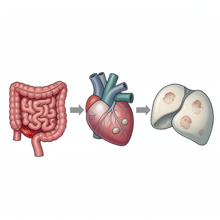
Pathogenesis Flowchart
AI-generated conceptual illustration (cattle heart muscle and liver). It explains the idea and does not show study data.
View source evidence ↗Figures & tables
From the original paper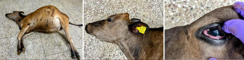 Figure 1
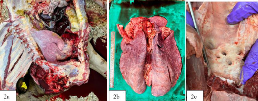 Figure 2
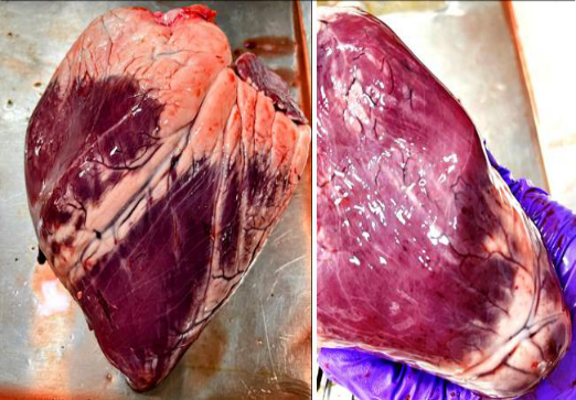 Figure 3
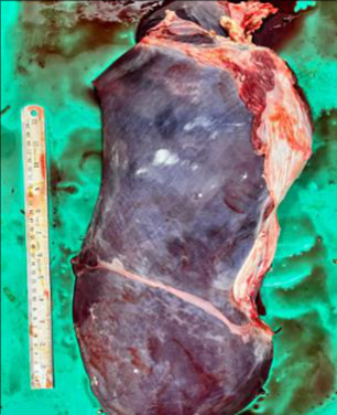 Figure 4
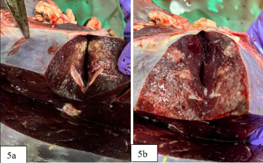 Figure 5A
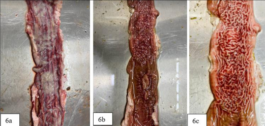 Figure 6
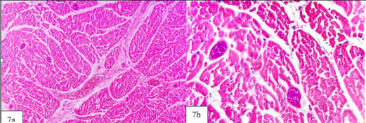 Figure 7
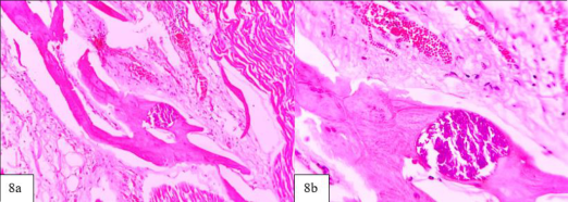 Figure 8
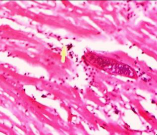 Figure 9
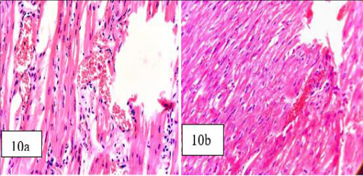 Figure 10
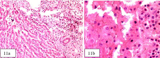 Figure 11
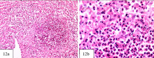 Figure 12
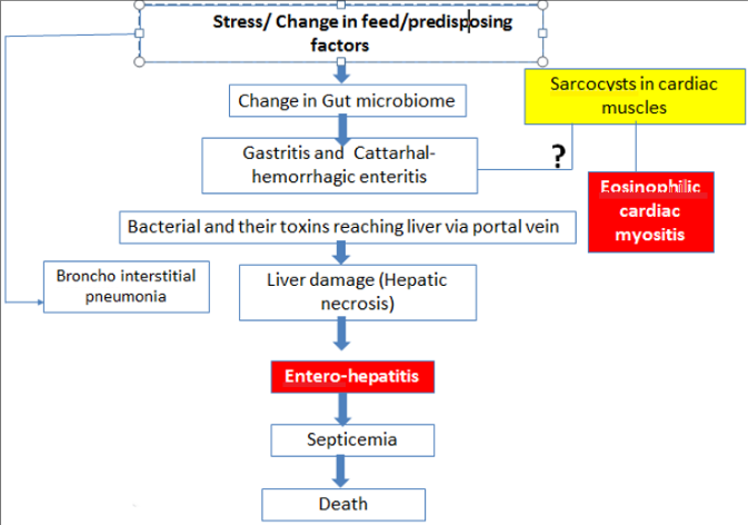 Figure 13
Introduction Background
Sarcocystis (Derived from the Greek word: Sarkos means flesh and Kystis means cyst), belongs to the family Sarcocystidae. Sarcocystis species are unicellular parasites that belong to cystogenic coccidia (Apicomplexa: Sarcocystidae). Members of this family are tissue cyst forming coccidia. They are heterogeneous parasites. The sexual stage is found in the final host and the asexual stage is found in intermediate hosts. During their life cycle, they require both an intermediate and a final host, the former usually a herbivorous and the latter a carnivorous vertebrate animal. Cattle are long-known intermediate hosts of S. Cruz, S. hirsuta and S. hominin, with canids, felids and humans as final hosts, respectively (Dubey and Lindsay, 2006) [1] . Sarcocystis cruzi is the most pathogenic and most prevalent of all bovine sarcocysts, with canids as its definitive hosts; it is not transmissible to humans (Dubey et al., 2016) [3] . Sarcocysts are found in virtually all striated muscles of the body including the tongue, esophagus and diaphragm as well as cardiac muscle and, to a lesser extent, smooth muscle. Sarcocystis is generally asymptomatic but can cause mild gastro-intenstinal signs such as diarrhea in cattle. Sarcocystis species infections are quite prevalent in farm animals (higher than 70% in some herbivore populations); however, there have been few outbreaks of clinical disease. Most animals are asymptomatic, and the tissue cysts (chronic stage) are discovered only at slaughter. Sarcocystis species infections are probably most important in growing ruminants in which they can result in subclinical anemia and reduced weight gain. Clinical signs such as Anorexia and fever are also encountered in some cases. Cases of necrotic encephalitis and fatal myocarditis in heifers have also been reported (Dubey et al., 2016) [3] .
Pathologic changes in myocardium and skeletal muscles were more pronounced in cows with lymphatic leukemia (Dubey et al., 2015) [2] .
Case presentation
One-year-old heifer of Vrindavani breed from Cattle & Buffalo farm under Livestock Production and Management division, IVRI was presented for post mortem with a brief history of diarrhea, anorexia and ruminal tympany, lack of response to treatment and a 3-day history of anorexia. The cow was in poor general health and carcass condition was bloated (Fig. 1). Rigor Mortis was present in both hind limbs and forelimbs and the conjuctival mucus membrane was slightly pale Fig. 1).
Results
Gross findings: There were numerous haemorrhages on the surface of the viscera (Fig. 2a). Moderate congestion and uncollapsed pulmonary lobes with thickened interlobular septa were observed (Fig. 2b) Diffused petechial haemorrhages over tracheal and bronchial mucosa were observed (Fig. 2c). Gross examination of heart revealed congested myocardium with multifocal petechiae at the apex, interventricular groove and on epicardial fat deposit (Fig. 3). Other prominent gross findings were enlarged liver with mild congestion seen on cut section and thickened hepatic capsule (Fig. 4) and cut section of liver showing congested parenchyma with pale grey areas of necrosis (Fig. 5a andb). Intestinal segments filled with watery blood-tinged diarrhoeic contents. A segment of colon with congested, corrugated mucosa with mucoid exudate (Fig. 6a) and severe transverse corrugations of intestinal mucosa of jejunum with multifocal irregular to elliptical raised mucosal thickenings were observed (Fig. 6b &c). Microscopic findings: Tissue sections from heart were evident of multifocal sarcocystic schizonts within cardiac muscle fascicles with congested blood vessels (Fig. 7a &b).
Micrograph through the muscle of a carcass affected with eosinophilic myositis with diffuse leukocytic infiltration and the S. cruzi, like sarcocysts (Fig. 9). Sarcocysts were also observed in purkinje fibres (Fig. 8) and endocardium. Myocardial necrosis and cardiac muscle degeneration were evident. Myositis and myocarditits were prominent microscopic findings in heart associated with sarcocysts. Elongated Sarcocyst containing bradyzoits in striated cardiac muscles and liberation of bradyzoites from cysts were observed (Fig. 9). Heart sections showing congested vessels and eosinophilic fibrinous substance around the blood vessels (Fig. 10). Focal area of necrosis infiltrated with numerous PMNLs in liver (Fig. 11 & 12) along with dilated sinusoids infiltrated with neutrophils and cells with intense eosinophilic cytoplasm (Fig. 11 &12) leading to hepatitis.
Discussion
Stress conditions or change in feed as possible predisposing factors leading to catarrhal enteritis and parasitic infestation of sarcocyst in cardiac muscles leading to eosinophilic myositis and cardiac muscle inflammation (Fig. 7, 9 & 10). Hepatic necrosis was evident in gross and histopathological findings along with enteritis causing septicemia and possibly leading to death. Eosinophilic myositis with diffused lymphocytic infiltration associated with Sarcocystis cruzi in muscles was reported by Gajadhar et al., (1992) [5] . Sarcocysts were observed in Purkinje fibres by Fayer (2004) [4] . Sarcocyst infestation in muscles causing widespread myositis and inflammation of cardiac muscle was reported by Fayer (2004) [4] .
Conclusions
Multiple possible causes leading to death (Fig. 13) with cardiac myositis particularly due to multiple sarcocysts in myocardium and hepatic necrosis were confirmed by histopathological examination. Chronic condition of Sarcocystis is diagnosed as multiple tissue cysts were evident in cardiac muscles during microscopic examination.
Acknowledgement
AcknowledgementsThe authors are thankful to the Head, Division of Pathology, Indian Veterinary Research Institute for providing the necessary facilities for this work and also gratitude for his whole-hearted encouragement, advice, expert suggestions, support and inspiration from all corners in completing this case report and for providing opportunity, scope and requisite administrative facilities for carrying out this work.
Figures and tables from the paper
References
1 in the original paper- Dubey JP, Lindsay DS. Neosporosis, toxoplasmosis, and sarcocystiosis in ruminants. Vet Clin North Am Food Anim Pract. 2006;22:645-71.