
Research article · 2023 · Full text
International Journal of Agriculture and NutritionSevere acute respiratory syndrome coronavirus two (Sars-Cov-2) review
Volume 5(1) · Pages 99–113 · DOI: 10.33545/26646064.2023.v5.i1b.105 ↗
Research areasSARS-CoV-2 and COVID-19 ResearchAnimal Virus Infections StudiesViral gastroenteritis research and epidemiology
Abstract
Author-writtenCovid-19 is a highly contagious zoonotic disease of humans and animals caused by SARS-CoV-2. The WHO has declared the enduring outbreak of COVID-19 is a global public health emergency and pandemic. SARS-CoV-2 is a spherical positive sense, single‐stranded RNA viruses that belong to beta coronavirus, subgenus sarbecovirus. The genomes of the virus are mainly composed of four types of structural proteins that are spike glycoprotein, envelope protein, nucleocapsid protein, and membrane protein. The genetic sequence of the SARS-CoV-2 showed more than 80% identity to SARS-CoV and 50% to the MERS-CoV. RaTG13 is the only BatCoV that shows as high as 96% full genome homology with SARS-CoV-2. The virus is suggested to be originated from bats and most closely relating to beta coronavirus of bat origin since it has 87.9% and more nucleotide similarity. The mutation is one of the driving factors for the evolution of the organisms. Community transmission, antiviral treatments, and ecological changes along with the genetic plasticity of RNA viruses facilitate the emergence of several new RNA viruses and the development of more virulent strains. The evolved strains may cause a high mortality rate and resistant to treatments. Therefore, systematic tracking of demographic, clinical patient information, and strain information is crucial to successfully combat COVID-19. Molecular methods, serology, and viral culture are used as diagnostic tools for isolation and identification of newly emerged viral disease. The RT-qPCR assay is regarded as the gold standard method for identification and surveillance of SARS-CoV-2 target sequences.
Summary
Source-checkedPlain-language reading of the full paper, prepared with AI and checked against the text. Each point links to the authors' own words.
Why it matters
- COVID-19 is a highly contagious zoonotic disease caused by SARS-CoV-2, which the World Health Organization has designated as a global pandemic and public health emergency. View evidence ↗
- The emergence of virulent RNA virus strains is driven by mutations, genetic plasticity, ecological changes, antiviral treatments, and community transmission. View evidence ↗
- Combating COVID-19 requires the systematic tracking of strain information as well as clinical and demographic patient data. View evidence ↗
The question
- The objective of this review is to systematically examine SARS-CoV-2, specifically focusing on its diagnosis, phylogenetic analysis, and genomic organization. View evidence ↗
What they did
- The review identifies viral culture, serology, and molecular methods as the primary diagnostic tools for identifying the virus. View evidence ↗
- The RT-qPCR assay is considered the gold standard for the surveillance and identification of SARS-CoV-2 target sequences. View evidence ↗
What they found
- Genomic comparisons indicate that SARS-CoV-2 has a 96% resemblance to the Rhinolophus affinis bat and an 80% resemblance to the Rhinolophus sinicus bat. View evidence ↗
- Pangolins show 91.02% or more genomic similarity to the virus, suggesting they may have served as an intermediate host. View evidence ↗
- Experimental studies show that cats and ferrets are susceptible to SARS-CoV-2, while pigs, ducks, and chickens are not. View evidence ↗
- The mortality rate for COVID-19 (3.4% to 6.6%) is lower than that of SARS-CoV (9.6%) and MERS-CoV (34.3%). View evidence ↗
- SARS-CoV-2 has a higher reproductive number (2.9) compared to SARS-CoV (1.77), indicating greater transmissibility. View evidence ↗
- As of September 30, 2020, Ethiopia had reported 74,584 confirmed COVID-19 cases and 1,191 deaths. View evidence ↗
- A developed IgM kit for SARS-CoV-2 has demonstrated a sensitivity of 86.6% and a specificity of 90.63%. View evidence ↗
What it means
- Because there are currently no effective vaccines or therapeutic agents, the authors recommend prioritizing surveillance, risk assessment, rapid diagnostics, and the investigation of intermediate hosts. View evidence ↗
- There is currently insufficient proof to precisely identify the exact source or the specific route of transmission from the animal reservoir to humans. View evidence ↗
Visual summary
1 visualsIllustrations explain the paper; they do not show study data.
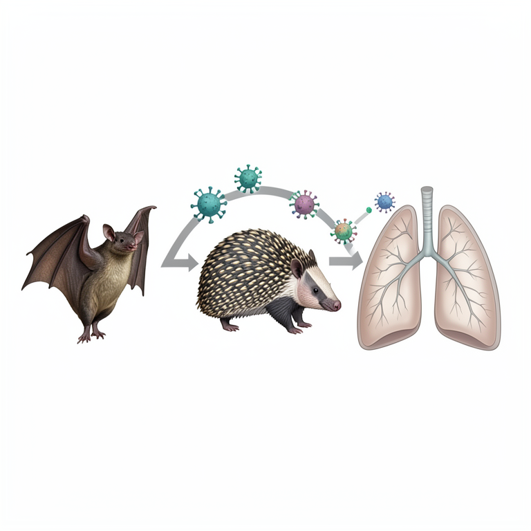
Zoonotic Origin Concept
AI-generated conceptual illustration (Horseshoe bats, pangolins, and human lungs). It explains the idea and does not show study data.
View source evidence ↗Figures & tables
From the original paper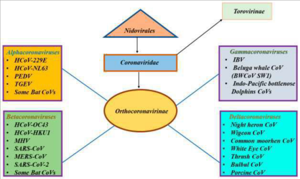 Figure 1
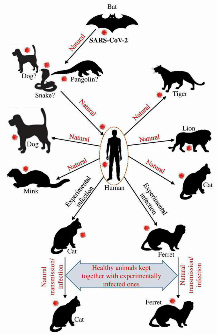 Figure 2
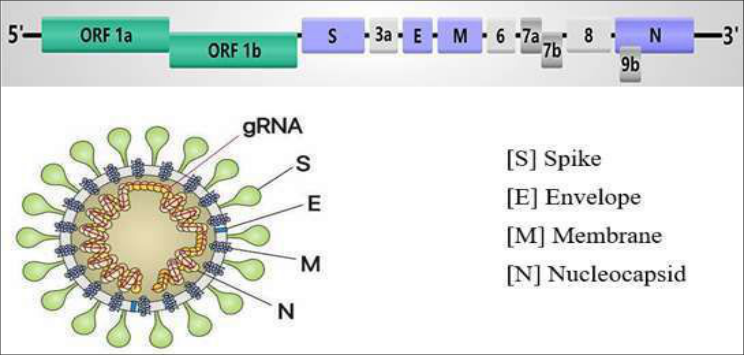 Figure 3
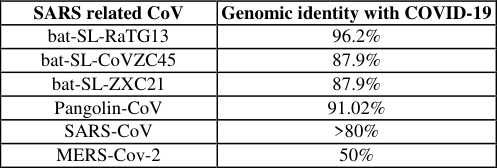 Table 1
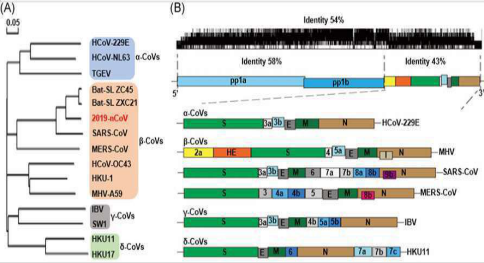 Figure 4
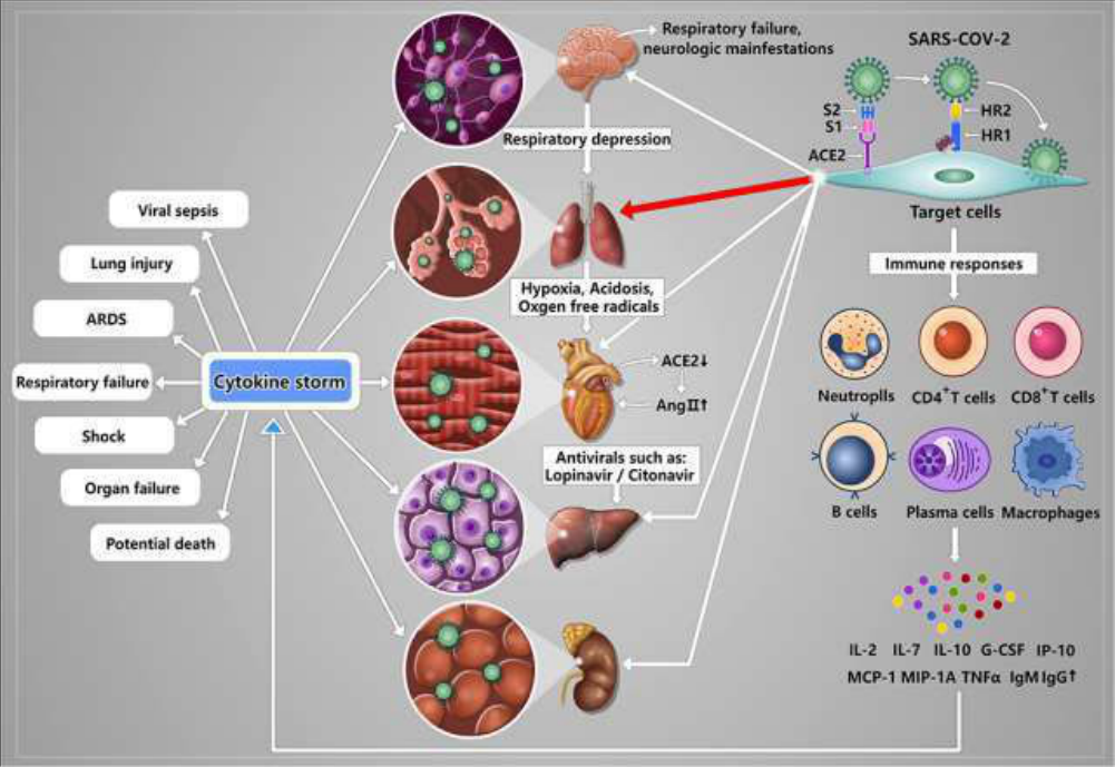 Figure 5
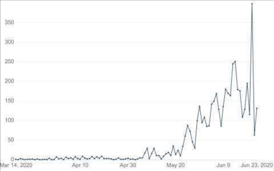 Figure 6
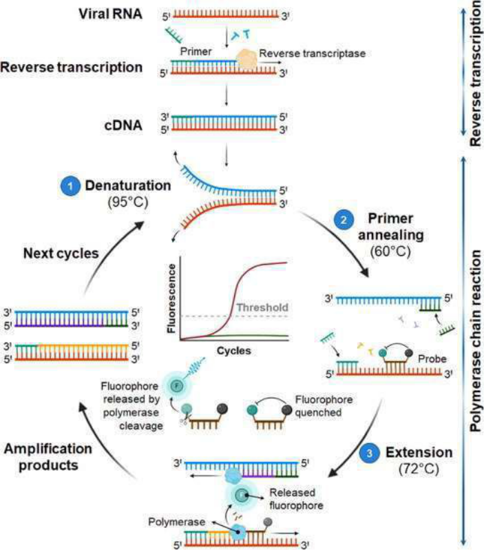 Figure 7
Introduction
Coronaviruses are zoonotic pathogens that infect both humans and animals (Sahin et al., 2020) [59] . Coronaviruses are known for their capacity to mutate quickly, change tissue tropism, traverse the species barrier, and adapt to a variety of epidemiological situations (Helmy et al, 2020) [32] . Since the 1960s, seven different kinds of human coronaviruses have been identified as respiratory pathogens. These are HCoV-229E, HCoV-NL63, HCoV-OC43, KHU1, Middle East Respiratory Syndrome Coronavirus (MERS-CoV), Severe Acute Respiratory Syndrome Coronavirus (SARS-CoV), and Severe Acute Respiratory Syndrome Coronavirus 2 (SARS-CoV-2). MERS-CoV, SARS-CoV, and SARS-CoV-2 belong to the genus Betacoronavirus (Ortiz-prado et al., 2020) [52] . Among these human coronaviruses, four of them cause mild illness which is similar to the common cold and gastrointestinal tract infection. Whereas severe acute respiratory syndrome coronavirus (SARS-CoV), Middle East respiratory syndrome coronavirus (MERS-CoV), and severe acute respiratory syndrome coronavirus (SARS-CoV-2) are highly pathogenic and have significant public health concerns due to their zoonotic emergence and crossing of the species barrier (Wu et al., 2020) [69] . Coronaviruses are enveloped single-strand RNA viruses that can infect a wide range of hosts including avian, wild, domestic mammalian species, and humans. The SARS-CoV-2 virus causes Coronavirus Disease 2019 (COVID-19), a newly emerging epidemic pathogen of humans and animals (Mason, 2020) [47] . Humans suffer a severe acute respiratory infection caused by the virus, which can be spreed from person to person. (Tang et al, 2020) [70] . SARS-CoV-2 was first detected in Wuhan, China, causing outbreaks of the coronavirus disease COVID-19 that has now spread globally (Jaimes, et al., 2020) [35] . The World Health Organization (WHO) temporarily named the newly emerged pathogenic virus like the 2019 novel coronavirus (2019-nCoV (Tahir et al., 2020) [63] .
International Committee of Taxonomy of Viruses (ICTV) announced "severe acute respiratory syndrome coronavirus 2 (SARS-CoV-2)" as the name of the new virus on 11 February 2020 based on phylogeny, taxonomy, and established practice (Zu et al., 2019b) [75] . The virus was given this name because it is genetically linked to the coronavirus that responsibl the SARS pandemic in 2003 (Li et al., 2020) [45] . COVID-19 was announced a Public Health Emergency of International Concern by the World Health Organization on January 31, 2020. (PHEIC). The WHO declared the COVID-19 outbreak a pandemic on March 11, 2020, since the virus is spreading globally (Ramphul and Mejias, 2020) [56] . The emergence of SARS-CoV-2 has been marked as the third introduction of a highly pathogenic coronavirus into the human population after the severe acute respiratory syndrome coronavirus (SARS-CoV) and the Middle East respiratory syndrome coronavirus (MERS-CoV) in the twenty-first century (Li et al., 2020) [45] . The newly emerged coronavirus is mainly caused respiratory and gastrointestinal tract infections which made similar to highly pathogenic human coronaviruses. Droplets and close contact are the most common routes of transmission of SARS-CoV-2 generated during coughing and sneezing by symptomatic and asymptomatic patients (Singhal, 2020) [62] . Source of infection, route of transmission, and susceptibility were reported as important factors in the viral spreading mechanism. Bats are considered to be the natural host of the virus and serve as the sources of infection. Pangolins, snakes, and other animals are thought to be intermediate hosts (Wang et al., 2020) [67] . SARS-CoV-2 forms a different lineage with Bats SARS-like CoV that is belonging to the Coronaviridae family, Betacoronavirus genus, and Sarbecovirus subgenus based on phylogenetic analysis. It shares 96.3%, 89%, and 82% nucleotide resemblance with bat CoV RaTG13, SARS-like CoV ZXC21, and SARS-CoV respectively. This genomic resemblance of the virus confirms its zoonotic origin (Helmy et al., 2020) [32] . Viral infections are the most frequent infectious diseases and are common triggers for constituting major biological, clinical, and socioeconomic problems worldwide. SARS-CoV-2, SARS-CoV, and MERS-CoV are the major global outbreaks infections with alarming health and economic concerns in their respective periods (Meo et al., 2020) [49] . The first outbreak of SARS-CoV occurred in Saudi Arabia in 2002, the second MERS-CoV in Asia in 2012, and followed by the novel coronavirus 2019 in Wuhan, China which has become the third coronavirus to appear in the human population and threatened the entire world (Helmy et al., 2020) [32] . These coronaviruses have zoonotic importance and infect different species of wild and domestic animals such as camels, cattle, cats, and bats. The viruses are transmitted from animal to animal, animal to human, and human to human. MERS-CoV transmission originated from bats to camels and then to humans. Genetic sequence data suggested that the SARS-CoV-2 virus originated from an animal source, but currently, there is no sufficient proof to identify exactly either the source or the route of transmission from the original animal reservoir to a proposed intermediate host and then to humans (European Commission, 2020) [79] . But bats are considered as the possible origin of the newly emerged SARS-CoV-2 virus. Ones COVID-19 disease transmitted in either source to human in turn human to human transmission has been confirmed (Meo et al., 2020) [49] . All the sequences from United States patients are similar to the one that China initially posted which suggesting that the likely origin of the novel coronavirus is from the animal reservoir. This is, therefore; the objective of this seminar is to systematically review newly emerged SARS-CoV-2 via focusing on genomic organization, phylogenic analysis, diagnosis, and other features based on recent research progress.
History and origin of Sars-COV-2
China notified the outbreak of COVID-19 disease to the World Health Organization on December 31 st , 2019. China shuttered the Huanan seafood market on January 1st. The virus is originated from the Huanan seafood market since environmental samples taken from there show positive results (Singhal, 2020) [62] . The massive migration of Chinese during the Chinese New Year increases the epidemic. Cases of COVID-19 in countries outside china were reported with no history of travel to china suggesting that the occurrence of human to human transmission. SARS-CoV-2 has infected several thousands of people and has caused many fatal cases. The cases of the virus are increasing exponentially across the world. Determining the origin and evolution of 2019-nCoV is important for the surveillance, drug and vaccine discovery, and prevention of the epidemic (Zhang et al., 2020) [71] . The zoonotic source of SARS-CoV-2 is not confirmed, however, the sequence-based analysis suggested bats are as the main reservoir (Hafeez et al., 2020) [29] . The studies showed that SARS-CoV-2 has an 80% resemblance to Rhinolophus sinicus bat and 96% resemblance with the Rhinolophus affinis bat based on the genomic comparison. Based on this evidence SARS-CoV-2 originated from bats. The novel coronavirus had 91.02% or more genomic similarity with pangolins as different findings indicated and suggested that the animal may be an intermediate host of the virus (Ramphul and Mejias, 2020) [56] . Generally, research evidence suggests that SARS-CoV-2, MERS-CoV, and the original SARS-CoV all originated from bats. SARS-CoV and MERS-CoV then spread from infected civets and dromedary camels to people respectively. Scientists are trying to determine how SARS-CoV-2 spread to people (Tahir et al., 2020) [63] . Extensive recombination among bat coronaviruses and strong purifying selection pressure among viruses from humans, bats, and pangolin may allow jumping between new hosts (Akram and Mannan, 2020a) [4] .
Classification of sars-cov-2
The genus Coronaviruses are the largest group of viruses belongs to the order Nidovirales in the Coronaviridae family (Sahin et al., 2020) [59] . The family Coronaviridae contains two subfamilies: Orthocoronavirinae and Torovirinae. The subfamily Orthocoronavirinae is further classifiedon basis of phylogenic analysis and genomic struct ure into four genera: alphacoronavirus, Betacoronavirus, ga mma coronavirus, and delta coronavirus (Ashour et al, 2020) [7] . Alpha and beta coronaviruses circulate in mammals and bats. Gamma coronaviruses mostly infect avian species and a few mammalian species, whereas delta coronaviruses infect birds and mammals. SARS-CoV-2 represents a distinct lineage in the subgenus Sarbecovirus. Corona in Latin means crown and gets its name due to the presence of spike projection from an enveloped virus. Nido refers to the ability of the order to make a nested set of subgenomic mRNA (Helmy et al., 2020) [32] .
Host diversity
There is a limited study on the investigation of SARS-CoV-2 in animals. SARS-CoV-2 can distinguish the host cells receptor ACE2 of pigs, ferrets, cats, and monkeys in similar efficiency with humans as different studies confirmed. Cats and ferrets are well susceptible to SARS-CoV-2 in an experimental study (Hossain et al., 2020) [33] . Identifying the host range of SARS-CoV-2 is important since some domestic species might harbor the virus and transmit it back to humans (Li et al., 2020) [45] . Cats, dogs, ferrets, lions, tigers, and other animals can contract the virus from a different source. Cat, dog, and ferrets are susceptible to SARS-CoV-2 according to natural infections history and experimental results. Whereas pigs, chickens, and ducks are not susceptible to the virus (Hossain et al., 2020) [33] . The susceptibility of the host to coronavirus infection is depending on the affinity between the viral receptor-binding domain and host ACE2 in the initial attachment step. The spike protein receptor-binding domain (RBD) is the critical determinant of viral tropism and infectivity. Infected cats and ferrets may transmit SARS-CoV-2 into the healthy groups lived together through direct or indirect contact and/or airborne routes. There is no current evidence of transmission from cats to humans. ACE2 molecules responsible for the binding of the SARS-CoV-2 spike are almost identical in pigs, ferrets, cats, orangutans, monkeys, and humans (Wan et al., 2020) [66] . Phylogenetic clustering and sequence alignment study showed that ACE2 of several domestic animals (cat, cow, buffalo, goat, sheep, and pigeon) could be used by SARS-CoV-2 (Qiu et al., 2020) [55] . Therefore, a wide range of domestic and wild animals may support the entry of the virus to humans.
Molecular characterization of sar-CoV-2
The genomic sequence of SARS-CoV-2 is almost identical to more than 99.9% sequence identity among patients. The length of the genome is 29, 891bases. It has six major open reading frames and encodes accessory proteins (Artika et al., 2020) [6] . Some of the SARS-CoV-2 has less than 80% nucleotide sequence identity to the analogous genes of SARS-CoV. However, there is 94.4% sequence identity between SARS-CoV-2 and SARS-CoV when compared with the amino acid sequence of conserved replaces domain in ORF1ab. Therefore, SARS-CoV-2 and SARS-CoV are considered as the same species which are SARS-related coronavirus as different findings indicated (Zhou et al., 2020) [74] .
Genetic structure of the SARS-CoV-2
SARS-CoV-2 are enveloped icosahedral, spherical, positive sense, single-stranded RNA viruses with non-segmented genome that are characterized by spike proteins projecting from the virion surface (Helmy et al., 2020) [32] . The name coronavirus is given due to the appearance of the virus as a royal crown under an electron microscope. Spike glycoprotein on the envelope leads to the formation of a royal crown shape. The viral structure formed primarily from four structural proteins such as spike (S), membrane (M), envelope (E), matrix, and nucleocapsid (N) proteins (Ashour et al., 2020) [7] . Nucleocapsid protein holds the RNA genome. The remaining protein S, M, and E together create a viral envelope (Akram and Mannan, 2020b) [5] . COVID-19 differs from other coronaviruses by encoding an additional glycoprotein that has acetyl esterase and Hemagglutination (HE) properties (Kannan et al, 2020) [37] . HE found on the surface of some beta coronavirus that binds to sialic acid on host cell surface glycoprotein and possesses acetyl-esterase activity. It enhances the entry and pathogenesis of coronavirus (Ashour et al., 2020) [7] . ) [6] . The nucleocapsid (N) protein of novel coronavirus has almost 90% amino acid sequence identity with SARS-CoV (Kannan et al., 2020) [37] . N protein contains two noninteracting structural domains that are the N-terminal RNAbinding domain (RBD) and the C-terminal dimerization domain (DD), surrounded by flexible linkers. The Cterminal domain presents completely as a dimer in solution.
The flexible linkers are essentially interaction sites with other protein and protein-RNA partners (Wan et al., 2020) [66] .
The membrane protein is found in higher quantities than other proteins. It has a critical role in virion assembly via M-M, M-S, and M-N protein interactions. The M proteins have the same basic structure as other coronaviruses even though their amino acid contents are different (Artika et al., 2020) [6] . M protein has a triple-helix bundle which forms a single three-transmembrane domain(TM). It has also a short aminoterminal domain outside of the membrane and a long carboxy terminal domain inside the membrane (Thomas, 2019) [64] . A mutation may influence host cell attachment and entry of the virus when M protein cooperates with spike protein.
Spike surface glycoprotein (S)
Spike protein is critical for SARS-CoV-2 infection. Glycosylated spike protein coat the surface of SARS-CoV-2 and bind to host cell receptor ACE2 which is mediating the entry of the virus into the host cell. S protein consists of an extracellular N-terminus, a transmembrane (TM) domain attached in the viral membrane, and a short intracellular Cterminal segment (Berend et al., 2007) [9] . Transmembrane protease serine two (TMPSS2) is located on the host cell membrane which promotes entry of the virus when it binds to the spike surface glycoprotein of the virus. The SARS-CoV-2 S protein is highly conserved among all human coronaviruses (HCoVs) and is involved in receptor recognition, viral attachment, and entry into host cells (Huang et al., 2020) [34] . The Spike protein consists of receptor-binding S1 and membrane-fusion S2 subunits, which is responsible for attachment to the host cell receptor and fusion with the cell membrane (Wu et al., 2020) [69] . Therefore, it is one of the most important targets for the SARS-CoV-2 vaccine and therapeutic developments.
The spike surface glycoprotein and its RBD in the SARS-CoV-2 virus is a principal determinant of the host range, tissue tropism, and pathogenicity. It is the major antigens that stimulate neutralizing antibody and important targets of cytotoxic lymphocytes. Spike protein(S gene) is highly important for molecular characterization of SARS-CoV-2 (Artika et al., 2020) [6] .
Envelope protein (E)
The envelope protein is present in a small amount within the virion that determines the shape of the virus jointly with membrane protein (Schoeman and Fielding, 2019) [60] . It is highly divergent which has N-terminal ectodomain and Cterminal endodomain. Envelope protein plays a significant role during the replication of coronavirus which facilitating assembly and release, budding, envelope formation, and pathogenesis aspects (Fehr and Perlman, 2015) [26] . The primary and secondary structure of envelope proteins indicates that as it has short hydrophobic N-terminus of 7-12 amino acids. The envelope protein of SARS-CoV-2 has long amino acid (76 aa) and possesses three critical domains. They are N-terminus, transmembrane (25 amino acid), and C-terminus. The long amino terminus has hydrophilic ends. The C-terminus domain of novel corona virus is essential for the establishment and maintenance of epithelial polarity in mammals (Hassan et al., 2020) [31] .
Genomic organization of SARS-COV-2
Coronaviruses are viruses whose genome structure is best known among all RNA viruses. There are four major structural proteins encoded by the corona viral genome on the envelope (Zu et al., 2019b) [75] . The structural proteins are spike (S), envelope (E), membrane (M), and nucleocapsid (N). The spike protein allows the virus to bind to the host's cell membrane (Ramphul and Mejias, 2020) [56] . The typical features of the genus coronavirus are a highly conserved genomic organization with 27-32kb positive polarity (Sahin et al., 2020) [59] . A SARS-CoV-2 virion is a monopartite, an enveloped, positive-sense, single-stranded RNA virus with a genome size of 29,903 nucleotides (Helmy et al., 2020) [31] . SARS-CoV-2 has a similar genomic organization to other beta-coronaviruses. The viral genome contains two untranslated regions (UTRs) that are 5'-untranslated region and 3'untranslated region or 3' poly (A) tail. It also contains 11
open reading frames (ORF) that encodes 27 proteins. The ORF1ab and ORF1a genes are located at the 5'-terminus of the genome that encodes the PP1ab and pp1a proteins respectively. The 3'-terminus of the genome holds four structural proteins and eight accessory proteins. The UTR acts as mRNA for the translation of Replicase polyproteins (Wu et al., 2020) [6] .
The two large open reading frame genes (ORF1ab and ORF1a) or replicase gene contain two-third of the genomes (20 kb) which encodes 16 non-structural proteins.
The remaining one-third of viral genomes comprise a structural gene unit that encodes four structural proteins(S, E, M, and N) and eight accessory proteins. Eight accessory proteins are orf3a, orf3b, orf6, orf7a, orf7b, orf8, orf14, and orf10) (Helmy et al., 2020) [32] . The structural and accessory proteins contain only 10kb of the viral genome. The 5' cap of the genome contains a leader sequence and untranslated region (UTR) that contains multiple stem-loop structures required for RNA replication and transcription (Fehr and Perlman, 2015) [26] . The 3′UTR also contains RNA structures required for replication and synthesis of viral RNA. Some accessory proteins have a significant role in viral pathogenesis. But it is nonessential for replication in tissue culture. Nonstructural proteins are essential for virus replication and pathogenesis. The four structural proteins are important for virus subtyping and response to vaccines.
Phylogenetic Analysis
Phylogenetic analysis is a technique to elucidate the evolutionary narration and relationships among a group of organisms. It is usually demonstrated in the form of a phylogenic tree to decide the evolutionary relationship between species clearly (Dayu et al., 2020) [17] . Singlestranded RNA viruses demonstrate a faster biological mutation rate due to the lack of proofreading activity of viral RNA polymerases (Elena and Sanjua, 2005) [25] . Phylogenetic analyses of coronaviruses have confirmed that the immediate ancestor of SARS-CoV-2 is most likely originated in a bat species. Though, whether SARS-CoV-2 or the progenitor of this virus is transmitted directly to humans or via an intermediate host is not yet determined. Deep comparative genomic analysis, evolutionary understanding, and structural analysis of ACE2 play a significant role to identify candidate intermediate host species and species at risk for SARS-CoV-2 (Damas et al., 2020) [16] . SARS-CoV-2 is a beta coronavirus, subgenus Sarbecovirus, lineage B, and genetically related to SARS related coronaviruses found in Chinese horseshoe bats (Chan et al., 2020) [11] . The genome sequence of COVID-19 isolates showed that 99.98 and 99.9% sequence identity among patients as different studies reported. The genetic sequence of the SARS-CoV-2 showed more than 80% identity to SARS-CoV and 50% to the MERS-CoV (Rothan and Byrareddy, 2020) [57] . MERS-CoV and human CoV HKU1 are very distant from SARS-CoV-2 (Wu et al., 2020) [69] .
Based on the genetic sequence identity and the phylogenetic reports, SARS-CoV-2 is sufficiently different from SARS-CoV and it can be considered as a new beta coronavirus that infects humans. The amino acid sequence of the virus differs from other coronaviruses specifically in the regions of ORF1ab polyprotein and surface glycoprotein (Kannan et al., 2020) [37] .
Discovering of the possible intermediate hosts of SARS-CoV-2 has a significant role in blocking of its interspecies transmission. The full-length genome of SARS-CoV-2 shows that as there is a high similarity to the genome of bat coronavirus, detected in Rhinolophus affinis (horseshoe bats). It is 96.2% sequence identity with bat coronavirus (BatCoVRaTG13). RaTG13 is the only BatCoV that shows as high as 96% full genome homology with SARS-CoV-2.
The phylogenic analysis of the spike gene and RdRp gene also shows RaTG13 is the first closest relative of the SARS-CoV-2 and they form a distinct lineage from the other SARS-related CoVs. It is also 98.7% nucleotide similarity to the partial RdRp gene of the bat coronavirus strain BtCoV/4991 and 87.9% nucleotide similarity to bat coronavirus strain bat-SL CoVZC45 and bat-SL-CoVZXC21 (Lai et al., 2020) [42] . These two bats SARS-like CoV are the second closest virus from bats to SARS-CoV-2. Therefore, based on sequence genome and phylogenic analysis, SARS-CoV-2 is suggested to be originated from bats and most closely relating to Betacoronavirus of bat origin (Zhou et al., 2020; Zhang and Holmes, 2020) [74,73] .
The phylogenetic analysis indicates that the SARS-CoV-2 belongs to subgenus Sarbecovirus, genus Betacoronavirus, and family Coronaviridae, which includes SARS-CoV and MERS-CoV (Dayu et al., 2020) [17] . Pangolin-CoV isolated from Malayan pangolins demonstrates that 91.02% genomic distinctiveness to SARS-CoV-2 and 90.55% identity to the BatCoV RaTG13 (Zhang et al., 2020) [71] based on research findings. The pangolin coronavirus spike protein sequences shared five key amino acids in the RBD with that of SARS-CoV-2 whereas RaTG13 shared only one key amino acids in its RBD. And only SARS-CoV-2 has a G-C rich polybasic furin cleavage site at the S1/S2 junction in the spike protein. Polybasic cleavage site (RRAR) is the second distinct characteristic of SARS-CoV2 located at the junction of S1 and S2 subunit of the spike protein. RaTG13 and pangolin coronaviruses do not have G-C rich cleavage site (Lin et al., 2020) [46] . Pangolin-CoV may be the second closest relative of SARS-CoV-2 next to RaTG13. The subunit of the S1 protein of Pangolin-CoV more similar to SARS-CoV-2 than RaTG13 (Artika et al., 2020) [6] . Generally, Pangolins may be considered as possible hosts of SARS-CoV-2 and play critical roles in the emergence of novel coronavirus since they have many similar identities to novel coronavirus as different findings reported Snakes, birds, and other small mammals also suggested being the origin of the newly emerged novel coronavirus since there is no specific animal associated with SARS-CoV-2 identified (Prompetchara et al., 2020) [78] . The existence of a higher degree of homology of the ACE2 receptor from a diversity of animal species is indicating that animal species can be a possible intermediate host or animal model for COVID-19 infections (Rothan and Byrareddy, 2020) [57] . The S proteins of SARS-CoV-2 and SARS-CoV have an amino-acid sequence identity of around 77% as studies reported. The Spike gene of SARS-CoV-2 and RaTG13 is longer than SARS-related coronavirus. It has three short insertions in the N terminal domain and changes in four out of five key residues in receptor binding motif (RBM) compared with the S gene sequences of SARS-CoV (Wu et al., 2020) [69] .
Fig 4:
The genomic structure and phylogenetic tree of coronaviruses. B, The genome structure of four genera of coronaviruses. Pp1a and pp1b represent the two long polypeptides that are processed into 16 non-structural proteins. HKU, coronaviruses identified by Hong Kong University; HCoV, human coronavirus; IBV, infectious bronchitis virus; MHV, murine hepatitis virus; TGEV, transmissible gastroenteritis virus (Mousavizadeh and Ghasemi, 2020) [50] Table 1: Genomic comparison of SARS-CoV-2 with other CoV strains as a summary
| SARS related CoV | Genomic identity with COVID-19 |
|---|---|
| bat-SL-RaTG13 | 96.2% |
| bat-SL-CoVZC45 | 87.9% |
| bat-SL-ZXC21 | 87.9% |
| Pangolin-CoV | 91.02% |
| SARS-CoV | >80% |
| MERS-Cov-2 | 50% |
Table text extracted from the original PDF; superscript letters are shown inline. The original table image is below.
View original table image
Genetic diversity and evaluation of sars-cov-2
Evolutionary changes in an organism occur as a result of mutations. The mutation is a modification of nucleotide sequences in the genome of organisms. Community transmission, antiviral treatments, and ecological changes along with the genetic plasticity of RNA viruses facilitate the emergence of many new RNA viruses. These results developments of new viral pathogens that affect potential hosts and stimulate host jumping (Kaur et al, 2020) [51] . In addition to the emergence of new pathogens, mutation of the viruses can result in the development of more virulent strains with a high mortality rate and appearance of resistant strains to treatment. Therefore, systematic tracking of demographic, clinical patient information, and strain information is crucial to successfully combat COVID-19 (Koyama and Parida, 2020) [38] . Mutations at S1 and S2 junction of the spike protein of SARS-CoV-2 are also responsible for evolutionary changes. RNA viruses usually have a relatively high mutation rate than DNA viruses and a million times greater than their hosts. This is due to the absence of proofreading RNA polymerase enzymes in RNA viruses. The advantages of high mutation rates are for better viral adaptability and evolvability of RNA viruses (Duffy, 2018) [22] . On the other hand, Coronaviruses which have the largest known genome size show relatively low rates of mutations than other RNA viruses. The slower mutations rates or conservation of genome associated with especial characteristics of the coronavirus replication-transcription complex which contains 3'-5'exonuclease activity. The exonuclease enzymes provide proofreading functions to the viruses (Chen et al., 2020) [12] . Annotation of the genome is the first step to explain the genetic dissimilarity of the organism since genetic information of any life is preserved in the genome (Junejo et al., 2020) [36] . Genetic variation can be assessed by retrieving complete or nearly complete genomes of SARS-CoV-2 from GISAID. All SARS-CoV-2, SARS-CoV, and MERS-CoV have high mutation rates that result in viral genetic diversity, flexibility, and adaptability to invade a wide range of hosts. SARS-CoV-2 genome has 80% similarity to the previous human coronavirus (SARS-like bat CoV) as different studies reported (Adnan et al., 2020) [3] . Nucleotide substitution is one of the driving forces for the occurrence of viral evolution in nature. The rapid spread of SARS-CoV-2 raises stimulating questions such as whether its evaluation is driven by mutation (Phan, 2020) [54] . Open reading frame 1ab, spike surface glycoprotein, matrix, and nucleoside protein are considered as a site of mutation.
The spike surface glycoprotein plays an essential role in binding to receptors on the host cell, which determines host tropism and major targets of neutralizing antibodies (Fei et al., 2020) [27] . Mutations in the spike surface glycoprotein might encourage its conformational changes, which probably led to the changing antigenicity (Phan, 2020) [5] .
Variants of SARS-COV-2
Different types of mutations such as missense mutations, synonymous mutations, and deletion mutation in the non-coding regions are major causes for the occurrences of genetic variants. As different studies indicate that there are many variants of SAS-CoV-2 found across different parts of the world. Currently, there are six stains of SARS-Cov-2. These are L, G, V, S, GR, and GH (https://www.sciencedaily. com/releases/2020/08/200803105246.htm).
The D614G strain has occurred where the aspartic acid residue at position 614 is replaced by glycine. This is done at the carboxy-terminal end of the S1 domain of the SARS-CoV-2 spike protein. The strain was first found in Europe in January 2020. D614G stain is the most widespread strain across the world and mutated into GR and GH after a while. It has five sublineages which are located in B-cell epitopes. D614G may influence the effectiveness of the vaccine. The other major lineage/strain is the L84S lineage with two subclades which is frequently observed in the United States. D448del and G392D lineages are small and they are without any significant sub lineages (Koyama et al., 2020) [38] . Mutations in the receptor-binding domain (RBD) of the spike protein suggest that variants are unlikely to reduce binding affinity with ACE2 because that would decrease the fitness of the virus. The V367F and D364Y variants increase the structural stability of spike protein that facilitating efficient binding to the ACE2 receptor as reported in studies (Ou et al., 2020) [53] .
In general, the genomic analysis showed that the presence of a high rate of non-synonymous mutation which can result in non-conservative substitution. This is indirect evidence of the intensive circulation of the virus in the human population, increased transmissibility of the virus, and its adaptation process to a new host (Kozlovskaya et al., 2020) [40] .
Pathogenic mechanism of sars-cov-2
The pathogenesis of the virus is poorly understood. The pathogenic mechanisms of SARS-CoV and MERS-CoV can give us a lot of information on the pathogenesis of SARS-CoV-2 infection to facilitate our recognition of COVID-19 (Li et al., 2020) [45] . SARS-CoV-2 uses ACE2 as its receptor which is similar to SARS-CoV.
Acute Respiratory Distress Syndrome (ARDS)
ARDS is a life-threatening lung condition that prevents enough oxygen from getting to the lungs and into the circulation, accounting for mortality of most respiratory disorders and acute lung injury. Genetic susceptibility and inflammatory cytokines are closely related to the occurrence of ARDS as different studies reported previously. ACE2, interleukin 10 (IL-10), tumor necrosis factor (TNF), and vascular endothelial growth factor (VEGF) are associated with the development of ARDS (Wu et al., 2020) [69] .
Spectrum of clinical symptom
Clinical manifestations of SARS-CoV-2 have similarities with SARS-CoV and MERS-CoV infections. The clinical picture of the virus ranges from very mild to life-threatening (Chen et al., 2020) [12] . Most cases of the virus are mild as the report suggests in china. Common symptoms are fever, nonproductive cough, dyspnea, and fatigue. Extrapulmonary feature of the virus includes nausea, vomiting, diarrhea, and acute renal failure (Meo et al., 2020) [49] . Fever is the dominant symptom of infection. The other clinical findings are increase white blood cell counts (neutrophils), decrease in lymphocytes, platelets, and red blood cells. Pneumonia, metabolic acidosis, septic shock, and bleeding are recognized in severe cases of the disease (Helmy et al., 2020) [32] . The mortality rate of SARS-CoV-2 is lower compared to SARS and MERS coronavirus. The mortality rate of COVID-19 is 3.4% to 6.6% while SARS or MERS CoV mortality rate is 9.6% and 34.3% respectively.
Epidemiology
The first cases of the acute respiratory syndrome of unknown etiology were reported in Wuhan City, Hubei Province, China among people linked to a local seafood market on 29 December 2019. Most of the early cases had some sort of contact history with the original seafood market. A secondary source of infection was found to be human to human transmission via close contact with no history of exposure to wildlife or visiting Wuhan (Adhikari et al., 2020) [1] . The virus rapidly spread in china and other countries around the world (Zu et al., 2019a) [75] . The novel coronavirus is suggested to be originated from wild bats and belongs to beta coronavirus.
As different studies reported that the virus mainly occurred among old aged people with a median age of 59 years. The gender-based analysis demonstrates that the cases consisted mostly of men with a median age range of 50-65 years (Meo et al., 2020) [49] . SARS-CoV-2 has a greater level of transmissibility and pandemic risk than SARS-CoV. Since the reproductive number of (R) of SARS-CoV-2(2.9) which is higher than the reported effective reproduction number of SARS-CoV (1.77). The mean incubation period of the virus is 7 days, ranging from 2 to 14 days as Chinese health authorities stated (Adhikari et al., 2020) [1] .
There is seasonal variation patterns of 2019-nCoV, SARS-CoV, and MERS-CoV infections. MERS-CoV infection outbreak has occurred in the summer season. 2019-nCoV and SARS-CoV infection outbreak took place in the winter season in contrast to the outbreaks of MERS-CoV (Dean, 2020) [18] . The discharged droplets of the virus can spread until 1-2 meter, and remain viable on the surface for a day in a favorable atmospheric environment. The virus can be destroyed within a minute via common disinfectants like sodium hypochlorite and hydrogen peroxide (Singhal, 2020) [62] . COVID-19 disease spread across many geographical regions of the globe. By September 30, 2020, there were 33, 852, 048 confirmed cases and more than 1, 012, 743 deaths of people worldwide with the novel coronavirus. Every hour the number of cases and deaths is increasing across the world. The United States of America is recording the maximum number of positive cases and deaths. Italy, Spain, Germany, and France in Europe continue to be the most affected ones (Kumar et al., 2020) [41] .
Status of COVID-19 in Ethiopia
On February 14, 2020, COVID-19 was reported in Africa for the first time in Egypt. The first case of COVID-19 was reported in Ethiopia on March 13, 2020. A range of control strategies was implemented to compact the advance spread of newly emerged pandemic disease. The strategies were quarantine people for at least 14days coming from abroad, closure of schools, suspending public gathering, closures of charges, and mosques. A state of emergency was also declared and peoples were advised to avoid unnecessary travel. Although all of the above and other prevention and control measures are taken, the number of cases is not decreasing rather it increases exponentially day over a period of time (Baye, 2020) [8] . The number of case increase exponentially and high cases are reported in Addis Ababa. Ethiopia reported a total of 74,584 COVID-19 cases and 1,191 deaths on September 30, 2020 (Biadgilign and Yigzaw, 2020) [10] .
Source: https://rstmh.org/news-blog/news/covid-19-in-ethiopia-status-and-responses [62] . Researchers have also detected SARS-CoV-2 in samples of stool, gastrointestinal tract, saliva and urine, tears, and conjunctively secretion of patients with COVID-19. The cluster of infected family members and medical workers confirmed the presence of human to human transmission by droplets, contacts, and fomite (Zu et al., 2019a) [75] . A retrospective study of pregnant women with COVID-19 indicated that there is no possibility of intrauterine vertical transmission between mothers and infants during late pregnancy (Wang et al., 2020) [67] . And also it is not certain that the consumption of viruscontaminated foods will cause infection and transmission (Duan, 2020a) [20] .
Diagnosis of sars-cov-2
Rapid and accurate identification of pathogenic viruses plays a vital role in selecting appropriate treatments, saving people's lives, understanding epidemiology, suppression transmission, and preventing epidemics (Hafeez et al., 2020) [29] . A quick standard diagnostic test for the detection of infectious diseases (COVID-19) is important to prevent subsequent secondary spread (Shen et al., 2020) [61] .
Diagnosis of SARS-CoV-2 infection is based on a history of detail contact, travel, and precise laboratory test. Molecular methods, serology, and viral culture are used as diagnostic tools for isolation and identification of newly emerged viral disease. Real-time reverse transcription Polymerase chain reaction and isothermal nucleic acid amplification are the golden standard molecular method for the diagnosis of COVID-19 with high sensitivity and specificity (Sahin et al., 2020) [59] .
Specimens
The precise samples for the detection of the virus are taken from upper and lower respiratory tracts. Nasopharyngeal and oropharyngeal swab or wash in ambulatory patients are taken as upper respiratory specimens. Sputum, endotracheal aspirates, and bronchoalveolar lavage or deep tracheal aspirate from patients are considered as a lower respiratory specimen with more severe respiratory disease (East, 2020) [23] . There are significantly higher viral load and genome fraction in the lower respiratory tract than upper respiratory tract samples (Sahin et al., 2020) [59] . The virus may also be detected in the stool and blood as an additional clinical specimen. Specimens should be kept refrigerated at 4-8 °C and sent to the laboratory where they will be processed within 24-72 hrs of collection. The samples should be refrigerated at 70 °C if not send within this period until shipment to the reference laboratory (East and States, 2020) [23] .
Diagnosis based on clinical symptoms
Diagnosis of COVID-19 is mainly based on epidemiological history and clinical manifestations such as coughing, fever, dyspnea, and history of travel from an epidemiological area. However; diagnosis based on clinical symptoms is atypical. Therefore; the auxiliary examination is necessary for the diagnosis of COVID-19 (Li et al., 2020) [45] .
Cultural isolation and identification of SARS-CoV-2
Viral culture is a more time-consuming method compare to other methods. The virus can grow in Vero E6 cells and observe the cytopathic effect (Kozlovskaya et al., 2020) [40] . Cultures used in in vitro and in vivo antiviral treatment and vaccine evaluation trials. [11] . Routine confirmations of the cases are based on the recognition of unique sequences of the virus via nucleic acid amplification technique.
Transcribing and amplifying specific SARS-CoV-2 genomic sequences are the basic principles of RT-qPCR. RNA extraction should be done in biosafety level two cabinets. Heat treatment of samples before RNA extraction is not recommended. The viral RNA is first extracted from the biological specimen collected from the nasal or nasopharyngeal swabs and is purified. The purified RNA template is transformed into a cDNA (complementary DNA) by reverse transcriptase (an RNA-dependent DNA polymerase enzyme). The cDNA is subsequently amplified by the PCR (Afzal, 2020) [2] . Sequence-specific forward and reverse primers and labeled probes are used in RT-qPCR diagnostic techniques according to the target genes of interest.
The viral genes that are conserved or plentifully expressed genes such as N, S, E, nonstructural RdRp, and ORF1a/b genes are preferred targets for SARS-CoV-2 RT-qPCR assay. The full genome directly amplified from RNA extracted from specimens using gene-specific primers for open reading frame 1b and N to produce overlapping PCR products (Sah, 2020) [58] .
The positive result of RT-qPCR for the presence of the SARS-CoV-2 is further confirmed via partial or wholegenome sequencing of the virus. Sequencing can be done using the Illumina MiSeq sequence method (East et al., 2020) [23] . Complementary DNA is amplified in PCR in three steps as indicated in the figure (Afzal, 2020) [2] 12.5 Serological diagnosis Serological tests are rapid and simple alternatives for screening infected individuals. Serological methods do not directly detect the presence of the virus but instead detect evidence of viral infection at some time point in the past. Serologic methods detect and/or measure the amount of antibodies to SARS-CoV-2 or antigen present quantitively or qualitatively (Zhan et al., 2020) [71] . Serological surveys can aid in investigation of an ongoing outbreak and retrospective assessment of the attack rate or extent of an outbreak. There are various types of serological tests are present such as enzyme-linked immunosorbent assay (ELISA), indirect immune fluorescent test assay (IIFA), lateral flow immune assay, and neutralization tests (Ortizprado et al., 2020) [52] . A new kit is developed using SARS-CoV-2 IgM with 86.6% and 90.63% sensitivity and specificity respectively as studies indicated (Li et al., 2020) [45] .
There is no cross-protection between SARS-CoV and COVID-19. The Challenge of serological test/diagnosis is Cross-reactivity of COVID 19 to other coronaviruses. The N protein antibodies of SARS-CoV cross-react with COVID-19 but not provide cross-immunity (Kannan et al., 2020) [37] . The sensitivity of antibody detection is generally lower than the molecular method of diagnosis.
Treatments and prevention mechanisms of sars-cov-2
To date, there are no specific antiviral drugs or vaccines available against COVID-19 infection for potential therapy and prevention of human's infection (Adhikari et al.; Gennaro et al., 2020) [1] . General treatment of COVID-19 infected patients needs complete bed rest and supportive treatment, ensuring adequate calorie and water intake to reduce the risk of dehydration. Water electrolyte balance and homeostasis need to maintain along with monitoring of vital signs and oxygen saturation; keeping the respiratory tract unobstructed and inhaling oxygen in more severe cases (Hafeez et al., 2020) [29] . The effective option of antiviral therapy and vaccination is currently under evaluation and development (Lai et al., 2020) [42] . Infected patients will receive supportive care including oxygen therapy, fluid therapy, and antibiotics for treating secondary bacterial infections according to WHO guidelines management strategies (Dhama et al., 2020) [19] . In the case of hypoxia nasal catheter and mask oxygen should be immediately provided to the patients. The CDC recommends multiple steps to prevent the transmission and risk of SARS-CoV-2 (Ramphul and Mejias, 2020) [56] . Frequent hand washing lasting at least 20 seconds by using soap and water, use of hand sanitizers with at least 60% alcohol, refrain from touching eyes, nose, and mouth with unwashed hands, and wear a face mask is advised (Unhale et al., 2020) [65] . Maintain social distancing and avoid close contact with people who are coughing or sneezing (Hafeez et al., 2020) [29] . Early detection, diagnosis, treatment, quarantine, border closure, and screening at the airport are classical effective prevention and control measures to block human to human transmission as well as secondary infections among close contact and health care worker. The major challenges for the prevention and treatment of SARS-CoV-2 viral infection are due to the presence of many potential natural hosts, intermediate hosts, and final hosts. And also asymptomatically infected persons and patients in the incubation period or recovered from COVID-19 may pose serious challenges for disease prevention and control (Duan, 2020b) [21] . In general, epidemiologic investigations, risk assessment, surveillance, rapid diagnostic tests, and vigilance are required by all countries to combat the potential outbreak of SARS-CoV-2 (Dagur and Dhakar, 2020) [15] .
Conclusion and Recommendation
Conclusion and Recommendation
Figures and tables from the paper
References
69 in the original paper- Adhikari SP, Meng S, Wu Y, Mao Y, Ye R, Wang Q, et al. Epidemiology, causes, clinical manifestation and diagnosis, prevention, and control of coronavirus disease (COVID-19) during the early outbreak period: A scoping review. Infectious Diseases of Poverty. 2020;9(29):1-12.
- Afzal A. Molecular diagnostic technologies for COVID-19. Limitations and challenges. Journal of Advanced Research. 2020;3(4):1-11.
- Adnan M, Khan S, Kazmi A, Bashir N, Siddique R. COVID-19 infection: Origin, transmission, and characteristics of human coronaviruses. Journal of Advanced Research. 2020;1(24):91-98.
- Akram A, Mannan N. Molecular Structure, Pathogenesis and Virology of SARS-CoV-2 : A Review. Journal of Infection Disease. 2020b;7(2003):536-540.
- Artika IM, Kresno A, Wiyatno A. Molecular biology of coronaviruses: Current knowledge. Heliyon. 2020;6(6):e04743.
- Ashour HM, Elkhatib WF, Rahman M, Elshabrawy HA. Insights into the Recent 2019 Novel Coronavirus (SARS-CoV-2) in Light of Past Human Coronavirus Outbreaks. Pathogens. 2020;9(1):1-15.
- Baye K. COVID-19 prevention measures in Ethiopia Current realities and prospects. International Food Policy Research Institute, ESSP. 2020;5:14.
- Bosch BJ, Van der Zee R, Cornelis AM, de Haan, PJMR. The Coronavirus Spike Protein is a class I Virus Fusion Protein. Journal of Virology. 2007;77(16):88018811.
- Biadgilign S, Yigzaw M. COVID-19 in Ethiopia: current situation, missed opportunities, and the risk of health system disruptions. Pan African Medical Journal. 2020;35(2):1-2.
- Chan JFW, Yuan S, Kok KH3, To KKW, Chu H, Yang J, et al. A familial cluster of pneumonia associated with the 2019 novel coronavirus indicating person-to-person transmission: A study of a family cluster. The Lancet. 2020;395(10223):514-523.
- Chen D, Xu W, Lei Z, Huang Z, Liu J, Gao Z, et al. Recurrence of positive SARS-CoV-2 in COVID-19: A case report. International Journal of Infectious Diseases. 2020;12(3):4009.
- Chen Y, Liu Q, Guo D. Emerging coronaviruses : Genome structure, replication, and pathogenesis. Journal of Medical Virology. 2020;92(1):418-423.
- Dagur HS, Dhakar SS. Genome Organization of Covid19 and Emerging Severe Acute Respiratory Syndrome (COVID-19 Outbreak): A Pandemic. European Journal Medical oncology. 2020;4(12):107-115.
- Damas J, Hughes GM, Keough KC, Painter CA, Persky NS. Broad host range of SARS-CoV-2 predicted by comparative and structural analysis of ACE2 in vertebrates. Proceedings of the National Academy of Sciences. 2020;117(36):22311-22322.
- Dayu D, Turista R, Islamy A, Kharisma VD, Nur A, Ansori M. Distribution of COVID-19 and Phylogenetic Tree Construction of SARS-CoV-2 in Indonesia. Journal Pure Applied Microbiol. 2020;14(5):10351042.
- Dean V. COVID-19 Update. Hachken Meridian School of Medicine. 2020;1(3):1-26.
- Duffy S. Why are RNA virus mutation rates so damn high ? Plos Biology. 2018;16(8):1-6.
- East M, Committee I, Who E, Surveillance G. Laboratory testing for coronavirus disease 2019 (COVID-19) in suspected human cases. WHElab @ who. Int. 2020;2019(3):1-10.
- East M, States M. Laboratory Guidelines for Detection and Diagnosis of the Novel Coronavirus (2019-nCoV). Infection Sample collection and proper shipment. Pan American Health Organization. 2020;01(2):1-5.
- Elena SF, Sanjua R. Guest commentary adaptive value of high mutation rates of RNA Viruses: Separating Causes from Consequences. Journal of Virology. 2005;79(18):11555-11558.
- Fehr AR, Perlman S. An Overview of Their Replication and Pathogenesis. Molecular Biology. 2015;7(2):1-24.
- Fei Yu, Lanying Du, Ojcius DM, Chungen Pan SJ. Measures for diagnosing and treating infections by a novel coronavirus responsible for a pneumonia outbreak originating. Journal Microbes and Infection. 2020;22(2):74-79.
- Gennaro FDi, Pizzol D, Marotta C, Antunes M, Racalbuto V, Veronese N, et al. Coronavirus Diseases (COVID-19) Current Status and Future Perspectives: A Narrative Review. International Journal of Environmental Research and Public Health. 2020;17(11):2690.
- Hafeez A, Ahmad S, Siddqui SA, Ahmad M. Treatments and Prevention. European Journal of Medical Oncology. 2020;4(2):116-125.
- Harapan H, Itoh N, Yufika A, Winardi W, Keam S, Te H, et al. Infection and public health coronavirus disease 2019 (COVID-19): A literature review. Journal of Infection and Public Health. 2020;13(5):667-673.
- Hassan SS, Choudhury PP, Roy B. SARS-CoV2 envelope protein: Non-synonymous mutations and their consequences. Journal of Genomics. 2020;112(6):38903892.
- Helmy YA, Fawzy M, Elaswad A, Sobieh A. The COVID-19 Pandemic: A comprehensive review of taxonomy, genetics, epidemiology, diagnosis, treatment and control. Journal of Clinical Medicine. 2020;9(4):1225.
- Hossain G, Javed A, Akter S. SARS-CoV-2 host diversity: An update of natural infections and experimental evidence. Journal of Microbiology, Immunology and Infection. 2020;3(4):4-10.
- Huang Y, Yang C, Xu X, Feng Xu, W, Liu SW. Structural and functional properties of SARS-CoV-2 spike protein: Potential antivirus drug development for COVID-19. Acta Pharmacologica Sinica. 2020;41(9):1141-1149.
- Jaimes JA, André NM, Chappie JS, Millet JK, Whittaker GR. Phylogenetic Analysis and Structural Modeling of SARS-CoV-2 Spike Protein Reveals an Evolutionary Distinct and Proteolytically Sensitive Activation Loop. Journal of Molecular Biology. 2020;432(10):3309-3325.
- Junejo Y, Ozaslan M, Safdar M, Khailany RA, Rehman S, Yousaf W, et al. Gene Reports Novel SARS-CoV-2 / COVID-19: Origin, pathogenesis, genes and genetic variations, immune responses, and phylogenetic analysis. Journal of Gene Reports. 2020;20(6):100752.
- Kannan S, Ali PSS, Sheeza A, Hemalatha K. COVID19 (Novel Coronavirus 2019) -Recent trends. Medical and Pharmacological Sciences. 2020;19(02):2006-2011.
- Koyama T, Parida L. Variant analysis of SARS-CoV-2 genomes. IBM Journal. 2020;2(6):495-504.
- Koyama T, Weeraratne D, Snowdon JL, Parida L. Pathogens emergence of drift variants that may affect COVID-19 Vaccine Development. Multidisciplinary Digital Publishing Institute journal. 2020;9(4):1-7.
- Kozlovskaya L, Piniaeva A, Ignatyev G, Selivanov A, Shishova A, Kovpak A, et al. Isolation and phylogenetic analysis of SARS-CoV-2 variants collected in Russia during the COVID-19 outbreak. International Journal of Infectious Diseases. 2020;2(7):1-21.
- Kumar GV, Jeyanthi V, Ramakrishnan S. A short review on antibody therapy for COVID-19. New Microbes and New Infections. 2020;35(4):100682.
- Lai C, Shih T, Ko W, Tang H, Hsueh P. Acute respiratory syndrome coronavirus 2 (SARS-CoV-2) and coronavirus disease-2019 (COVID-19): The epidemic and the challenges. International Journal of Antimicrobial Agents. 2020;55(3):105924.
- Li J, Dong S, Sun J, Mao Z, Wang L, Lu Y, et al. A guideline for homology modeling of the proteins from newly discovered Betacoronavirus, 2019 novel coronavirus (2019-nCoV). Journal of medical virology; c2019. p. 0-2.
- Li X, Geng M, Peng Y, Meng L, Lu S. Molecular immune pathogenesis and diagnosis of COVID-19. Journal of Pharmaceutical Analysis. 2020;20(3):1-17.
- Lin XS, Chen S, Diego S. Major Concerns on the identification of bat coronavirus strain RaTG13 and Quality of Related Nature Paper. Nature. 2020;1(6):124.
- Mason RJ. Biology perspective. European Respiratory Journal. 2020;55(3):9-11.
- Khan TM, Musa TH, Aldana BDK, Morales RAJ. COVID-19: Zoonotic aspects. Travel Medicine and Infectious Disease. 2020;04(3):1-3.
- Meo SA, Alhowikan AM, Meo IM, Halepoto DM. Novel coronavirus 2019-nCoV: Prevalence, biological, and clinical characteristics comparison with SARS-CoV and MERS-CoV. Medical and Pharmacological Sciences. 2020;24(1):2012-2019.
- Mousavizadeh L, Ghasemi S. Genotype and phenotype of COVID-19: Their roles in pathogenesis. Journal of Microbiology, Immunology and Infection. 2020;4(4):04.
- Kaur N, Singh R, Dar Z. Genetic comparison among various coronavirus strains for the identification of potential vaccine targets of SARS-CoV2Infection. Genetics and Evolution. 2020;6(1):1-63.
- Prado OE, Rivera SK, Barreno LG, Neira RM, Guaman LP, Kyriakidis NC, et al. Clinical, molecular, and epidemiological characterization of the SARS-CoV-2 virus and the Coronavirus Disease 2019 (COVID-19), a comprehensive literature review. Diagnostic Microbiology and Infectious Disease. 2020;98(1):115094.
- Ou J, Zhou Z, Dai R, Zhang J, Lan W, Zhao S, et al. The emergence of RBD mutations in circulating SARSCoV-2 strains enhancing the structural stability and human ACE2 receptor affinity of the spike protein. International License. 2020;3(4):1-30.
- Phan T. Infection, Genetics, and Evolution Genetic diversity and evolution of SARS-CoV-2. Genetics and Evolution. 2020;81(2):104260.
- Qiu Y, Zhao YB, Wang Q, Li JY, Zhou ZJ, Liao CH, et al. Predicting the angiotensin-converting enzyme 2 (ACE2) utilizing capability as the receptor of SARSCoV-2. Microbes and Infection. 2020;22(4-5):221-225.
- Ramphul K, Mejias SG. Coronavirus Disease: A Review of a new threat to public health. Cureus. 2020;19(3140):7276.
- Rothan HA, Byrareddy SN. The epidemiology and pathogenesis of coronavirus disease (COVID-19) outbreak. Journal of Autoimmunity. 2020;3(2):102433.
- Sah R, Morales RAJ, Jha R, Chu DKW, Gu H, Peiris M, et al. Complete genome sequence of a 2019 novel coronavirus (SARS-CoV-2) strain isolated in Nepal. Microbiology Resource announcement Journal. 2020;9(2):19-21. 9:e00169-20.
- Sahin AR, Erdogan A, Agaoglu PM, Dineri Y, Senel ME, Okyay RA, et al. 2019 Novel Coronavirus (COVID-19) Outbreak: A Review of the Current Literature. European Journal of Medical Oncology. 2020;4(1):1-7.
- Schoeman D, Fielding BC. Coronavirus envelope protein: Current knowledge. Virology Journal. 2019;16(1):1-22.
- Shen M, Zhou Y, Ye J, Al-Maskri AAA, Kang Y, Zeng S, et al. Recent advances and perspectives of nucleic acid detection for coronavirus. Journal of Pharmaceutical Analysis. 2020;24(2):1-13.
- Singhal T. A Review of Coronavirus Disease-2019 (COVID-19). The Indian Journal of Pediatrics. 2020;87(4):281-286.
- Tahir M, Alqahtani SM, Alamri MA, Chen L. Structural basis of SARS-CoV-2 3CL pro and antiCOVID-19 drug discovery from medicinal plants. 2020;1(2):1-26.
- Thomas S. The structure of the membrane protein of SARS-CoV-2 resembles the sugar transporter. Journal of Chemical Information and Modeling. 2019;1(4):117.
- Unhale SS, Ansar QB, Sanap S, Thakhre S, Wadatkar S. A Review on Corona Virus (COVID-19). International Journal of Pharmaceutical and Life Sciences. 2020;6(4):109-115.
- Wan Y, Shang J, Graham R, Baric RS, Li F. Receptor recognition by the Novel Coronavirus from Wuhan: An Analysis assessed on Decade-Long Structural Studies of SARS Coronavirus. Journal of Virology. 2020;94(7):1-25.
- Wang L, Wang Y, Ye D, Liu Q. Review of the 2019 novel coronavirus (SARS-CoV-2) based on current evidence. International Journal of Antimicrobial Agents. 2020;3(4):1-7.
- Wu A, Peng Y, Huang B, Ding X, Wang X, Niu P, et al. Genome Composition and Divergence of the Novel Coronavirus (2019-nCoV) Originating in China. Cell Host and Microbe. 2020;27(3):325-328.
- Wu J, Yuan X, Wang B, Gu R, Li W, Xiang X, et al. Severe Acute Respiratory Syndrome Coronavirus 2: From gene structure to pathogenic mechanisms and potential therapy. Frontiers in Microbiology. 2020;11(7):1-13.
- Tang X, Wu C, Xiang Li, Song Y, Yao X, Wu X, et al. On the origin and continuing evolution of SARS-CoV2. Journal of Microbiology. 2020;4(2):1-26.
- Zhang L, Fu-ming Shen Fei Chen ZL. Clinical Infectious Diseases. Clinical Infectious Diseases Journal. 2020;2(Ml):2019-2020.
- Zhang Y, Holmes EC. Commentary A Genomic Perspective on the Origin and Emergence of SARSCoV-2. Elsevier. 2020;181(1):223-227.
- Zhou P, Lou YX, Wang XG, Hu B, Zhang L, Zhang W, et al. A pneumonia outbreak is associated with a new coronavirus of probable bat origin. Nature. 2020;579(7798):270-273.
- Zu ZY, Jiang MDi, Xu PP, Chen W, Ni QQ, Lu GM, et al. Coronavirus Disease 2019 (COVID-19): A Perspective from China. Radiology Journal; c2019a. p. 1-29.
- Prompetchara E, Ketloy C, Palaga T. Immune responses in COVID-19 and potential vaccines: Lessons learned from SARS and MERS epidemic. Asian Pacific Journal of Allergy and Immunology. 2020;38(1):1-9.