
Research article · 2020 · Full text
International Journal of Biology SciencesTwo new species of Lucanus Scopoli, 1763 from Yunnan, Southwest China (Coleoptera, Lucanidae, Lucaninae)
Volume 2(2) · Pages 68–79 · DOI: 10.33545/26649926.2020.v2.i2a.93 ↗
Research areasPlant and animal studiesForest Ecology and Biodiversity StudiesScarabaeidae Beetle Taxonomy and Biogeography
Abstract
Author-writtenThe stag beetle genus Lucanus Scopoli, 1763 (Coleoptera, Lucanidae, Lucaninae, Lucanini) includes 118 species (Without subspecies) worldwide with more than half of them, 64 species, known from China. Members of this genus are very charismatic that they can work their magics on both amateurs and professionals. Two new species of the genus Lucanus Scopoli, 1763, L. niu sp. n. and L. zenghuae sp. n., are described both from Yunnan Province, Southwest China. Diagnostic characters of the two new species are illustrated and comparisons with closely-related congeners are provided in tables.
Summary
Source-checkedPlain-language reading of the full paper, prepared with AI and checked against the text. Each point links to the authors' own words.
Why it matters
- The stag beetle genus Lucanus is globally distributed with 118 species, and China is a major center of diversity, hosting 64 of these species. View evidence ↗
- While found across the Nearctic, Oriental, and Palearctic regions, the genus Lucanus reaches its peak diversity in eastern Asia. View evidence ↗
The question
- The study aims to describe and illustrate two previously unknown species of Lucanus from Yunnan Province, Southwest China, named L. niu and L. zenghuae. View evidence ↗
What they did
- Specimens were prepared for study by being softened in a 44.4°C water bath for 12 hours and cleaned with distilled water. View evidence ↗
- To analyze the genitalia, the authors detached the abdomens and treated them with a 10% potassium hydroxide solution for 12 hours. View evidence ↗
- Morphological imaging was performed using Canon DSLR cameras with specialized macro lenses and twin lite flash for lighting. View evidence ↗
- The researchers used Zerene Stacker 1.04 to combine images from different focal planes and Adobe Photoshop CS6 for final processing. View evidence ↗
What they found
- The holotype male of L. niu is a large specimen with a total body length of 57.4 mm. View evidence ↗
- L. niu is distinguished from L. vitalisi by size, as large L. niu males are shorter than 60 mm, while large L. vitalisi males are generally longer than 70 mm. View evidence ↗
- The mandibles of L. niu are shorter relative to the head (about 2.3 times the head length) compared to L. vitalisi (about 3.0 times the head length). View evidence ↗
- The holotype male of L. zenghuae is a large specimen with a total body length of 48.6 mm. View evidence ↗
- L. zenghuae is distinguished by its reddish elytra, compared to the orange-red elytra of L. pesarinii and the reddish-brown to blackish-brown elytra of L. derani fukinukiae. View evidence ↗
- The metaventrite pubescence of L. zenghuae is short and sparse, unlike that of L. derani fukinukiae, which is longer and denser than on other body parts. View evidence ↗
- In the male genitalia of L. zenghuae, the basal stalk of abdominal sternite IX is not extended laterally, whereas it is extended in both L. pesarinii and L. derani fukinukiae. View evidence ↗
What it means
- With the description of L. niu and L. zenghuae, the authors conclude that the total number of Lucanus species known from China is now 66. View evidence ↗
Figures & tables
From the original paper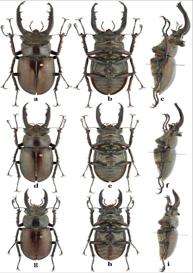 Figure 1
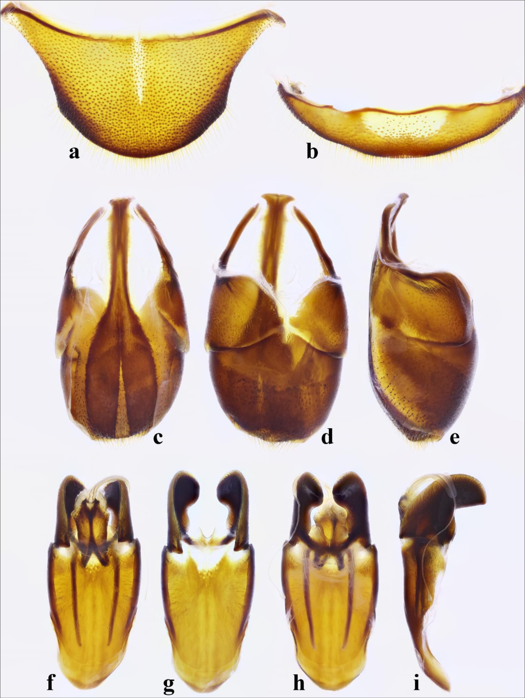 Figure 2
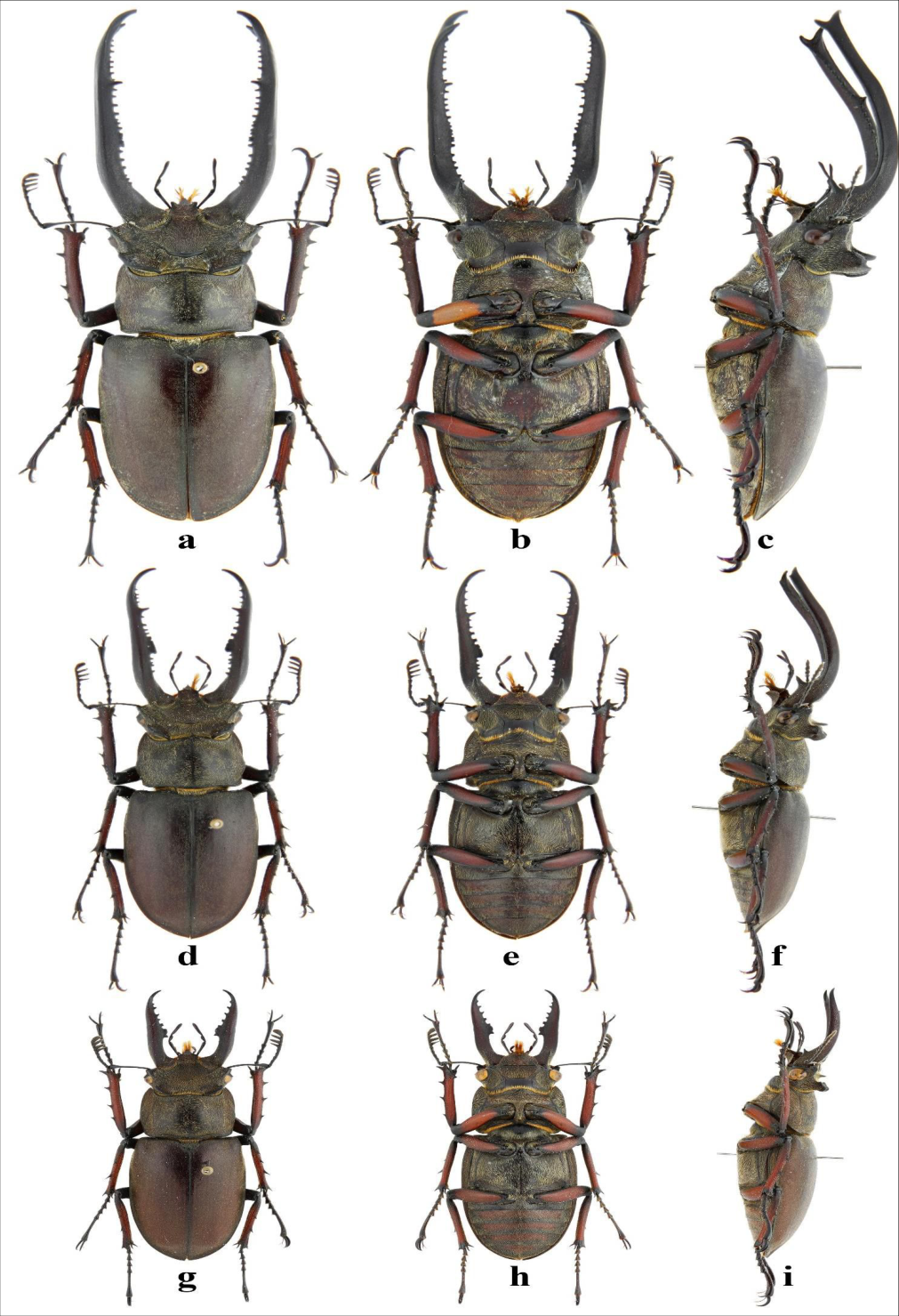 Figure 3
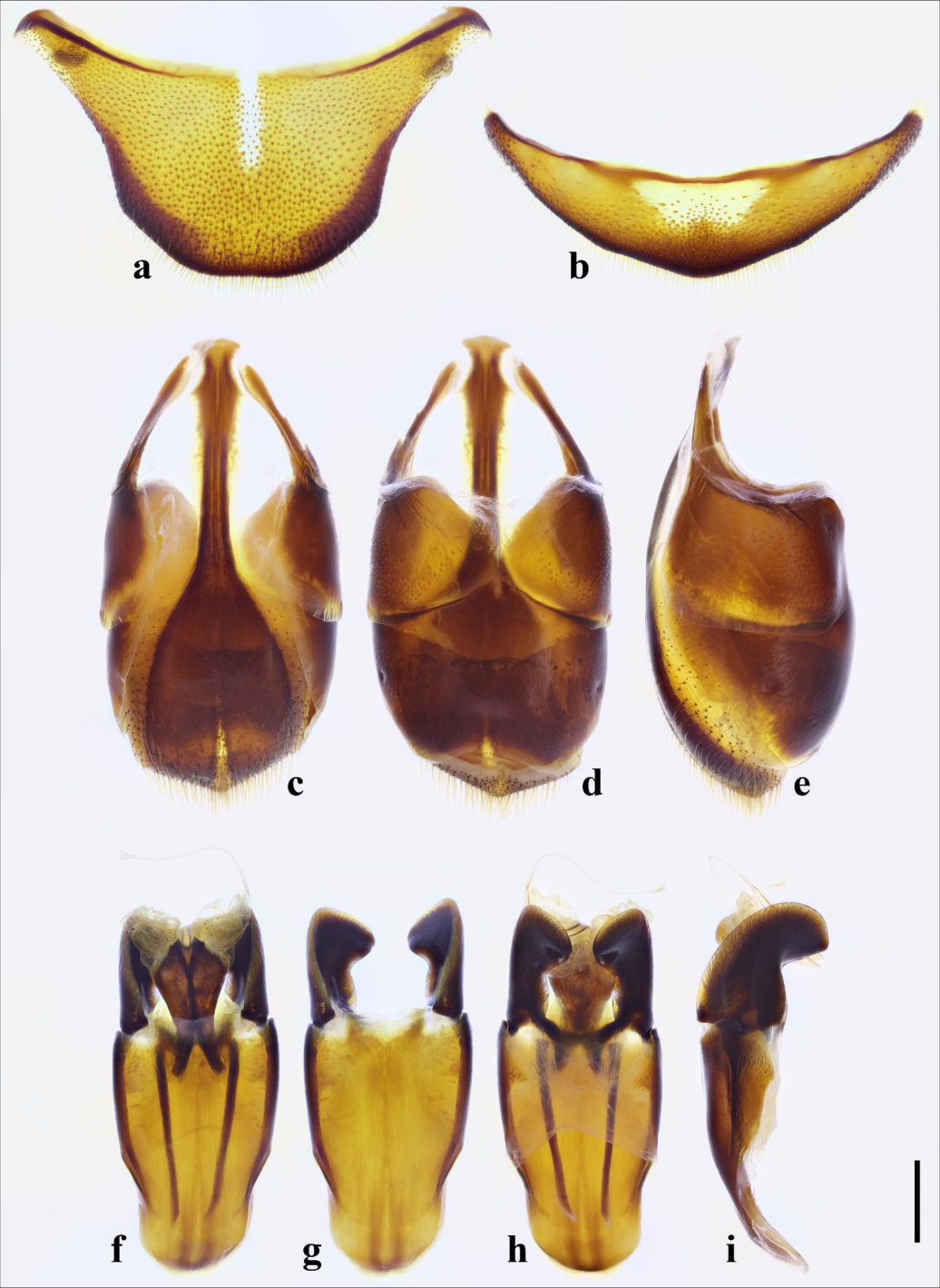 Figure 4
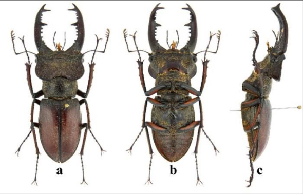 Figure 5
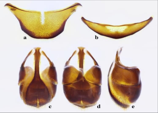 Figure 6
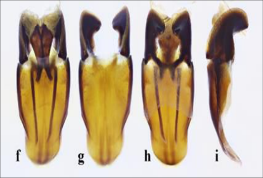 Figure 6
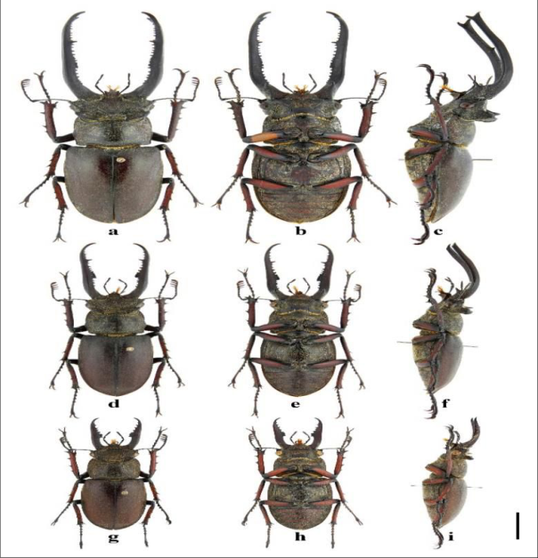 Figure 7
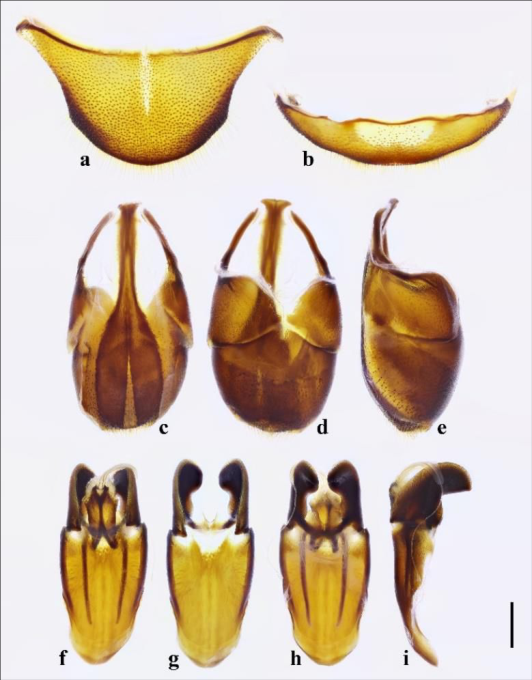 Figure 8
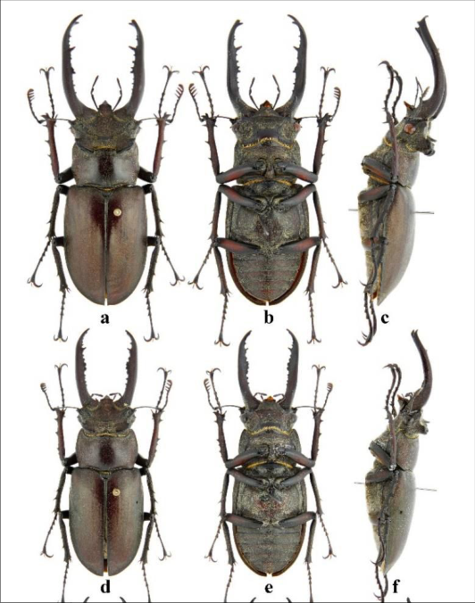 Figure 8
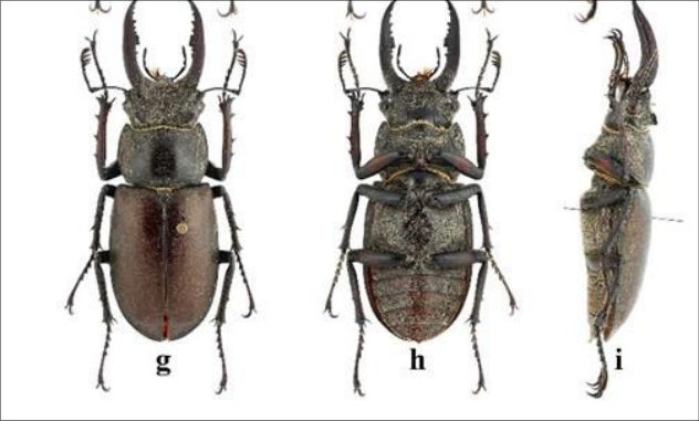 Figure 9
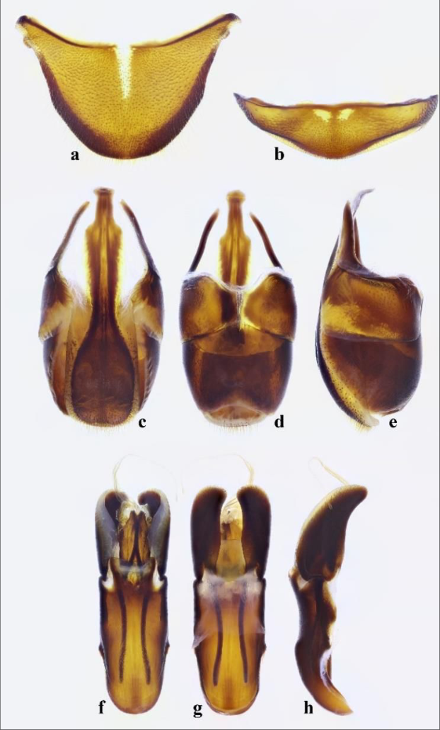 Figure 10
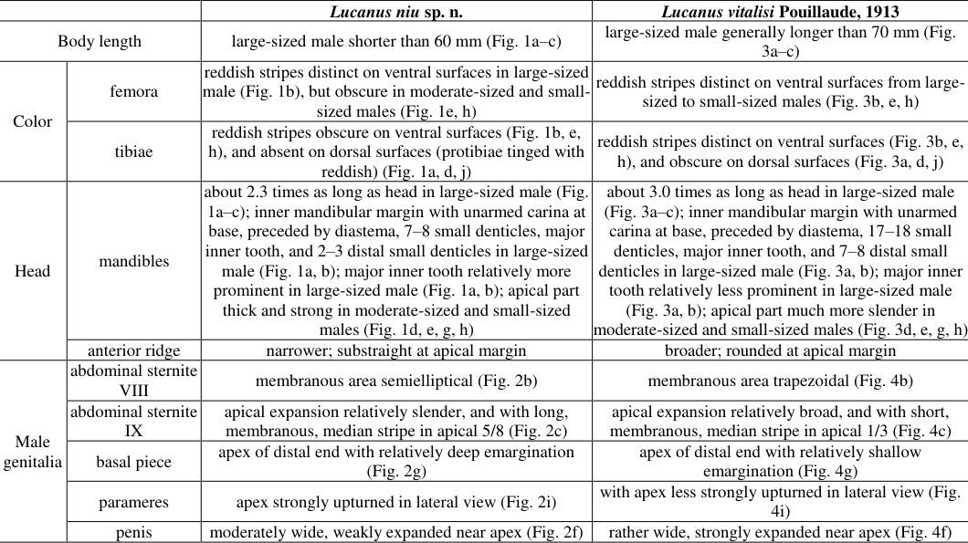 Table 1
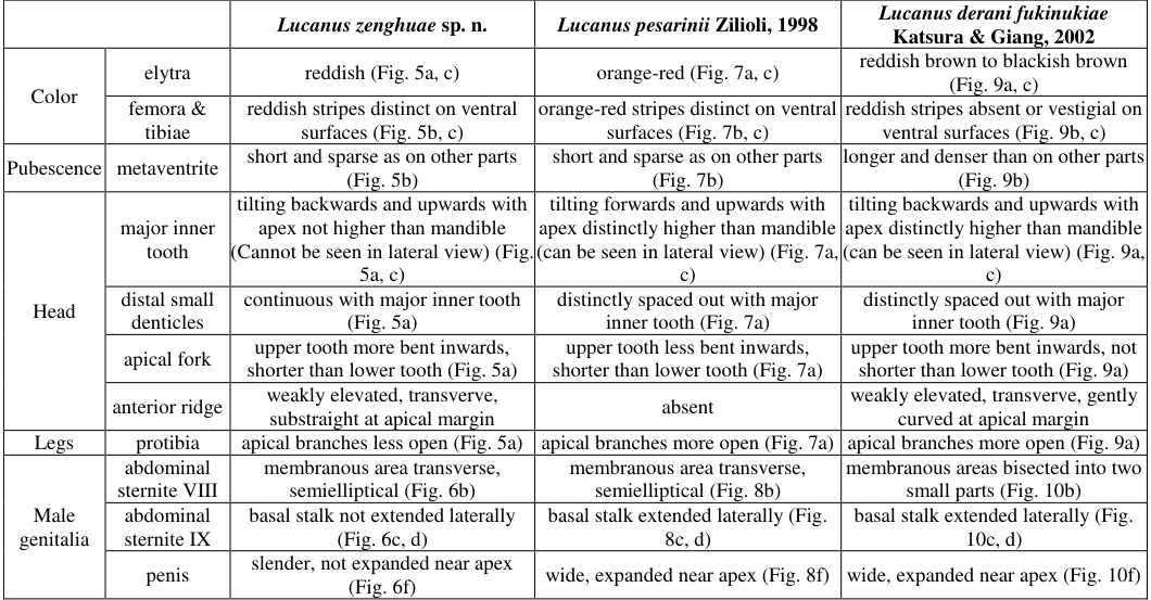 Table 2
Introduction
Lucanus Scopoli, 1763 is the type genus of Lucanidae (Coleoptera) with members occurring in the Nearctic, Oriental and Palearctic Regions; however, it reaches its highest diversity in eastern Asia. Since the three milestone books of "Stag beetles of China" by Huang & Chen Morphological terminology follows Holloway (2007) [4] , Huang & Chen (2010) [5] , Huang & Chen (2013) [6] and Huang & Chen (2017) [7,9] . The material examined for this study is deposited in the following institutional and private collections: CCZC: collection of Chao Zhou, Chengdu, China; CTLH: Collection of Tian-Long He, Huainan, China; CYCX: collection of Yun-Chuan Xu, Yuxi, China; MAHU: Museum of Anhui University, Hefei, China; MYNU: the invertebrate collection of Mianyang Normal University, Mianyang, China. The following material was studied for comparison: Lucanus vitalisi Pouillaude, 1913 (vs V.2020. Measurement criteria in millimetres (mm) are as follows: body length: length between the apex of mandible and the elytral apex along the midline; elytral length: length between the basal border and the apex of elytra along suture; elytral width: widest part of both elytra combined; head length: length between the anterior apex of clypeus and the posterior margin of occiput along the midline; head width: widest part of head; mandible length: length from the apex of mandible to its base; pronotal length: length of the pronotum along the midline; pronotal width: widest part of pronotum. (right, basal one tiny) teeth along outer margin; apex bifurcate with branches narrowly rounded at tips. Except apical spurs and spines, mesotibia with 3 (right) or 4 (left, basal one tiny) small lateral spines and metatibia with 3 lateral spines (basal one tiny). Surfaces finely punctate, intervals matt and finely granulose on femora and tibiae, feebly sheen and microreticulate on tarsi. Male genitalia. Abdominal tergite VIII (Fig. 2a) with subround lateral angles and with longitudinal, thin, membranous stripe in middle of basal 5/9; sternite VIII (Fig. 2b) with transverse, semielliptical, membranous area in middle of basal half. Abdominal sternite IX (Fig. 2c) with apical expansion relatively slender, and with long, membranous, median stripe in apical 5/8; basal stalk not extended laterally. Aedeagus (Fig. 2f) about 2.3 times as long as wide in ventral view. Basal piece (Fig. 2f-h) constricted in basal part, about 1.8 times as long as parameres, with a pair of sclerotized dorsal plates (Fig. 2h); ventral plate (Fig. 2g) bifurcate at apex of distal end, with relatively deep emargination. Paramere with basal process (Fig. 2g-h); apex strongly upturned in lateral view (Fig. 2i). Penis (Fig. 2f-h) moderately wide, weakly expanded near apex, 0.2 times shorter than parameres. Flagellum relatively short, about 2.2 times as long as parameres, apex distinctly enlarged. Male paratypes. Body 43.5-58.0 mm long. All male paratypes consistent with holotype except for usual allometric differences (Fig. 1d-i). Large-sized male like holotype with more pronounced morphological characters, while in small-sized male, mandible strongly shorter, apical fork of mandible less opened to vestigial, number of inner teeth fewer, major inner tooth ill-developed, clypeolabrum shorter, anterior and lateral ridges of head weaker. Female. Unkown. Etymology. The specific epithet is from the Chinese Pinyin "niú [牛]", which means ox, indicating this species was identified as new in 2021, the year of the ox. The name is a noun in apposition. Distribution. China (Yunnan). Differential diagnosis. This new species should be assigned to the Lucanus laminifer group (sensu Huang & Chen 2010) [5] . It most resembles Lucanus vitalisi Pouillaude, 1913, but can be distinguished by the combination of characters shown in Table 1. 6a) without lateral angles and with longitudinal, thin, membranous stripe in middle of basal 5/8; sternite VIII (Fig. 6b) with transverse, semielliptical, membranous area in middle of basal part. Abdominal sternite IX (Fig. 6c,e) with apical expansion relatively slender, without membranous area; basal stalk not extended laterally. Aedeagus (Fig. 6f) about 3.0 times as long as wide in ventral view. Basal piece (Fig. 6f,g) weakly constricted in basal part, about 1.4 times as long as parameres, with a pair of sclerotized dorsal plates (Fig. 6g); ventral plate (Fig. 6f) bifurcate at apex of distal end, with relatively deep emargination. Paramere with basal process (Fig. 6g); apex moderately upturned in lateral view (Fig. 6h). Penis (Fig. 6f) slender, 0.4 times shorter than parameres. Flagellum relatively short, about 2.2 times as long as parameres, apex distinctly enlarged. Male paratypes. Body 34.5-49.1 mm long. All male paratypes consistent with holotype except for usual allometric differences. Large-sized male like holotype with more pronounced morphological characters, while in smallsized male, mandible shorter, apical fork of mandible less opened, number of inner teeth fewer, major inner tooth illdeveloped, clypeolabrum shorter, anterior and lateral ridges of head weaker. Female. Unkown. Etymology. The new species is dedicated to Zeng-Hua Tang, the mother of the collector Tian-Long He. The name is a noun in the genitive case. Distribution. China (Yunnan). Differential diagnosis. This new species should be assigned to the Lucanus fortunei group (sensu Huang & Chen 2010) [5] . It is most similar to L. pesarinii Zilioli, 1998 and L. derani fukinukiae Katsura & Giang, 2002, but can be distinguished by the combination of characters shown in Table 2. Body length large-sized male shorter than 60 mm (Fig. 1a-c) large-sized male generally longer than 70 mm (Fig. 3a-c)
| — | — | Lucanus zenghuae sp. n. | Lucanus pesarinii Zilioli, 1998 | Lucanus derani fukinukiae Katsura & Giang, 2002 |
|---|---|---|---|---|
| Color | elytra | reddish (Fig. 5a, c) | orange-red (Fig. 7a, c) | reddish brown to blackish brown (Fig. 9a, c) |
| femora & tibiae | reddish stripes distinct on ventral surfaces (Fig. 5b, c) | orange-red stripes distinct on ventral surfaces (Fig. 7b, c) | reddish stripes absent or vestigial on ventral surfaces (Fig. 9b, c) | |
| Pubescence | metaventrite | short and sparse as on other parts (Fig. 5b) | short and sparse as on other parts (Fig. 7b) | longer and denser than on other parts (Fig. 9b) |
| Head | major inner tooth | tilting backwards and upwards with apex not higher than mandible (Cannot be seen in lateral view) (Fig. 5a, c) | tilting forwards and upwards with apex distinctly higher than mandible (can be seen in lateral view) (Fig. 7a, c) | tilting backwards and upwards with apex distinctly higher than mandible (can be seen in lateral view) (Fig. 9a, c) |
| distal small denticles | continuous with major inner tooth (Fig. 5a) | distinctly spaced out with major inner tooth (Fig. 7a) | distinctly spaced out with major inner tooth (Fig. 9a) | |
| apical fork | upper tooth more bent inwards, shorter than lower tooth (Fig. 5a) | upper tooth less bent inwards, shorter than lower tooth (Fig. 7a) | upper tooth more bent inwards, not shorter than lower tooth (Fig. 9a) | |
| anterior ridge | weakly elevated, transverve, substraight at apical margin | absent | weakly elevated, transverve, gently curved at apical margin | |
| Legs | protibia | apical branches less open (Fig. 5a) | apical branches more open (Fig. 7a) | apical branches more open (Fig. 9a) |
| Male genitalia | abdominal sternite VIII | membranous area transverse, semielliptical (Fig. 6b) | membranous area transverse, semielliptical (Fig. 8b) | membranous areas bisected into two small parts (Fig. 10b) |
| abdominal sternite IX | basal stalk not extended laterally (Fig. 6c, d) | basal stalk extended laterally (Fig. 8c, d) | basal stalk extended laterally (Fig. 10c, d) | |
| penis | slender, not expanded near apex (Fig. 6f) | wide, expanded near apex (Fig. 8f) | wide, expanded near apex (Fig. 10f) |
Table text extracted from the original PDF; superscript letters are shown inline. The original table image is below.
View original table image
Taxonomy
Taxonomy
Lucanus niu
Lucanus niu
Lucanus zenghuae
Color femora reddish stripes distinct on ventral surfaces in large-sized male (Fig. 1b), but obscure in moderate-sized and smallsized males (Fig. 1e,h) reddish stripes distinct on ventral surfaces from largesized to small-sized males (Fig. 3b,e,h) tibiae reddish stripes obscure on ventral surfaces (Fig. 1b, e, h), and absent on dorsal surfaces (protibiae tinged with reddish) (Fig. 1a,d,j) reddish stripes distinct on ventral surfaces (Fig. 3b, e, h), and obscure on dorsal surfaces (Fig. 3a,d,j)
Head mandibles about 2.3 times as long as head in large-sized male (Fig. 1a-c); inner mandibular margin with unarmed carina at base, preceded by diastema, 7-8 small denticles, major inner tooth, and 2-3 distal small denticles in large-sized male (Fig. 1a,b); major inner tooth relatively more prominent in large-sized male (Fig. 1a,b); apical part thick and strong in moderate-sized and small-sized males (Fig. 1d, e, g, h) about 3.0 times as long as head in large-sized male (Fig. 3a-c); inner mandibular margin with unarmed carina at base, preceded by diastema, 17-18 small denticles, major inner tooth, and 7-8 distal small denticles in large-sized male (Fig. 3a,b); major inner tooth relatively less prominent in large-sized male (Fig. 3a,b); apical part much more slender in moderate-sized and small-sized males (Fig. 3d,e,g,h) anterior ridge narrower; substraight at apical margin broader; rounded at apical margin Male genitalia abdominal sternite VIII membranous area semielliptical (Fig. 2b) membranous area trapezoidal (Fig. 4b) abdominal sternite IX apical expansion relatively slender, and with long, membranous, median stripe in apical 5/8 (Fig. 2c) apical expansion relatively broad, and with short, membranous, median stripe in apical 1/3 (Fig. 4c) basal piece apex of distal end with relatively deep emargination (Fig. 2g) apex of distal end with relatively shallow emargination (Fig. 4g) parameres apex strongly upturned in lateral view (Fig. 2i) with apex less strongly upturned in lateral view (Fig. 4i) penis moderately wide, weakly expanded near apex (Fig. 2f) rather wide, strongly expanded near apex (Fig. 4f) Color elytra reddish (Fig. 5a,c) orange-red (Fig. 7a,c) reddish brown to blackish brown (Fig. 9a,c) femora & tibiae reddish stripes distinct on ventral surfaces (Fig. 5b,c) orange-red stripes distinct on ventral surfaces (Fig. 7b,c) reddish stripes absent or vestigial on ventral surfaces (Fig. 9b,c)
Pubescence metaventrite short and sparse as on other parts (Fig. 5b)
short and sparse as on other parts (Fig. 7b) longer and denser than on other parts (Fig. 9b) Head major inner tooth tilting backwards and upwards with apex not higher than mandible (Cannot be seen in lateral view) (Fig. 5a,c) tilting forwards and upwards with apex distinctly higher than mandible (can be seen in lateral view) (Fig. 7a,c) tilting backwards and upwards with apex distinctly higher than mandible (can be seen in lateral view) (Fig. 9a,c) distal small denticles continuous with major inner tooth (Fig. 5a) distinctly spaced out with major inner tooth (Fig. 7a) distinctly spaced out with major inner tooth (Fig. 9a) apical fork upper tooth more bent inwards, shorter than lower tooth (Fig. 5a) upper tooth less bent inwards, shorter than lower tooth (Fig. 7a) upper tooth more bent inwards, not shorter than lower tooth (Fig. 9a) anterior ridge weakly elevated, transverve, substraight at apical margin absent weakly elevated, transverve, gently curved at apical margin Legs protibia apical branches less open (Fig. 5a) apical branches more open (Fig. 7a) apical branches more open (Fig. 9a) Male genitalia abdominal sternite VIII membranous area transverse, semielliptical (Fig. 6b) membranous area transverse, semielliptical (Fig. 8b) membranous areas bisected into two small parts (Fig. 10b) abdominal sternite IX basal stalk not extended laterally (Fig. 6c,d) basal stalk extended laterally (Fig. 8c,d) basal stalk extended laterally (Fig. 10c,d) penis slender, not expanded near apex (Fig. 6f) wide, expanded near apex (Fig. 8f) wide, expanded near apex (Fig. 10f)
Acknowledgement
AcknowledgementsWe appreciate Gui-Qiang Huang (Liupanshui Normal University, Liupanshui, China), who rekindling our
Figures and tables from the paper
References
15 in the original paper- Adachi N. A new species of the genus Lucanus from Szechuan province, southwest China. Kogane. 2020;23:8-10.
- Bartolozzi L, Sprecher-Uebersax E, Bezděk A. Family Lucanidae Latreille, 1804. In: Löbl, I. & Löbl, D. (Eds.), Catalogue of Palaearctic Coleoptera.
- Scarabaeoidea-Scirtoidea-Dascilloidea-BuprestoideaByrrhoidea. Revised and Updated Edition. Brill, Leiden and Boston. 2016;3:58-84. https://doi.org/10.1163/9789004309142_003
- Bian CZ, Zhan ZH. A new species of Lucanus Scopoli, 1763 (Coleoptera, Lucanidae) from Tibet, China. Faunitaxys. 2021;9(27):1-4.
- Holloway BA. Lucanidae (Insecta: Coleoptera). Fauna of New Zealand. 2007;61:1-254.
- Huang H, Chen CC. Stag beetles of China I. Formosa Ecological Company, Taipei, 2010, 288.
- Huang H, Chen CC. Stag beetles of China II. Formosa Ecological Company, Taipei, 2013, 716.
- Huang H, Chen CC. Stag beetles of China III. Formosa Ecological Company, Taipei, 2017, 524. International Journal of Biology Sciences https://www.biologyjournal.net/
- Huang H, Chen CC. Lucanus liuweii sp. nov. (Coleoptera: Lucanidae: Lucaninae) from Zhejiang, China. Beetles World. 2019;19:2-6.
- Huang H, Chen CC. New or little-known stag beetles (Coleoptera: Lucanidae: Lucaninae) from China. Beetles World. 2017;21:2-11.
- Lin JZ. Description of Lucanus yulaoensis sp. nov., a new species stag beetle from northern Taiwan (Coleoptera, Lucanidae). Faunitaxys. 2021;9(2):1-5.
- Qi ZH. Lucanus moae sp. nov., A new species from Sichuan, China (Coleoptera: Lucanidae: Lucaninae). Faunitaxys. 2021;9(23):1-7.
- Wang CB, Zhu X. Lucanus zhanbishengi sp. n., a new species from Hunan, central China (Coleoptera: Lucanidae: Lucaninae). International Journal of Research Studies in Zoology. 2017;3(4):54-69. http://dx.doi.org/10.20431/2454-941X.0304008
- Wang CB, Zhan BS. Lucanus zhuxiangi sp. n., a new species from southeast China (Coleoptera: Lucanidae: Lucaninae). International Journal of Research Studies in Zoology. 2018;4(3):1-11. https://doi.org/10.20431/2454-941X.0403001
- Wang LJ, Ko HP. Description of Lucanus chengyuani sp. nov. from Taiwan, with a key to the species of Taiwanese Lucanus Scopoli (Coleoptera: Lucanidae). Japanese Journal of Systematic Entomology. 2018;24(2):257-263.
Cited by 4
From OpenAlex, OpenCitations and reference lists read by Scinovex
- In vivo antioxidant and immunomodulatory effect of the aqueous extract of Mimela sp. on cyclophosphamide induced immunocompromised mice ↗
Sonam Drema Tukshipa, Jaya Chakravorty, Prveen Prveen · International Journal of Pharmacognosy and Life Science · 2022
Insect and their products have been used as an integral part of local therapies around the globe since decades. Several studies on insects have found it to be highly nutritious and rich in several bioactive compounds possibly attributing to its anti-oxidative, anti-carcinogenic, anti-inflammatory, anti-microbial, anti-diabetic and immune regulating properties. Mimela sp. is a small shiny green beetle highly preferred as…
- Feeding potential of ladybird beetle, Cheilomenes sexmaculata (Fabricius) on different species of aphids
Singh NA, Dabhi MR, Rojasara YM, Varma CB · International Journal of Advanced Biochemistry Research · 2024
A study was carried out to explore the feeding capabilities of the zig-zag beetle, Cheilomenes sexmaculata (Fabricius), a commonly found species of ladybird beetle, at Department of Agricultural Entomology, B. A. College of Agriculture, Anand Agricultural University, Anand (Gujarat) during the year 2019-20. The feeding potential of the predator was calculated on four hosts i.e., mustard aphid, Lipaphis erysimi (Kaltenbach), cotton aphid, Aphis gossypii (Glover), maize aphid, Rhopalosiphum maidis (Fitch) and cowpea aphid, Aphis craccivora (Koch). Results revealed that highest number of aphids c
- Studies on biology of Callosobruchus chinensis (L.) on Chickpea seed in storage ↗
Prateek Kumar Singh, M. K. Tripathi, Bal Mukund Pandey · Journal of Pharmacognosy and Phytochemistry · 2021
Studies carried out in Laboratory condition on the biology of the pulse beetle, Callosobruchus chinensis (L.) (Coleoptera: Bruchidae) on the stored chickpea at department of Entomology, T.D. college, Jaunpur revealed that the insect completed one generation from July to September, 2017. The study on biology of C. chinensis on chickpea indicate 5.16 ± 0.87 days eggs incubation period, larval+ pupal
- A critique of a practice: Withdrawing from authorship or ceding first authorship to a non-taxonomist ↗
Cheng-Bin Wang · Ecologica Montenegrina · 2026
N/A
❝ 0PDF