
Research article · 2023 · Full text
International Journal of Veterinary Sciences and Animal HusbandryCongenital Chondrodysplasia in a Punganur Dwarf Cow: A case report
Volume 8(3) · Pages 28–30 · DOI: 10.22271/veterinary.2023.v8.i3a.515 ↗
Research areasAnimal Disease Management and EpidemiologyAnimal Genetics and ReproductionCancer-related gene regulation
Abstract
Author-writtenCongenital anomalies are not uncommon in domestic animals, and they can have various causes, including genetic and environmental factors. This case study reports the occurrence of a rare congenital anomaly in a Punganur breed calf, which was born lifeless with a bull-dog like face and unnaturally rotated limbs. The breeding records and pedigree of the cow were not available, and nutritional deficiencies and ingestion of teratogenic plants were ruled out as potential causes. The chondrodysplasia syndrome, caused by a homozygous type of Dexter breed, is known to be inherited and lethal. However, the exact cause of the anomaly in this Punganur calf remains uncertain. This case report highlights the importance of careful observation and examination of new-born animals, particularly in breeds with a known genetic predisposition to certain congenital conditions.
Summary
Source-checkedPlain-language reading of the full paper, prepared with AI and checked against the text. Each point links to the authors' own words.
Why it matters
- Punganur dwarf cattle are a small humped breed from southern India known for their climatic adaptation and disease resistance. View evidence ↗
- Congenital malformations in cattle are caused by gene or chromosomal abnormalities, leading to disproportionate dwarfism at birth. View evidence ↗
- Bull dog type chondrodysplasia is a form of brachycephalic dwarfism characterized by short legs, nose ridges, a short upper jaw, and a compressed cranium. View evidence ↗
The question
- The study aims to describe the clinical and morphological features of the first reported case of a purebred male Punganur fetus with bulldog-type chondrodysplasia produced via artificial insemination. View evidence ↗
What they did
- The subject was a four-year-old Punganur cow impregnated with Punganur semen via artificial insemination. View evidence ↗
- After 10 hours of unsuccessful labor and confirmation of fetal death, a Caesarean section was performed to remove the fetus. View evidence ↗
- Diagnosis of bulldog-type chondrodysplasia was made based on the phenotypic characteristics of the fetus compared to normal appearance. View evidence ↗
What they found
- The fetus measured 9 inches from head to foreleg toe and 17.5 inches from the tip of the nose to the tuber coxae. View evidence ↗
- The hind legs showed marked outward convexity with hooves rotated inwards and overlapping 3rd phalanges at their distal ends. View evidence ↗
- The fetus exhibited a straight spine, a flattened back, and a tail-head that originated far forward along the back and arched upwards. View evidence ↗
- The fetus had a shrunken thoracic region and a pot-belly appearance caused by excessive subcutaneous fat in the abdominal region. View evidence ↗
- Environmental causes, specifically intoxication from teratogenic plants and nutritional deficiencies, were ruled out as the cause of the anomaly. View evidence ↗
What it means
- The authors conclude that while the exact cause is uncertain, the available evidence suggests genetic factors likely played a role in the anomaly. View evidence ↗
- The authors recommend the use of genetic screening and proper breeding practices in livestock management to prevent such anomalies. View evidence ↗
- The lack of available pedigree information and breeding records limited the authors' ability to definitively determine the genetic cause. View evidence ↗
Visual summary
1 visualsIllustrations explain the paper; they do not show study data.
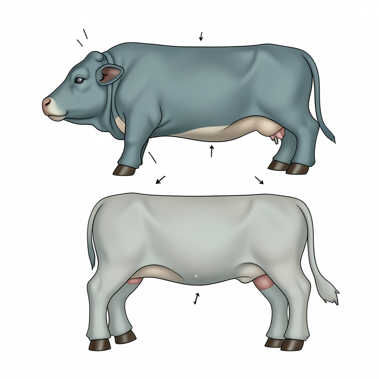
Morphological Features of Bulldog-Type Chondrodysplasia
AI-generated conceptual illustration (Punganur dwarf calf). It explains the idea and does not show study data.
View source evidence ↗Figures & tables
From the original paper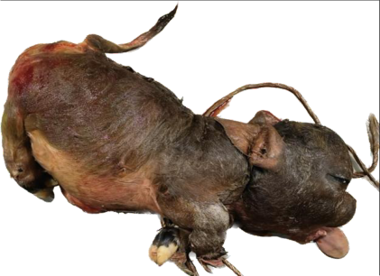 Figure 3
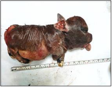 Figure 2
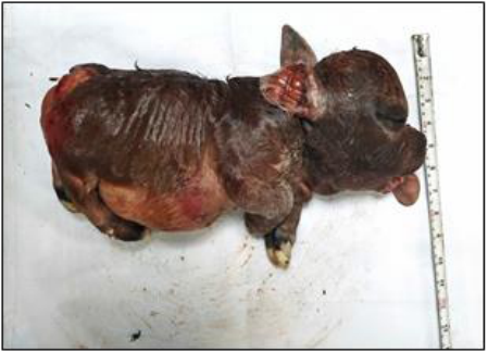 Figure 1
Introduction
Punganur dwarf cattle is a small humped cattle breed with exceptional disease resistance and climatic adaptation [1,2] , originating from the Chittoor district of Andhra Pradesh in southern India. This breed is known to have a low incidence of inherited congenital anomalies worldwide. However, some cases of congenital malformations have been reported in Punganur cattle, including chondrodysplasia. Congenital malformations are caused by chromosomal or gene abnormalities that result in disproportionate dwarfism present at birth. Chondrodysplasia is a complex vertebral malfunction that leads to irregular skeletal development [3] , including osteopetrosis and osteogenesis imperfecta [4] . Bull dog type chondrodysplasia is a specific type of chondrodysplasia that results in a compressed cranium with a short upper jaw, nose ridges, and short legs, consistent with brachycephalic dwarfism. This type is diagnosed when the cranial bones have a larger size than the rest of the body due to ineffective endochondral ossification, while intramembranous ossification augments beneath the periosteum, leading to abnormal bone growth. Although cows have a lower incidence of congenital malformations compared to buffaloes [5] , this paper details the clinico-morphological features of the first instance of bulldog-type chondrodysplasia male, purebred foetus produced by artificial insemination of a Punganur cattle.
Case Report
A four-year old Punganur cow was impregnated with semen straw of Punganur cattle using artificial insemination technique prior to transport and had an established pregnancy of seven months. After two months, the cow showed signs of labour about ten days premature to the estimated date of parturition. The straining continued for about 10 hours, but the cow was unable to deliver the foetus. The foetus did not show any body movements, and the head was deformed on per rectal examination. Dystocia due to cranial malformation consistent with bull dog type foetus has been reported in cows [6] . Due to the inconsistency in shape and size of the foetus's head and a relatively small birth canal, dystocia was inexorable. Obstetric manoeuvres were engaged after confirmation of foetal death for the expulsion of the foetus through the birth canal without success. Thereby, Caesarean section was performed to remove the dead foetus from the uterus as per the routine procedure. The height of the foetus from head to foreleg toe was 9 inches (Fig. 1), and the length from the tip of the nose to tuber coxae was 17.5 inches (Fig. 2). The hind legs had marked outward convexity, and the hooves were rotated and directed inwards (Fig. 2). The angle of the 3rd phalanx of each hind leg was positioned in a fashion that they overlapped each other towards their distal end. The dead foetus was determined to carry polymonstrosities. The monster appearance can be attributed to micromyelia and a stout body (Fig. 3). The skin was leathery, thick, and covered with hair. The face was dark brown with slightly faded brown on the body, white on the underbelly, and black on the tail tassel. A craniofacial disproportion included bulging cranium, depressed nose, protruding tongue, brachygnathia inferior described as the upper jaw longer than the lower jaw resulting in malocclusion of lower incisors with the dental pad [7] (Fig. 1). The foetus had a posteriorly pushed-out appearance of the buttocks, the tail-head originated far up and forward along the calf's back, and at the same time, it arched upwards (Fig. 3). It presented a straight spine and a flattened back devoid of usual curvatures.
The thoracic part looked shrunken, and the major portion of the body was occupied by the abdominal region. The foetus had a pot-belly appearance due to excessive accumulation of subcutaneous fat (Fig. 3). It was diagnosed to be a case of chondrodysplasia fetalis or bull dog type chondrodysplasia calf based on phenotypic characteristics and its variations from normal appearance [8,9,10] . The calf born was male, but earlier studies determined after much deliberation that there was an absence of sex-linkage to chondrodysplasia [8] . The absence of cleft palate and superior brachygnathism could be attributed to specific breed predisposition as both have not been reported in Punganur cattle earlier, and the findings of this paper supplement the said hypothesis [3,11] .
Based on the available information, it is difficult to pinpoint the exact cause of the congenital anomaly in the Punganur cow. While genetic factors cannot be ruled out, the lack of breeding records and pedigree information makes it difficult to determine if this is the case. Nutritional deficiencies and intoxication due to ingestion of teratogenic plants were also ruled out as possible causes. However, it is worth noting that the homozygous type of Dexter breed chondrodysplasia causes a congenitally fatal syndrome that is thought to be inherited. Punganur cattle are also considered to be pure homozygous bantam cattle from a genetic perspective [12] . Previous studies in Dexter breed cattle have shown that the chondrodysplasia or bull dog type monstrosity could have been caused by recessive genes. Additionally, it is important to note that this is the solitary chondrodysplastic condition that is lethal. Overall, while the exact cause of the congenital anomaly in the Punganur cow cannot be determined with certainty, the available information suggests that genetic factors may have played a role. Further studies and breeding records may be necessary to gain a better understanding of the underlying cause of this condition
Discussion
Congenital anomalies in cattle are a rare but serious issue that can lead to significant losses for cattle breeders. Numerous studies have explored the causes and effects of these anomalies, shedding light on the genetics, environmental factors, and other potential causes of such conditions. For instance, studies have indicated that genetic factors play a significant role in the development of chondrodysplasia in Dexter breed cattle, a condition which can be fatal for affected animals [13] . Similarly, nutritional deficiencies have also been linked to the development of congenital anomalies in cattle, in a study by Lorenz et al. (2011) suggesting that micronutrient deficiencies can lead to limb malformations in developing foetuses [14] .
Other studies have focused on the classification of congenital anomalies and their associated morbidity and mortality rates.
A study by Purohit et al. (2015) explored various types of congenital anomalies, including monstrosities, and found that the morbidity and mortality rates associated with such conditions were significantly higher compared to other anomalies [15] . The study also highlighted the importance of early detection and intervention in minimizing losses due to congenital anomalies. Additionally, studies have explored the potential for genetic testing and selective breeding as tools for preventing congenital anomalies in cattle. For example, a study by Weikard et al. (2015) investigated the genetic basis of chondrodysplasia in Dexter cattle and suggested that genetic testing and selective breeding could be used to prevent the condition from being passed on to future generations [16] .
Overall, these studies demonstrate the complex and multifactorial nature of congenital anomalies in cattle and highlight the need for further research into the underlying causes and potential prevention strategies.
Conclusion
The case study of the congenital anomaly in a Punganur cow highlights the importance of proper breeding practices and genetic screening in livestock management. While the exact cause of the chondrodysplasia in this particular case remains unknown, the presentation of morphological characteristics and lethal outcomes suggest a possible association with recessive genes. The exclusion of other potential causes such as nutritional deficiencies and intoxication due to teratogenic plants emphasizes the need for a thorough investigation and diagnosis of congenital anomalies in livestock. By understanding and addressing the underlying factors contributing to such conditions, it is possible to improve the health and welfare of animals and enhance the productivity and sustainability of the livestock industry.
References
11 in the original paper- Bramhaiah VK, Rao V, Reddy RY. Reproduction in Punganur cattle. The Indian Journal of Animal Reproduction. 2003;24(1):51.
- Thangamani A, Hussaini YD. The Native cattle breeds (Ongole and Punganur) of Andhra Pradesh-A white pearl and pride of Andhra Pradesh. The Science World a Monthly e-Magazine. 2022;2(12):2182-2187.
- Charmila D'Soares S, Muramoto C, Antonio Silva WO, Moisés Freitas D, Vitor Carvalho S, Ana Caroline Souza SN, et al. Peixoto: Chondrodysplasia Bulldog type in cattle in the state of Bahia, Brazil. Brazilian Journal of Pathology Veterinary. 2020;13(2):536-541.
- Gholap PN, Kale DS, Sirothia AR. Genetic Diseases in Cattle: A Review. Research Journal of Animal, Veterinary and Fishery Sciences. 2014;2(2):24-33.
- Purohit GN, Kumar P, Kanika S, Shekher C, Yadav SP. Perspectives of fetal dystocia in cattle and buffalo. Veterinary Science Development. 2012;2(e8):31-42.
- Kumar A, Honparke M, Dhaliwal GS. Dystocia due to Bulldog calf in a crossbred cow. Indian Journal Animal Reproduction. 2007;284:84-85.
- Muhammad SA, Sajid M, Shahzad AH, Asif AR, Ali M, et al. Delivery of Bulldog Type Achondroplasia Calf after Management of Dystocia in Sahiwal Cattle. Pakistan Journal of Life and Social Sciences. 2021;19(1):43-45.
- Mukherjee A, Anand MK, Arora AK, Singh A. Clinical and histopathological investigation of chondrodysplastic calves. Veterinary World. 2016;9(8):890-894.
- Lorenz MD, Kornegay JN, Boudrieau RJ. Congenital limb malformations in cattle: a review and assessment of their significance. Veterinary and Comparative Orthopaedics and Traumatology. 2011;24(5):297-304.
- Purohit GN, Kalita P, Saini M, Tyagi S. Congenital anomalies in bovines: A review. Journal of Animal Research. 2015;5(3):495-500.
- Weikard R, Altmaier E, Suhre K, Kühn C. Genetics of congenital monstrosities in cattle. Genetics Selection Evolution. 2015;47(1):1-8.