
Research article · 2019 · Full text
International Journal of Agriculture and Food ScienceDemonstration of pathogenicity in three different Magnaporthe oryzae isolates
Volume 1(4) · Pages 29–32 · DOI: 10.33545/2664844x.2019.v1.i4a.25 ↗
Research areasPlant Pathogens and Fungal DiseasesFungal Biology and ApplicationsPlant-Microbe Interactions and Immunity
Abstract
Author-writtenIn present study, three Magnaporthe isolates were analyzed for understanding pathogenicity of blast disease in rice. M oryzae 6808 and M oryzae ch1 isolates produced pyriform shaped macroconidia and were able to form appressoria under artificial condition. M oryzae 6926 produced crescent shaped microconidia but failed to produce appressoria in the similar condition. Pathogen inoculation assay results showed that all three Magnaporthe isolates were infective with varying degree of infection. Pathogen isolates produced spindle shaped lesion with necrotic borders on different cultivars of paddy lines with different disease intensity. Both types of sporulation contributed to disease incidence and disease severity in rice lines. Entry of pyriform shaped macroconidia and formation of appressoria in suitable host plant has been widely studied. Likely, there must be another mechanism for crescent shaped microconidia to make entry into host cell to cause disease symptoms in suitable host plant. It was observed that pathogenicity in rice was caused due to macroconidia and microconidia both. Microconidial infection needs to be further explored in understanding blast disease cycle.
Summary
Source-checkedPlain-language reading of the full paper, prepared with AI and checked against the text. Each point links to the authors' own words.
Why it matters
- Rice blast, caused by the fungus Magnaporthe oryzae, is a highly destructive disease that reduces both the quality and quantity of rice yields. View evidence ↗
- Globally, rice blast is estimated to cause a yield reduction of approximately 14-18%. View evidence ↗
- Most existing research on rice blast has focused on macroconidia, leaving a gap in the understanding of microconidia. View evidence ↗
The question
- The study aimed to explore appressoria formation in three Magnaporthe oryzae isolates to determine if this process is linked to pathogenicity. View evidence ↗
What they did
- The researchers used three isolates of M. oryzae: 6808, 6926, and a field isolate named ch1. View evidence ↗
- Appressoria formation was tested by placing conidial suspensions (1*103/ml) on sterile coverslips. View evidence ↗
- Pathogenicity was tested by spraying 4-week-old paddy plants of five cultivars (Phule Samruddhi, Indrayani, Chimansal, Ek-70, and Phule Radha) with a spore suspension (1*103/ml). View evidence ↗
What they found
- Isolates 6808 and ch1 produced dark pigmented mycelia due to melanin, while isolate 6926 produced white mycelia, indicating a lack of melanin. View evidence ↗
- Isolates 6808 and ch1 produced pyriform macroconidia and formed appressoria, whereas isolate 6926 produced crescent-shaped microconidia and failed to form appressoria. View evidence ↗
- Despite the differences in appressoria formation, all three isolates were found to be infective to the rice plants. View evidence ↗
- Isolate ch1 exhibited the highest percentage of disease incidence and caused more severe disease than the other two isolates. View evidence ↗
- Isolate 6926 had a disease incidence similar to the others, but the severity was lower, which the authors attribute to its inability to form appressoria. View evidence ↗
What it means
- The authors concluded that both macroconidia and microconidia are capable of causing pathogenicity in rice. View evidence ↗
- The authors state that the specific mechanism by which crescent-shaped microconidia enter the host plant needs further study. View evidence ↗
Visual summary
1 visualsIllustrations explain the paper; they do not show study data.
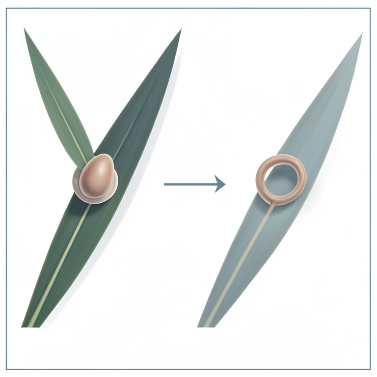
Comparison of Infection Pathways
AI-generated conceptual illustration (Rice plants (Oryza sativa) and Magnaporthe oryzae fungal spores). It explains the idea and does not show study data.
View source evidence ↗Figures & tables
From the original paper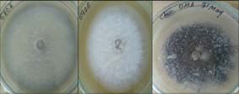 Fig1
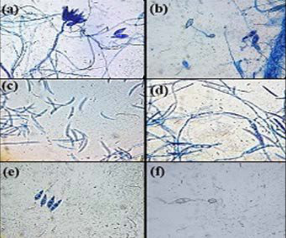 Figure 2
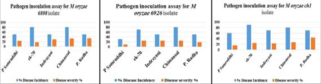 Figure 3
Introduction
Blast caused by Magnaporthe oryzae is the most devastating disease that occurs to rice at any stages of plant's growth. It causes heavy losses in both quality and quantity of the produce. It is estimated about 14-18% of the yield reduction worldwide (Hajano, Pathan, Rajput, & Lodhi, 2011) [7] . Blast infestations could lead to complete failure of the crop causing severe losses. (Gavhane, Kulwal, Kumbhar, Jadhav, & Sarawate, 2019) [6]. Magnaporthe oryzae (teleomorph) (and its anamorph, Pyricularia oryzae) is a filamentous heterothallic ascomycete that causes blast disease mainly on cultivated gramene plants. It produces three-celled, pyriform macrospore or macroconidia in the imperfect stage, and four-celled, spindle-shaped ascospore or microspore i.e. microconidia in the perfect stage. (Chuma et al., 2009) [4] ; (Kato et al., 1994) [9] ; (Yaegashi & Nishihara, 1976) [14].
In general, more detailed information is available for macroconidia as most of the studies have been dealt exclusively with germination of macroconidia only. Rice blast infections are initiated when an asexual tri septate pyriform spore lands on the surface of a rice leaf in presence of dewdrops. Spore attaches itself to the cuticle by release of an adhesive found in an apical compartment of the spore. Spore contains various types of endogenous reserves which are required for germination when there is no nutrient available. These include glycogen, trehalose, polyols such as glycerol, erythritol and mannitol etc. (Foster, Littlejohn, Soanes, & Talbot, 2016) [5] . In very short span after landing on the leaf, a polarized germ tube is formed from macrospore. Germ tube is formed from one of the apical cell of the conidium and extends only for short distance where pathogen is hooked on the surface then recognition of host occurs and then lastly appressorium formation takes place.
Developing appressorium has a cell wall which is made up of two layers i.e. outer layer of chitin and inner layer of melanin. These layers are required to generate turgor pressure of up to 8MPa. (Jenkinson et al., 2007) [8]. Cell wall acts as a barrier to the efflux of solute from the appressorium. The cellular turgor is then converted to mechanical force. This force is exerted by emerging penetration peg so as to forcibly get inside in to the leaf cuticle. (Wilson & Talbot, 2009) [12]. There are no reports of appressoria formation from microconidia as not much literature is available on microconidia In the present studies, three types of Magnaporthe oryzae isolates were explored for appressoria formation event as it is widely considered that appressoria synthesis occurs in pathogenic strains only. Cytological analyses of different sporulation pattern among Magnaporthe isolates were evaluated for their role in demonstrating pathogenicity.
Pathogen inoculation assay: Three blast isolates i.e. Magnaporthe oryzae ch1, 6808 and 6926 isolates were used as source of pathogen for inoculation against Paddy lines viz. Phule Samruddhi, Indrayani, Chimansal, Ek-70 and Phule Radha, obtained from Mahatma Phule Krishi Vidyapeeth (MPKV), Rahuri, Maharashtra. Paddy plants of 4 weeks old age were sprayed with spore suspension (1*10 3 /ml) of all three isolates on different lines. Experiment was done in duplicates for each cultivar with respect to each pathogen isolate. Control plants were kept along with experimental plants. After spraying, each cultivar was sealed by transparent polybags from top. (Nagao Hayashi et al, 2009) [11]. All the plants were kept in dark for 24 -48 hours which would favour disease setting on the plants.
Every day monitoring was done for disease symptom observations
Every day monitoring was done for disease symptom observations
Result and discussion
Mycelial growth of M oryzae 6808 and M oryzae ch1 isolates on oat meal agar was dark pigmented cottony mycelia due to secretion of melanine where as in M orzae 6926 isolate mycelia growth was white colored, which showed lack of melanine pigmentation as shown in Figure1. ) [15] (Babu et al., 2013) [2] (Chi, Park, Kim, & Lee, 2009) [3] All Magnaporthe isolates used in experiment i.e. Magnaporthe oryzae ch1, 6808 and 6926 isolates produced spindle shaped lesion with necrotic borders on different cultivars with varying disease intensity. Statistical analysis was performed. Percent disease incidence and percent disease severity were estimated according to (Asafaha M. et al., 2015) [1] and (Hajano et al., 2011) [7] as in Figure3 In general, M oryzae ch1 had more disease incidence and was more severe with different rice cultivars as compared to other two isolates. M oryzae 6926 produced microconidia but showed approximate similar percent disease incidence though severity of disease found to be towards little lower side, this might be due to lack of appressoria formation.
According to Hajime Kato et al., (1994) [9] . and Chuma et al.,(2009) [4] . M oryzae isolates from various graminous plants which possessed mating ability produced phialide and microconidia potentially. Microconidia were hyaline, cylindrical and rounded at end first then lunate with thin cell wall with 5-8 um long, 0.5 to 0.8 um wide with one nucleus. Microconidia were crescent shaped and more in number which forms globose mass.
In the present study, crescent shaped microconidia in M oryzae 6926 isolate and macroconidia in M oryzae 6806 and ch1 isolates were observed (as shown in figure 2). Thus macroconidia and microconidia production in present study found be specific to the type of isolate in controlled laboratory conditions. Appressoria formation was reported in Magnaporthe isolates producing macroconidia. Microconidia failed to produce appressorial structure which is considered to be important factor for breaching the host cell surface and allowing entry of pathogen into host cell. Pathogen inoculation assay results showed that all three types of isolates were infective with varyiing degree of infection. Thus, there must be another mechanism for microconidia to make entry in host cell to cause disease and produce symptoms. Zhang et. al., (2014) [16] . demonstrated germination and infectivity of microconidia and macroconidia. Colonies produced by microconidia were normal and pathogenic. In infection assays with rice and barley seedlings, microconidia could colonize and develop necrotic lesions on wounded leaves and stem. Microconidia were found to cause disease symptoms on inoculated spikelets in barley and brachypodium heads. These microconidia were detected inside the rice plants and produced blast lesions under laboratory and field conditions. Microconidia could germinate and become infectious, thus play important role in rice blast cycle. Due to their smaller size, microconidia may allow M. oryzae to spread readily through the vascular system, which could be an important but overlooked factor in the disease cycle and outbreaks of rice blast and other important diseases caused by Magnaporthe species.
Conclusion
Conclusion
References
22 in the original paper- Asfaha MG, Selvaraj T, Woldeab G. Assessment of disease intensity and isolates characterization of blast disease (Pyricularia oryzae CAV.) from South West of Ethiopia. International Journal of Life Sciences. 2015; 3(4):271-286.
- Babu TK, Thakur RP, Upadhyaya HD, Reddy PN, Sharma R, Girish AG, et al. Resistance to blast (Magnaporthe grisea) in a mini-core collection of finger millet germplasm. European Journal of Plant Pathology. 2013; 135(2):299-311. https://doi.org/10.1007/s10658-012-0086-2
- Chuma I, Shinogi T, Hosogi N, Ikeda KI, Nakayashiki H, Park P, et al. Cytological characteristics of microconidia of Magnaporthe oryzae. Journal of General Plant Pathology. 2009; 75(5):353-358. https://doi.org/10.1007/s10327-0090181-1
- Foster AJ, Littlejohn GR, Soanes DM, Talbot NJ. Strategies for Nutrient Acquisition by Magnaporthe oryzae during the Infection of Rice. Host-Pathogen Interaction. Microbial metabolism, pathogenicity, Antiinfectives, 2016, 93-108.
- Gavhane DB, Kulwal PL, Kumbhar SD, Jadhav AS, Sarawate Cataloguing of blast resistance genes in landraces and breeding lines of rice from India. Journal of Genetics, 2019, 98(5). https://doi.org/10.1007/s12041-0191148-4
- Hajano JJ, Pathan MA, Rajput QA, Lodhi MA. Rice blastmycoflora, symptomatology and pathogenicity. International Journal for Agro Veterinary and Medical Sciences. 2011; 5(1):53
- Asfaha MG, Selvaraj T, Woldeab G. Assessment of disease intensity and isolates characterization of blast disease ( Pyricularia oryzae CAV.) from South West of Ethiopia. International Journal of Life Sciences. 2015; 3(4):271-286.
- Babu TK, Thakur RP, Upadhyaya HD, Reddy PN, Sharma R, Girish AG, et al. Resistance to blast (Magnaporthe grisea) in a mini-core collection of finger millet germplasm. European Journal of Plant Pathology. 2013; 135(2):299-311. https://doi.org/10.1007/s10658-012-0086-2
- Chi M, Park S, Kim S, Lee Y. A Novel Pathogenicity Gene Is Required in the Rice Blast Fungus to Suppress the Basal Defenses of the Host, 2009, 5(4).https://doi.org/10.1371/journal.ppat.1000401
- Chuma I, Shinogi T, Hosogi N, Ikeda KI, Nakayashiki H, Park P, et al. Cytological characteristics of microconidia of Magnaporthe oryzae. Journal of General Plant Pathology. 2009; 75(5):353-358. https://doi.org/10.1007/s10327-0090181-1
- Foster AJ, Littlejohn GR, Soanes DM, Talbot NJ. Strategies for Nutrient Acquisition by Magnaporthe oryzae during the Infection of Rice. Host-Pathogen Interaction. Microbial metabolism, pathogenicity, Antiinfectives, 2016, 93-108.
- Gavhane DB, Kulwal PL, Kumbhar SD, Jadhav AS, Sarawate CD. Cataloguing of blast resistance genes in landraces and breeding lines of rice from India. Journal of Genetics, 2019, 98(5). https://doi.org/10.1007/s12041-0191148-4
- Hajano JJ, Pathan MA, Rajput QA, Lodhi MA. Rice blastmycoflora, symptomatology and pathogenicity. International Journal for Agro Veterinary and Medical Sciences. 2011; 5(1):53. https://doi.org/10.5455/ijavms.20110226124750
- Jenkinson JM, Wilson RA, Cartwright Z, Soanes DM, Kershaw MJ, Talbot NJ, et al. Functional genomics of plant infection by the rice blast fungus magnaporthe grisea. Fungi in the Environment, 2007, 227-254. https://doi.org/10.1017/CBO9780511541797.012
- Kato HY, Urashima S, Sekine R, Eiji H (n.d.). Microconidium formation in Magnaporthe grisea. Ann. Phytopath. Soc. Japan. 1994; 60:175-185.
- Kulkarni K, Peshwe S. Screening, Isolation and Molecular Identification of Rice Pathogen Magnaporthe Oryzae. International Journal of Advanced Research. 2019; 7(3):428433. https://doi.org/10.21474/ijar01/8653
- Hayashi Nagao, Li Cheng Yun Li, Ja Lui Naito, Hideki. In vitro production on rice plants of perithecia of Magnaporthe grisea from Yunnan, China Mycol. Res. 1997; 101(11):1308-1310
- Wilson RA, Talbot NJ. Under pressure: investigating the biol-ogy of plant infection by Magnaporthe oryzae. Nat. Rev. Microbiol. 2009; 7:185-195. https://doi.org/10.1038/nrmicro2032
- Xu F, Liu XH, Zhuang FL, Zhu J, Lin FC. Analyzing autophagy in Magnaporthe oryzae. Autophagy. 2011; 7(5):525-530. https://doi.org/10.4161/auto.7.5.15020
- Yaegashi H, Nishihara N. Production of the perfect stage in Pyricularia from cereals and grasses. Japanese Journal of Phytopathology. 1976; 42(4):511-515. https://doi.org/10.3186/jjphytopath.42.511
- Zellerhoff N, Jarosch B, Groenewald JZ, Crous PW, Schaffrath U. Nonhost resistance of barley is successfully manifested against Magnaporthe grisea and a closely related Pennisetum-infecting lineage but is overcome by Magnaporthe oryzae. Molecular Plant-Microbe Interactions. 2006; 19(9):1014-1022. https://doi.org/10.1094/MPMI-191014
- Zhang H, Wu Z, Wang C, Li Y, Xu JR. Germination and infectivity of microconidia in the rice blast fungus Magnaporthe oryzae. Nature Communications. 2014; 5:1-9. https://doi.org/10.1038/ncomms5518