
Research article · 2023 · Full text
International Journal of Biology SciencesThe association between reactive oxygen species and α-tocopherol treatment in a sample of infertile Iraq male
Volume 5(1) · Pages 104–108 · DOI: 10.33545/26649926.2023.v5.i1b.154 ↗
Research areasSperm and Testicular Function
Abstract
Author-writtenMale infertility estimated about 20-70% of couples or >30 million men worldwide. The goal of this study was to elucidate the interrelations between oxidative stress (O.S) in seminal fluid and antioxidant inducing disruption of spermatogenesis. Twenty four infertile patients aged (23-41) years were involved in this study. Which were received α- tocopherol (vitamin E). This group (vit E group) was sub classified according to the WHO criteria (2010) into three different subgroups: oligoasthenoteratozoospermia (OAT) Asthenoteratozoo- spermia (A.T), and Asthenozoospermia (Astheno.). The control group consist of samples obtained from healthy donors of proven fertility (n=23). The main parameters were determined in seminal fluid of infertile patients and healthy men are the sperm and seminal plasma Malondialdehyde (MDA) concentration, and vitamin E concentration in seminal plasma. The results showed that, there was a high significant decrease (p≤0.001) in sperm parameters (concentration, progressive motility and normal morphology), and vitamin E concentration in infertile group compared with that fertile group.After analyzing the statistically difference in the different measured parameters between the vit E group after treatment, in comparison with that of before treatment; the following was observed A high significant increase (p≤0.001) were found in progressive motility, sperm [MDA] (MDAp), vitamin E concentration, and significant increase (P ≤0.05) in the sperm concentration. From all of these observations, it can be concluded that O.S contributes most likely to sperm damage and may be responsible for poor seminal fluid characteristic of the infertile patients involved in the present study. Antioxidants α-tocopherol (vitamin E) plays an important role in the improvement of seminal fluid parameters.
Summary
Source-checkedPlain-language reading of the full paper, prepared with AI and checked against the text. Each point links to the authors' own words.
Why it matters
- Male infertility is a significant global health issue, affecting an estimated 20-70% of couples or more than 30 million men worldwide. View evidence ↗
- Oxidative stress in seminal fluid occurs when there is an imbalance between the production of reactive oxygen species (ROS) and the antioxidant capacity to manage them. View evidence ↗
- Alpha-tocopherol (vitamin E) acts as a membrane stabilizer and antioxidant that can scavenge various free radicals. View evidence ↗
The question
- The study aimed to investigate the relationship between oxidative stress in seminal fluid and the role of antioxidants in the disruption of spermatogenesis. View evidence ↗
What they did
- The study compared a control group of 23 healthy fertile donors with a patient group of 24 infertile men. View evidence ↗
- The infertile patient group received a daily dose of 400 mg of alpha-tocopherol (vitamin E) for a period of three months. View evidence ↗
- Researchers measured the concentration of Malondialdehyde (MDA) in both sperm and seminal plasma, as well as the concentration of vitamin E in the seminal plasma. View evidence ↗
What they found
- The concentration of vitamin E in the seminal plasma of the infertile group (2.13±0.11 µmol/L) was lower than that of the control group (3.41±0.12 µmol/L). View evidence ↗
- Following treatment, there was a highly significant increase in vitamin E concentration in the patient group, rising from 2.13±0.1 to 2.52±0.68 µmol/L (p≤0.001). View evidence ↗
- Treatment with vitamin E led to a significant increase in sperm concentration (p≤0.05) and a highly significant increase in progressive motility and vitamin E concentration (p≤0.001). View evidence ↗
What it means
- The authors conclude that oral administration of alpha-tocopherol likely improves sperm motility and concentration by reducing MDA levels in infertile patients. View evidence ↗
- The authors state that oxidative stress is most likely a contributing factor to sperm damage in the studied infertile patients. View evidence ↗
Visual summary
1 visualsIllustrations explain the paper; they do not show study data.
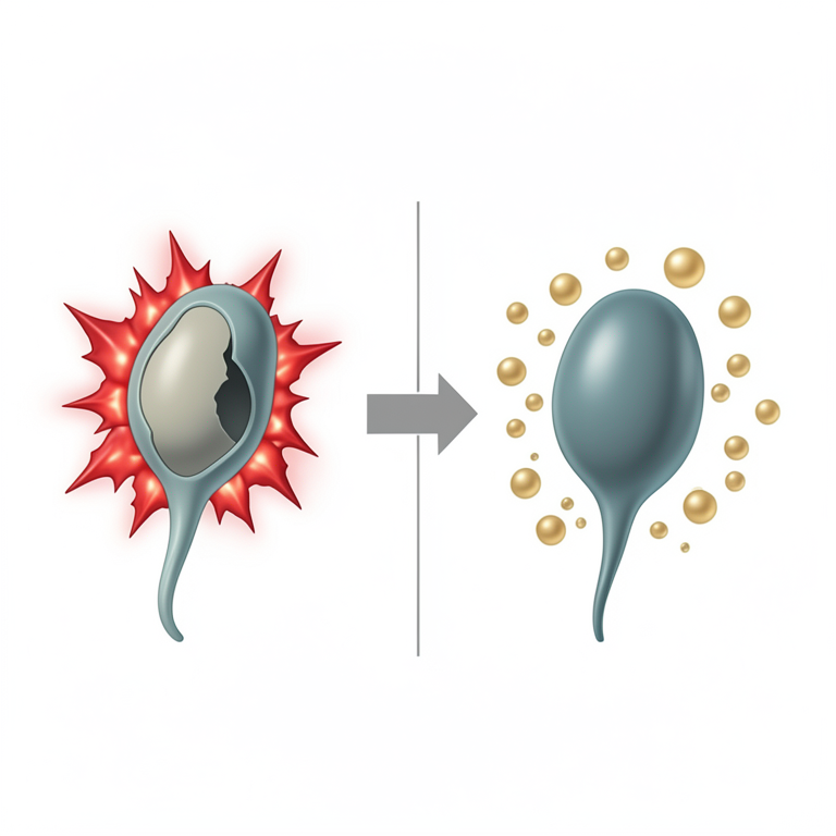
Mechanism of Vitamin E in Reducing Oxidative Stress
AI-generated conceptual illustration (human sperm cells). It explains the idea and does not show study data.
View source evidence ↗Figures & tables
From the original paper Table 1
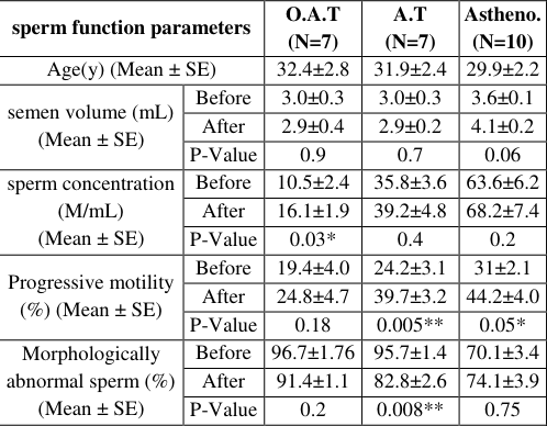 Table 2
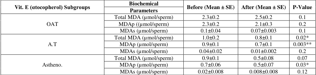 Table 3
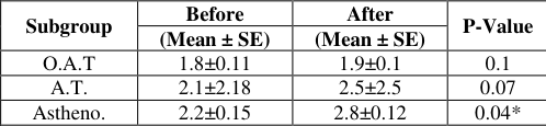 Table 5
 Table 4
 Table 6
 Table 6
Introduction
Infertility is a world health problem that affects an estimated 15% (1 out of 7) couples worldwide, and can be caused by abnormalities in generating of viable gametes, and defects in fertilization and embryonic development [1] Many studies in the area of male infertility have been focused on oxidative stress-related mechanisms of sperm damage, Oxidative stress occurs when cells no longer have the antioxidant capacity to manage the excess production of ROS [2] . However, high generation of ROS (superoxide, hydroxyl, hydrogen peroxide, nitric oxide, peroxynitrile) by immature and abnormal spermatozoa, contaminating leukocytes, sperm processing (excessive centrifugation, cryopreservation / thawing), accompanied by low scavenging and antioxidant levels in blood serum and seminal plasma, will induce a state of oxidative stress. Therefore, seminal oxidative stress (OS) develops as a result of an imbalance between ROS generating and scavenging activities [3][4] . On the other hand, human spermatozoa are capable of generating controlled low amounts of endogenous ROS, which play a significant role inducing sperm capacitation/acrosome reaction and acquisition of sperm-fertilizing ability [5] . The tocopherols, specifically α-tocopherol (vitamin E), have been studied extensively in mammalian researches as membrane stabilizers and antioxidants that scavenge oxygen free radicals, lipid peroxy radicals, and singlet oxygen. The antioxidant properties of α-tocopherol are the result of its ability to quench both singlet oxygen and peroxides [6][7] .
Eight fat-soluble substances, including tocopherols and tocrerinol, make up vitamin E. [8] . Tocopherol, one of these types, is popular in North American diet and is present in maize oil, soybean oil, ghee, and dressings. The second type of vitamin as a fat-soluble antioxidant, alpha-tocopherols is one of the most physiologically active tocopherols in North America because they prevent the formation of reactive oxygen species when lipids are subjected to oxidation [9] . The eight vitamin E forms are divided into two categories; four of them are tocopherols and the other four are tocotrienols, and they are identified by the prefixes α, β, γand δ [10] .
Materials and Methods
Materials and Methods
Sample collection:
The current study involved a control group (N=23), who consisted of sample obtained from healthy donors of proven fertility and normal genital examinations, and a patients group (N=24), who had been unable to initial pregnancy during a period of at least one year of unprotected sexual intercourse and diagnosed according to the protocol described by WHO (WHO, 2010). Both groups their ages ranged from (23-41) years. The patient' group (vit E group) were given a Tablet of vitamin E (αtocopherol) 400 mg/daily for three months. Which was subdivided into three different subgroups: oligoasthenoteratozoospermia (OAT), Asthenoteratozoospermia (A.T), and Asthenozoospermia (Astheno.). The exclusion criteria which were considered excluding patients with diabetes, high blood pressure or leucopenia in this study.
Standard Semen Analysis
Freshly ejaculated semen samples were obtained by masturbation after (3-5) days of sexual abstinence. The specimens were placed in an incubator at 37 °C for (15-30) minutes to allow liquefaction, after liquefaction, semen samples were evaluated for semen volume, appearance. pH and viscosity. Then specimens were analyzed for sperm concentration, progressive motility and normal morphology according to WHO criteria (WHO, 2010).
Measurement of Malondialdehyde (MDA) in Semen Fluid
Malondialdehyde (MDA) levels were measured in both sperm and seminal plasma according to method described by Rao et al., 1989; MDA was assessed using the thiobarbituric acid reaction (TBAR) method [11] , as follows:
Sperm MDA assay (MDAp)
A volume of 1 ml of seminal plasma was centrifuged at (400xg) for sperm 10 minutes. After washing twice in a solution of sodium chloride (Nacl), (0.9%), the sperm pellet was re-suspended in 1ml of distilled water into a glass tube.
Another 1 ml of distilled water was transferred to another glass tube that served as a blank. To each of the tubes (blank and assay) 500 µl of TBA (Thiobarbituric acid,) reagent was added. Each tube was then covered with foil and heated for 1h in a boiling water both. After cooling, each tube was centrifuged to 10 minute at (4000 xg) and the absorbance of the supernatant was read on a spectrophotometer at wavelength of 534 nm.
Seminal plasma MDA Assay (MDAs)
MDA level were analyzed using (Reo et al, 1989); method with a slight modification. This modification involved using 0.5 ml of seminal plasma rather than 0.1 ml as suggested in the original paper. A volume of 0.5 ml of seminal plasma was added to 0.5 ml distilled water in a glass tube. To each tube, 0.5 ml of thiobarbituric acid reagent was added and then heated for 1 hour in a boiling water bath. After cooling, each tube was centrifuged for 10 min. at 4000 xg and the supernatant absorbance was measured at of 534 nm.
Vitamin E (α-tocopherol) Measurement
In this study, a simple and rapid high performance liquid chromatography (HPLC) was used for determination of vitamin E (α-tocopherol) concentration in human semen plasma a according to Catignani and Bieri, 1983 method [12] . To prepare a 5 g/one litter stock standard vitamin E solution. 0.05 grams of standard vitamin E (α-tococpherol) was dissolved in 10 ml of ethanol. The solution was thoroughly mixed using vortex until complete dissolution of the vitamin E. A working solution was prepared by dilution of each of above standard solution 100-fold with ethanol. The concentration of the working solution was then calculated by HPLC.A series of calibrators was then prepared with the appropriate volume of ethanol to final vitamin E (αtocopherol) concentration of µmol/L. A standard curve was plotted between α-tocopherol standard concentrations against peak area of each standard concentration. The concentration of vitamin E is generally reported in µmol/L after appropriate correction for sample size dilution.
Statistical Analysis
Statistical analyses were performed using the SSPS for Windows release 21. All values were given as mean with corresponding standard error. (Mean ± S.E) student's depended up on tests, (Tukey's test for multiple comparisons) by ANOVA Table of variance were used for data analysis. Correlations between individual variables were examined by Pearson correlation coefficient.
Results
Results
Conventional semen Analysis
The results of certain sperm function parameters between the infertile and healthy control groups as shown in Table 1.
| Group | Age (y) (Mean ± SE) | Semen volume (mL), (Mean ± SE) | Sperm concentration (M/mL), (Mean ± SE) | Progressive motility (%), (Mean ± SE) | Morphologically abnormal sperm (%), (Mean ± SE) |
|---|---|---|---|---|---|
| Control group | 33.4±1.7 | 3.2±0.18 | 69.4±3.6 | 64.2±2.5 | 67±2.4 |
| Infertile group | 32.5±2.1 | 2.77±0.12 | 13.3±3.4 | 24.1±1.5 | 85.14±1.36 |
| P-Value | 0.7 | 0.8 | 0.001 | 0.001 | 0.001 |
Table text extracted from the original PDF; superscript letters are shown inline. The original table image is below.
View original table image
Measurement of Malondialdehyde (MDA)
Sperm and seminal plasma samples' MDA levels were assessed using the 2-thiobarbituric acid method (Rao et al., 1989), in order to evaluate the oxidative stress detected in patients with different types of infertility present in studied patients. The results of Total MDA, MDAp (In sperm) and MDAs (In semen plasma) in patients groups were (1.486±0.13, 1.42±0.12 and 0.05±0.01 respectively) whereas their concentration were found in control group (0.25±0.016, 0.243±0.015 and 0.009±0.002 respectively). It is clear that, there were a highly significant decrease in total MDA, MDAp values and a significant decrease in MDAs values of patient's group comparison with that of the control group. The results of vit E subgroups indicated presence of that no significant decrease in Total MDA, MDAp and MDAs in O.A.T. subgroup, while it was significantly decreased in Total MDA and a highly significant decrease of MDAp in A.T. of vit E subgroups after treatment when compared with that of before treatment. (Table : 1-3) Which the other subgroups of vit E group, no significant difference were found in Total MDA, and MDAs, whereas significant decrease was percent in MDAp in asthenospermia of vit. E subgroup after treatment compared with that of before treatment.
The result (Mean ± SEM) of MDAs percent in patients group were (3.42±0.8) and MDAp percent (96.5±0.8), whereas in control group the values of percent were found to be (3.52±0.5) in MDAs and (96.3±0.5) in MDAp. The results showed no significant difference between the percentage of MDA (%) of patients group and control group. The results of the MDAs and MDAp percentages of vit E group is presented in (Table 1234).
Discussion
In recent years the generations of ROS (reactive oxygen species) in male reproductive tracts have become a real concern because of their potential toxic effects at high levels. Which are short-lived, unstable, and highly reactive species containing at least one oxygen atom, are able to snatch electrons from other molecules to achieve an electronically-sTable state. In this process, the other molecule loses an electron following which a new radical is formed. Subsequently, this radical reacts with another neighboring molecule, thus passing on the radical status via a reaction called 'radical-chain reaction' until two radicals react with one another forming a stable bond. These reactions amplify the degree of alterations in the cellular structures [13] . These free radicals induced oxidative damage to spermatozoa; this in turn causes poor sperm function and infertility [13][14] . Polyunsaturated fatty acids (PUFAs) have been hypothesized to make up a significant portion of the plasma membrane of spermatozoa and their cytoplasm has a low level of scavenging enzymes. They are consequently more vulnerable to the harm caused by oxidative stress [15] . Increased cell permeability, enzyme deactivation, structural DNA damage, and cell death are all consequences of lipid peroxidation (LPO), which causes the loss of membrane integrity [16] . Malondialdehyde (MDA), one of the byproducts of LPO, has been utilized in biochemical assays to measure the level of oxidative damage to spermatozoa [17] . When the MDA concentration was measured throughout the current study in the sperm (MDAp) and seminal plasma (MDAs), the results (Table 1-3) indicated that MDA concentration (represented as percentage) was higher in the sperm, than in the seminal plasma in both fertile and infertile groups. The sperm plasma membrane contains a high amount of PUFA s, which is vulnerable to attack by ROS [18][19] . ROS in the seminal fluid has been reported to originate from two sources leukocytes & immature spermatozoa [19] . Since the patients with leukocytspermia were excluded from the present study, it can be concluded that the source of ROS in the semen sample of the present study was the immature spermatozoa α-tocopherol (vitamin E) which is well -documented antioxidant and has been shown to inhibit free radical-inducing damage to sensitive cell membrane [21] . In the other hand, the use of vitamin E in vitro has been also documented to improve sperm motility and viability [21] , Whereas in other studies there was no effect of even higher level of vitamin E (600-1200) mg/dl for three weeks [22] , alone or in combination with vitamin C [23] . To investigate the role of vitamin E in the O.S present in the studied infertile group (vit E group), the [MDA] level were measured in vit E group before and after oral administration of vitamin E in a dose of 400 mg/dl for 3 months. Vitamin E is considered as the major chain breaking antioxidant in the sperm plasma membrane. Because of its presence within plasma membrane, It neutrale hydrogen peroxide H2O2, superoxide anion (O2˙ֿ ) and hydroxyl radical (HO˙), and protect the plasma membrane from LPO by scavenging peroxyl (ROO˙) and alkoxyl (RO˙) radicals there, by breaking the chain reaction [21][22][23][24] .The observed highly significant increase (p≤0.001) in vitamin E concentration in vit E group and the significant increase observed (p≤0.05) in the Astheno. Subgroup (Table 1-3) after such treatment in comparison with that of before treatment improved semen parameters. Therefore, a highly significant increase in progressive motility, and significant increase in sperm concentration in vit E group was observed in the present study, with presence of non-significant difference in normal morphology. As well, positive correlation with a highly significant difference (p≤0.001) was detected between vitamin E concentration and each of: sperm concentration& progressive motility (Table : 1-5). Therefore, administration of vitamin E may enhance fertility by decreasing freeradical damage detected by the reduction of MDA concentration [25][26][27] .
Conclusion
Oxidative stress (O.S) contributes most likely to the damaged sperm in the patients included in the present study and the conclusion achieved is based on the following results; The higher level of ROS (MDA concentration) is present in infertile patients than normal group.as well as highly significant decrease in antioxidant capacity αtocopherol (vitamin E) in seminal plasma of infertile patients group when compared with that of control group, this contribution is through impairment of semen parameters which include (Sperm concentration, progressive motility and normal morphology) of infertile patients group. Oral administration α-tocopherol (vitamin E) contributed most likely to improved sperm motility and sperm concentration after treatment, by reducing MDA concentration in infertile patients.
Figures and tables from the paper
| sperm function parameters | — | O.A.T (N=7) | A.T (N=7) | Astheno. (N=10) |
|---|---|---|---|---|
| Age(y) (Mean ± SE) | 32.4±2.8 | 31.9±2.4 | 29.9±2.2 | |
| semen volume (mL) (Mean ± SE) | Before | 3.0±0.3 | 3.0±0.3 | 3.6±0.1 |
| After | 2.9±0.4 | 2.9±0.2 | 4.1±0.2 | |
| P-Value | 0.9 | 0.7 | 0.06 | |
| sperm concentration (M/mL) (Mean ± SE) | Before | 10.5±2.4 | 35.8±3.6 | 63.6±6.2 |
| After | 16.1±1.9 | 39.2±4.8 | 68.2±7.4 | |
| P-Value | 0.03* | 0.4 | 0.2 | |
| Progressive motility (%) (Mean ± SE) | Before | 19.4±4.0 | 24.2±3.1 | 31±2.1 |
| After | 24.8±4.7 | 39.7±3.2 | 44.2±4.0 | |
| P-Value | 0.18 | 0.005** | 0.05* | |
| Morphologically abnormal sperm (%) (Mean ± SE) | Before | 96.7±1.76 | 95.7±1.4 | 70.1±3.4 |
| After | 91.4±1.1 | 82.8±2.6 | 74.1±3.9 | |
| P-Value | 0.2 | 0.008** | 0.75 |
Table text extracted from the original PDF; superscript letters are shown inline. The original table image is below.
View original table image
| Vit. E (αtocopherol) Subgroups | Biochemical | Before (Mean ± SE) | After (Mean ± SE) | P-Value |
|---|---|---|---|---|
| Parameters | ||||
| OAT | Total MDA (µmol/sperm) | 2.3±0.2 | 2.5±0.2 | 0.1 |
| MDAp ((µmol/sperm) | 2.3±0.2 | 2.1±0.3 | 0.2 | |
| MDAs (µmol/sperm) | 0.1±0.04 | 0.07±0.003 | 0.1 | |
| A.T | Total MDA (µmol/sperm) | 1.0±0.2 | 0.8±0.1 | 0.02* |
| MDAp (µmol/sperm) | 0.9±0.1 | 0.7±0.1 | 0.003** | |
| MDAs (µmol/sperm) | 0.04±0.02 | 0.01±0.002 | 0.2 | |
| Astheno. | Total MDA (µmol/sperm) | 0.9±0.1 | 0.5±0.08 | 0.07 |
| MDAp (µmol/sperm) | 0.7±0.06 | 0.5±0.07 | 0.03* | |
| MDAs (µmol/sperm) | 0.02±0.008 | 0.008±0.008 | 0.12 |
Table text extracted from the original PDF; superscript letters are shown inline. The original table image is below.
View original table image
| Subgroup | Before | After | P-Value |
|---|---|---|---|
| (Mean ± SE) | (Mean ± SE) | ||
| O.A.T | 1.8±0.11 | 1.9±0.1 | 0.1 |
| A.T. | 2.1±2.18 | 2.5±2.5 | 0.07 |
| Astheno. | 2.2±0.15 | 2.8±0.12 | 0.04* |
Table text extracted from the original PDF; superscript letters are shown inline. The original table image is below.
View original table image
| Vit. E group | Before | After | P-Value |
|---|---|---|---|
| MDAp% | 95.9±1.17 | 97.9±0.9 | 0.9 |
| MDAs% | 4±1.17 | 2±0.9 | 0.9 |
Table text extracted from the original PDF; superscript letters are shown inline. The original table image is below.
View original table image
| Biochemical parameters | Sperm concentration (M/ml) | Progressive motility (%) | Normal morphology (%) |
|---|---|---|---|
| r p | r p | r p | |
| Total MDA (µmol/sperm) | - 0.562 0.004** | - 0.429 0.03** | - 0.318 0.3 |
| MDAp (µmol/sperm) | - 0.778 0.0001** | - 0.583 0.003** | - 0.405 0.05 * |
| MDAs(µmol/sperm) | - 0.562 0.004** | - 0.529 0.008** | - 0.231 0.2 |
| Vitamin E concentration (µmol/l) | 0.308 0.1 | 0.178 0.7 | 0.461 0.06 |
Table text extracted from the original PDF; superscript letters are shown inline. The original table image is below.
View original table image
| Biochemical parameters | Sperm concentration (M/ml) | Progressive motility (%) | Normal morphology (%) |
|---|---|---|---|
| r p | r p | r p | |
| Tota MDA(µmol/sperm) | 0.791 0.0001** | 0.695 0.0001** | 0.384 0.06 |
| MDAp(µmol/sperm) | 0.774 0.0001** | 0.624 0.003** | 0.395 0.07 |
| MDAs(µmol/sperm) | 0.629 0.01* | 0.557 0.005** | 0.202 0.3 |
| Vitamin E concentration (µmol/l) | 0.550 0.005** | 0.454 0.001** | 0.296 0.1 |
Table text extracted from the original PDF; superscript letters are shown inline. The original table image is below.
View original table image
References
27 in the original paper- Arhin SM, Mensah KB, Agbeno E. Pharmacotherapy of infertility in Ghana: retrospective study at the cape coast teaching hospital. Journal of Pharmaceutical Policy and Practice. 2019;12:28.
- Dutta S, Majzoub A, Agarwal A. Oxidative stress and sperm function: A systematic review on evaluation and management. Arab Journal of Urology. 2019;17(2):8797.
- Herwig R, Knoll C, Planyavsky M, Pourbiabany A, Greilberger J, Bennett K. Proteomic analysis of seminal plasma from infertile patients with oligoasthenoteratozoospermia due to oxidative stress and comparison with fertile volunteers. Fertility and Sterility. 2013;100(2):355-366.e2.
- Hosen M, Slam M, Begum F, Kabir Y, Howlader M. Oxidative stress induced sperm DNA damage, a possible reason for male infertility. Iranian Journal of Reproductive Medicine. 2015;13(9):525-532.
- Chakraborty S, Saha S. Understanding sperm motility mechanisms and the implication of sperm surface molecules in promoting motility. Middle East Fertility Society Journal. 2022;27(1):4. https://doi.org/10.1186/s43043-022-00094-7.
- Niki E, Saito T, Kawakami A, Kamiya Y. Oxidation of lipids. VI. Inhibition of oxidation of methyl linoleate in solution by vitamin E and vitamin C. Journal of Biological Chemistry. 1984;259(7):4177-4182. PMID: 6706998
- Morita M, Naito Y, Yoshikawa T, Niki E. Plasma lipid oxidation induced by peroxynitrite, hypochlorite, lipoxygenase and peroxyl radicals and its inhibition by antioxidants as assessed by diphenyl-1pyrenylphosphine. Redox Biology. 2016;8:127-135. https://doi.org/10.1016/j.redox.2016.01.005.
- Brigelius-Flohé B. Vitamin E, the shrew waiting to be tamed. Free Radical Biology & Medicine. 2009;46(5):543-554.
- Packer L, Weber SU, Rimbach G. Molecular aspects of α-tocotrienol antioxidant action and cell signaling. Journal of Nutrition. 2001;131(2):369S-73S.
- Mohammad K, Moslemi ST. Selenium-vitamin E supplementation in infertile men: effects on semen parameters and pregnancy rate. International Journal of General Medicine. 2011;4:99-104.
- Rao B, Souflir JC, Martin M, David G. Lipid peroxidation in human spermatozoa as related to midpiece abnormalities and motility. Gamete Research. 1989;24(2):127-134.
- Catignani GL, Bieri JG. Simultaneous determination of retinol and a-tocopherol in serum or plasma by liquid chromatography. Clinical Chemistry. 1983;29(4):708712.
- Shang YL, Zhu FX, Yan J, Chen L, Tang WH, Xiao S, et al. Novel DPY19L2 variants in globozoospermic patients and the overcoming this male infertility. Asian Journal of Andrology. 2019;21(2):183-189.
- Agarwal A, Virk G, Ong C, du Plessis SS. Effect of oxidative stress on male reproduction. World Journal Men's Health. 2014;32(1):1-17.
- Bisht S, Faiq M, Tolahunase M, Dada R. Oxidative stress and male infertility. Nature Reviews Urology. 2017;14(8):470-485.
- Lavranos G, Balla M, Tzortzopoulou A, Syriou V, Angelopoulou R. Investigating ROS sources in male infertility: A common end for numerous pathways. Reproductive Toxicology. 2012;34(3):298-307.
- Saleh RA, Agarwal A, Nada EA, El-Tonsy MH, Sharma RK, Meyer A, et al. Negative effects of increased sperm DNA damage in relation to seminal oxidative stress in men with idiopathic and male factor infertility. Fertility and Sterility. 2003;79(Suppl 3):1597-1605.
- Aitken RJ. Molecular mechanisms regulating human sperm function. Molecular Human Reproduction. 1997;3(3):169-173.
- Gautam S, Chawla B, Bisht S, Tolahunase M, Dada R. Journal of the Anatomical Society of India Impact of mindfulness based stress reduction on sperm DNA damage. Indian Journal of Rheumatology. 2018;67(2):124-129.
- Layek SS, Mohanty TK, Kumaresan A, Parks JE. Cryopreservation of bull semen: Evolution from egg yolk based to soybean based extenders. Animal Reproduction Science. 2016;172:1-9.
- Kowalczyk A. The Role of the Natural Antioxidant Mechanism in Sperm Cells. Reproductive Sciences. 2022;29(5):1387-1394. DOI: 10.1007/s43032-021-00795-w.
- Moilanen J, Hovatta O, Lindroth L. Vitamin E levels in seminal plasma can be elevated by oral administration of vitamin E in infertile men. International Journal of Andrology. 1993;16(2):165-1666.
- Rolf C, Cooper TC, Yeung CH. Antioxidant treatment of patients with asthenospermia or moderate oligoasthenospermia with high-dose of vitamin C and vitamin E, a randomized, placebo -controlled, doubleblind study Hum. Reproductive. 1999;14:1028-1033.
- Halliwell B. Free radicals and vascular disease: how much do we know? BMJ. British Medical Journal. 1993;307(6909):885-886. DOI: 10.1136/bmj.307.6909.885.
- Geva E, Bartoov B, Zabludovsky N. The effect of antioxidant treatment on human spermatozoa and fertilization rate in an in vitro fertilization program. Fertility and Sterility. 1996;66(3):430-434.
- Lavranos G, Balla M, Tzortzopoulou A, Syriou V, Angelopoulou R. Investigating ROS sources in male infertility: A common end for numerous pathways. Reproductive Toxicology. 2012;34(3):298-307.
- Ourique GM, Saccol EMH, Pês TS, Glanzner WG, Schiefelbein SH, Woehl VM, et al. Protective effect of vitamin E on sperm motility and oxidative stress in valproic acid treated rats. Food and Chemical Toxicology. 2016;95:159-167. DOI: 10.1016/j.fct.2016.07.011.