
Research article · 2023 · Full text
International Journal of Biology SciencesPrevalence of mastitis causing Streptococcus in selected farms in Bishoftu Town, East Shewa, Central Ethiopia
Volume 5(1) · Pages 82–86 · DOI: 10.33545/26649926.2023.v5.i1b.149 ↗
Research areasMilk Quality and Mastitis in Dairy CowsMicrobial infections and disease research
Abstract
Author-writtenStreptococci is among mastitis causing bacterial pathogens in dairy farming in Ethiopia. Accordingly, the cross-sectional study to isolate Streptococcus from two selected intensive dairy farms and mastitis cases from Veterinary Teaching Hospital clinic in Addis Ababa University, college of Veterinary Medicine; from July to September, 2021 in Bishoftu town, central Ethiopia. A total of 52 milk samples were collected from the individual lactating cows through randomly sampling from the farms apparently health and purposively from clinical case from veterinary teaching hospital. The milk samples from both clinically infected and apparently health lactating cows and conducting culturing characterization (colony morphology and hemolysis), Gram staining (shape, arrangement and color) and Biochemical tests (catalase and oxidase) to differentiate Streptococcus from Staphylococcus as Streptococcus is catalase negative while Staphylococcus is catalase positive. The overall prevalence of streptococci isolated from both milks collected from the clinical and subclinical cow was 18/52 (34.6%) in which the prevalence of Streptococcus in milks collected from clinical case and apparently health milk collected from farm were 41.7% and 32.5% respectively with no significant differences (P-value = 0.3). Also the prevalence of Streptococcus were 33.3%, 45.5% and 30% at early, mid and late lactating period respectively with significant differences (P-value = 0.05). This indicated that those cows at mid period were highly infected with the Streptococcus. Therefore, good farm managements, hygiene of animal’s house, maintenance of temperature and ventilation and good milking practice, such as teat disinfection and drying, as well as regular cleaning and checking of the milking machine should be recommended for all farms.
Summary
Source-checkedPlain-language reading of the full paper, prepared with AI and checked against the text. Each point links to the authors' own words.
Why it matters
- Ethiopia possesses the largest livestock population in Africa, with approximately 7.2 million dairy cows, but bovine mastitis serves as a primary economic obstacle for these cattle. View evidence ↗
- Streptococci are significant pathogens in bovine mastitis, negatively affecting the health of the cow as well as the quality and productivity of the milk. View evidence ↗
- In urban and peri-urban areas of Addis Ababa, economic losses from mastitis are estimated at US$58 and 78.65 per cow and per lactation, respectively, due to factors like reduced yield and treatment costs. View evidence ↗
The question
- The study aimed to isolate Streptococcus from dairy cow milk in the study area to estimate its prevalence and assess associated risk factors. View evidence ↗
What they did
- The researchers employed a cross-sectional study design conducted between July and September 2021. View evidence ↗
- A total of 53 milk samples were collected: 20 from the Ethiopian Meat and Milk Institute, 20 from the CVMA farm, and 13 purposively sampled clinical cases from the CVMA Teaching Hospital. View evidence ↗
- Streptococcus was identified using culturing characterization, Gram's staining, and biochemical tests, specifically using the catalase test to distinguish it from Staphylococcus. View evidence ↗
- The study used descriptive analysis with percentages to process the raw data. View evidence ↗
What they found
- The overall prevalence of streptococci in the sampled milk from both clinical and subclinical cows was 34.6% (18 out of 52 samples). View evidence ↗
- Prevalence was 41.7% in clinical cases and 32.5% in apparently healthy cows, a difference that was not statistically significant (P-Value = 0.3). View evidence ↗
- Cows in the mid-lactation period had the highest prevalence of Streptococcus at 45.5%, compared to 33.3% in early and 30% in late periods (P-value = 0.05). View evidence ↗
- Young cows showed a higher prevalence of Streptococcus (37.9%) compared to adult cows (30.4%), with a significant P-value of 0.03. View evidence ↗
- The prevalence of Streptococcus was higher at the CVMA farm (50%) than at the EDIDI farm (15%). View evidence ↗
What it means
- The authors conclude that poor hygiene and management practices within the dairy farms allowed for the spread of environmental and contagious infections. View evidence ↗
- The authors recommend implementing good milking practices, including regular machine maintenance and teat disinfection and drying. View evidence ↗
- The authors state that further detailed studies are needed to specifically identify the different groups of Streptococci involved. View evidence ↗
Visual summary
3 visualsIllustrations explain the paper; charts are replotted from the paper's own table values.
Bacterial Identification Process
AI-generated conceptual illustration (cow milk and Streptococcus bacteria). It explains the idea and does not show study data.
View source evidence ↗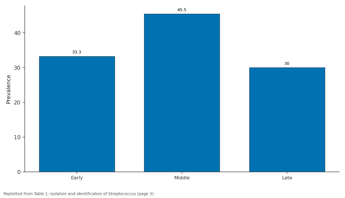
Prevalence: Early vs Middle vs Late
Replotted from Table 1 (page 3) of the original paper. Every bar is the paper's own table value.
View source evidence ↗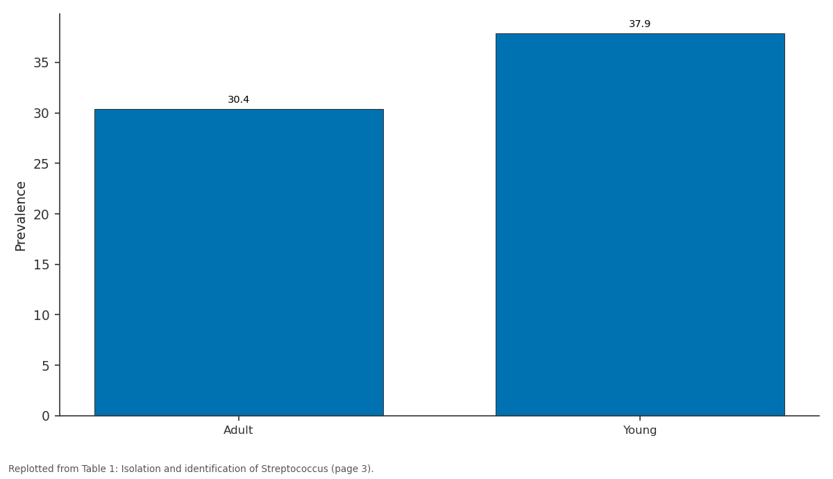
Prevalence: Adult vs Young
Replotted from Table 1 (page 3) of the original paper. Every bar is the paper's own table value.
View source evidence ↗Figures & tables
From the original paper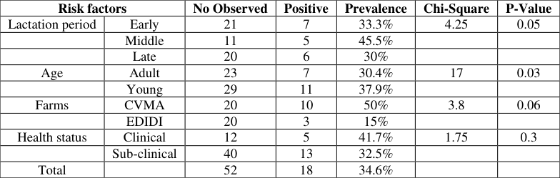 Table 1
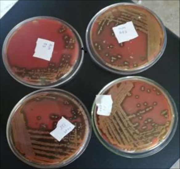 Figure 1
Introduction
Ethiopia has the largest livestock population of African country with estimated 7.2 million dairy cows that represents a major national resource and form an integral part of the agricultural production system, however, the infectious disease including bovine mastitis is the main economic obstacle disease of dairy cattle.in developing countries, including Ethiopia [10,22] . It is categorized as the second infectious disease loss directly milk production with higher occurrence frequency the farmer to culling of dairy cows [20] . Bovine mastitis, a complex multi-factorial disease resulted from pathogenic agents invade the udder, multiplying, and often producing toxins that have a significant detrimental effect on the mammary tissue itself and the general health status of the animal [1] . Over 130 pathogens are known to be associated with bovine mastitis, some of them belonging to the Streptococcus genus [13] . Streptococci are among the major mastitis pathogens which have a considerable impact on cow health, milk quality, and productivity [18] . Streptococcus uberis is one of the most common mastitis pathogens found in dairy herds throughout the world and is responsible for a significant proportion of clinical and subclinical mammary gland infections [11] . The disease in dairy herds is of great economic importance due to a reduction in the milk yield, the change in milk quality, the possibility of permanent change to one or more quarters, or even to the entire udder, and death of the cow as well as loss due to increased premature culling rate and cost of treatment [17] . Streptococci are Gram-positive bacteria of spherical shape (0.5-2 µm) that usually form pairs or chains.
They are classified on the basis of colony morphology, hemolysis, and serologic specificity into the Lancefield group taxonomic system [13] . S. agalactiae, S. dysgalactiae, and S. uberis is usually associated with bovine [13] . Streptococcus uteri is commonly isolated from the dairy farms with clinical and subclinical mastitis infections [15] . Acquaintance through environmental streptococci arises during and between milking, during the dry period, or prior parturition of heifers [13] . Chronically affected animals could contribute particularly to the transmission of the bacterium during milking [15] . It can persist in the environment up to months, particularly under moist cool conditions. It can be transmitted by direct contact, fomites, or ingestion of not well-cocked meat, or unpasteurized cow milk of infected cows. These organisms classically inhabit as commensals and can cause opportunistic infections in those hosts. It can be found occasionally in humans [21] . Group B Streptococci is a bovine pathogen causing inframammary infections. The likelihood of GBS interspecies transmission is largely unknown. On-farm interspecies transmission of GBS between people and cattle is possible and a strain is capable of colonizing different hosts and environments, including the human throat and rectum, the bovine udder and rectum, and the farm environment. Other strains, which are currently mainly found in cattle, such as members of CC61/67 and CC103, could emerge from the bovine reservoir as a commensal or pathogen of humans [5] . Mastitis is an important in Ethiopia where no routine prevention and control practices are in place [2] . In Ethiopia, subclinical mastitis is considered to be an important challenge for the dairy development [5] . Mastitis has been contributing to reduce milk production and a major source of economic loss to the dairy industry, through reduced milk yield and quality, cost of drugs and veterinary treatment, discarded milk, and forced culling estimated the economic losses from urban and peri-urban areas of Addis Ababa, to be US$58 and 78.65 per cow and per lactation, respectively. In addition to its economic impact, Streptococcus agalactiae, Group B Streptococcus (GBS), is the major etiologic agent of invasive neonatal infections in humans in industrialized countries, causing sepsis, pneumonia, meningitis, Osteomyelitis, and soft tissue infections [16] . In Ethiopia, a few studies have been conducted with the purpose of estimating the occurrence of bovine mastitis due to Streptococcus. So that information about the prevalence of the Streptococcus and factors associated with the disease as well as the pathogen involved is essential in designing prevention and control measures against the disease. Therefore, the current study was conducted to estimate the prevalence, to assess the risk factors, and to isolate the Streptococcus from the dairy cow milk in the study area through the Morphological characterization of the colony on Blood agar culture, Gram's staining and Biochemical characterization of the isolates.
Materials and Methods
Materials and Methods
Study design, sample size determination and study population
A cross-sectional study was conducted from July to September 2021. Milk samples were collected from the individual lactating cows through randomly sampling from the farms and purposively from clinical cases from veterinary teaching hospitals. Sample size was determined based on time availability and accessibility of dairy farms. Totally, 53 samples were collected from apparently lactating cows from CVMA farm and Ethiopian Meat and Dairy Industry Development Institute farm randomly and clinical case with mastitis's sign from Veterinary teaching hospital in CVMA purposively. Accordingly, 20 milk samples from Ethiopian meat and Milk Institute, 20 samples from CVMA farm, and 13 clinical cases from CVMA Teaching Hospital. Specifically, the study was target to isolate and identifying the streptococci that could cause the mastitis in lactating cows by taking the milk samples from both clinically infected and apparently healthy lactating cows. All milk samples were collected from cows with clinical mastitis together with samples without clinical signs. Milk samples were collected promptly into sterile tubes, and then the samples were stored on ice and immediately transported to our laboratory for analysis within 6 hours of sampling.
Study methodology 2.2.1 Clinical examination of the udder
The udders were examined visually and then palpated to detect any possible fibrosis, inflammatory swelling, and atrophy of the tissue by a veterinary practitioner. The size and consistency of the mammary quarter were inspected for the presence of any abnormalities such as disproportional symmetry, swelling, firmness, and blindness of the teat canal. In addition, two streaks of milk from each quarter in a strip cup were inspected visually for the presence of any flakes, clots, pus, watery appearance, blood, and color change.
Bacterial isolation and identification
Streptococcus was isolated by conducting culturing characterization (colony morphology and hemolysis), Gram's staining (shape, arrangement, and color) and Biochemical tests (catalase and oxidase) to differentiate Streptococcus from Staphylococcus as Streptococcus is catalase negative while Staphylococcus is catalase positive. 10 μl was streaked onto 5% sheep blood agar (Blood agar plate, Creative Life Science, Taiwan) and incubated aerobically at 35-37 °C for 18-24 hours. After incubation, bacteria were recognized as streptococci based on colony morphology and Gram's staining [7] . Firstly, identification of the bacteria was performed by appreciating the growth on a blood agar plate. Then followed by colony morphology gross examinations (colony size, shape, and color), hemolysis (presence or absence and type of hemolysis), gram stain staining (Gram-positive or negative, bacterial shape, structure, arrangement), then Gram-positive culture on nutrient agar which then were incubated aerobically at 37 °C for 24 to 48 hours for growth of bacteria. Identification of the bacteria on primary culture was done on the basis of colony morphology, hemolytic characteristics, and Gram's stain reaction including shape and arrangements of the bacteria. The small-medium sized colonies that were hemolytic or non-hemolytic on 5% sheep blood agar and yielding gram-positive cocci were sub-cultured onto nutrient agar to obtain a pure isolate for further identification and subjected to catalase and oxidase tests.
Data analysis
The raw data generated from the study were arranged, organized, coded, and entered into an Excel spreadsheet (Microsoft Office Excel 2010). Then the data was analyzed through descriptive analysis with percentages. The results of analyses were mostly described in proportion. Proportion was estimated as the numbers of samples detected positive to Streptococcus from the total sample tested.
Results and Discussion
Results and Discussion
Prevalence of Streptococci species
Streptococci are reported to be among the main pathogens causing bovine mastitis all over the world [13] . Streptococci are regularly isolated from mastitis infected in dairy cows with only incomplete information about the antimicrobial susceptibility of these organisms [14] . The most common risk factors associated with mastitis caused by Streptococcus is milking hygiene and practical management [7] . Isolation and identification of Streptococcus causing mastitis is important for a rapid treatment, control, and prevention of bovine mastitis circulating in dairy farms by giving information on the prevalence of Streptococcus to decision-makers and stakeholders (Kabelitz et al., 2021) [31] . Therefore, the aim of the current study was to determine the prevalence of Streptococcus causing mastitis in Bishoftu Town, central Ethiopia. The overall prevalence of streptococci isolated from both milks collected from the clinical and subclinical cows was 18/52 (34.6%) (Table 1). The prevalence of streptococci obtained in the current study was higher than that reported by Fesseha et al., (2022) [10] who identified streptococci as a cause of bovine mastitis in central Ethiopia with the prevalence of Streptococcus species (24.3%) and Abebe et al., (2020) [2] who identified 18.6% Streptococcus spp as a risk factor for mastitis in his study conducted on the Prevalence, risk factors and bacterial causes of bovine mastitis in southern Ethiopia but the prevalence of Streptococcus spp obtained in the current study was lower than that obtained by Abd et al., (2020) [1] who reported Streptococcus dysagalactiae (23%) and Streptococcus agalactiae (20.1%) in his study of "Antimicrobial profile of multidrug-resistant Streptococcus spp. isolated from dairy cows with clinical mastitis. In the current study, the prevalence of streptococci isolated from the milk collected from clinical cases from Veterinary teaching hospital and apparently health cows in the College of Veterinary Medicine farm and Ethiopian Dairy Industry Development Institute farm were 41.7% and 32.5% respectively with no significant differences (P-Value = 0.3) between clinical and subclinical cases from which samples were collected. These results indicated the great differences with the report of Tora et al., (2022) [20] who reported the prevalence of Streptococci species as risk factor of Bovine mastitis with the prevalence of 10.6% and 8.1% for clinical and subclinical mastitis, respectively according to his report of the Bacterial profile of bovine mastitis in Ethiopia: A systematic review and meta-analysis. The prevalence of Streptococcus between the farm and clinical case at VTH and also among the farms were different in which CVMA farm (20%), followed by Ethiopian meat and milk institute (15%) and lower prevalence in clinical case at VTH (7.69%). The differences in environment between dairy farms, for instance, temperature, humidity, and even the design of barns, results in the difference in the dominant agents that cause mastitis. Moreover, several previous studies have determined numerous risk factors of streptococcal mastitis, such as high yields, long days in milk, multiparous cows, concave teat ends, poor sanitation, liner slips during milking and the use of straw or manure solids as bedding materials [7] . The effect of poor sanitation and improper hygienic measures inside the two dairy farms under study was clear in the current results of microbial isolation from the examined milk samples as high incidence rate of udder co-infections and high incidence rate of environmental microbes. Furthermost of the farm hygienic management of the milking areas, milk containers sanitation, udder and teats washing, separate use of towels for each dairy cow, and miller hygiene were not fully applied by most of the farm owners [13] .
| Risk factors | — | No Observed | Positive | Prevalence | Chi-Square | P-Value |
|---|---|---|---|---|---|---|
| Lactation period | Early | 21 | 7 | 33.3% | 4.25 | 0.05 |
| Middle | 11 | 5 | 45.5% | |||
| Late | 20 | 6 | 30% | |||
| Age | Adult | 23 | 7 | 30.4% | 17 | 0.03 |
| Young | 29 | 11 | 37.9% | |||
| Farms | CVMA | 20 | 10 | 50% | 3.8 | 0.06 |
| EDIDI | 20 | 3 | 15% | |||
| Health status | Clinical | 12 | 5 | 41.7% | 1.75 | 0.3 |
| Sub-clinical | 40 | 13 | 32.5% | |||
| Total | 52 | 18 | 34.6% |
Table text extracted from the original PDF; superscript letters are shown inline. The original table image is below.
View original table image
When comparing the number of samples expected to be positive by the lactating period the prevalence was 33.3%, 45.5%, and 30% at early, mid, and late lactating periods respectively with significant differences (P-value = 0.05) (Table 1). This indicated that those cows at mid-period were highly infected with the Streptococcus. Also, when the results obtained are compared by the age of the cow the obtained results indicate the prevalence of 29.7% and 37.93% for young and adults respectively. Higher incidence at early lactation period than late in the present study is in agreement with the findings of Abureema et al., (2014) [3] who indicated S. uberis to be the most common isolate at early lactation. However, Chairman et al. (2012) [6] noted S. uberis to be dominant pathogen at all stages of lactation since S. uberis mastitis is mainly the result of heavy contamination of the teats and udder with water, mud, and faecal matter at any stage during lactation [19] . however, the prevalence of lactating periods at the middle and late periods were different, the prevalence at early period was agreed with the report of Lakew et al., (2019) [16] who found the early, middle, and late (35%, 13.02% and 14.84% respectively) with the isolation and identification of Streptococcus agalactiae from farms in and around Haramaya district, eastern Ethiopia. The prevalence of streptococci in young is lower than that of adult cows. So that the result in current study was agreed with the review of Kromker et al., (2014) [15] who stated that S. uberis infections are exhibited as acute mastitis, usually during the later lactation, and showed that in older cows, prevalence increased with the lactation stage, while during early lactation, there were no significant differences between primiparous and multiparous cows. (Kromker et al., 2014) [33] . Contagious bacteria have the potential to spread within a herd easily and widely. In contrast, environmental pathogens are able to survive outside the host and are part of the normal microflora of the cow's vicinity.
Exposure through environmental streptococci occurs during and between milking, during the dry period, or prior parturition of heifers. The pathogen exposure is related to their environmental abundance, which is influenced, e.g., by humidity and temperature. Environmental pathogens invade the udder when the teat channel is opened after milking or after damage (Kabelitz et al., 2021) [31] . (Eg & Km, 2021) [9] revealed that mastitis is the major problem in smallholder dairy farms, especially the sub-clinical form, so that creation of awareness about the importance and prevention of subclinical mastitis among smallholder dairy farmers, milking infected animals and their respective quarters at last and periodic monitoring of infection status of the udder is recommended (Eg & Km, 2021) [9] . Subclinical mastitis is a complex disease and the prevalence could be affected due to variations in management system, age, milk yield, body condition, parity, environment, and other conditions. Thus, these factors might have contributed to the observed differences in prevalence of subclinical mastitis (Birhanu et al., 2017) [27] .
Conclusion and Recommendation
The overall prevalence of streptococci isolated from both milks collected from the clinical and subclinical cow was 18/52 (34.6%). The prevalence of Streptococcus between the farm and clinical case at VTH and also among the farms were different in which CVMA farm (20%), followed by Ethiopian meat and milk institute (15%) and lower prevalence in clinical case at VTH (7.69%). The results indicated that high prevalence at the farm level which are managed intensively and at the clinic level from clinical mastitis cases. Hence, poor standard hygienic and management practices inside the dairy farms which permitted the spread of both contagious and environmental infections.
4.1 Based on the above conclusion, the following recommendations were forwarded Cows are preventively treated with antibiotics during the dry-off period and infected cows should be rapidly detected and separated to avoid bacterial spreading Physical barrier that helps to prevent entrance of pathogens into the mammary gland. Should be used Good milking practices, such as teat disinfection and drying, as well as regular cleaning and checking of the milking machine. Detailed study should be conducted to investigate and identify the different groups of Streptococci
Figures and tables from the paper
References
7 in the original paper- Beilharz K, Novakova L, Fadda D, Branny P, Massidda O, Veening J. Control of cell division in Streptococcus pneumoniae by the conserved Ser/Thr protein kinase StkP. Proceedings of the National Academy of Sciences. 2012;109(15):E905-E913. http://dx.doi.org/10.1073/pnas.1119172109
- Chuang JHYYS. (N.D.). Identification of Streptococcus spp. Isolated from bovine milk and characterization of their antimicrobial susceptibility profiles in Taiwan. The Thai Journal of Veterinary Medicine. 2019;49(1):57-63.
- Feyisa A, Hassen A, Tamerat N, Abraha A. East African Journal of Veterinary and Animal Sciences. Isolation and Identification of Streptococcus uberis in Lactating Cows of Haramaya University Dairy Farm Study Area. 2017 Jun 25;1(1):7-12.
- Genzebu D, Tamir B, Berhane G. Study of reproductive and production performance of cross breed dairy cattle under Smallholders management system in Bishoftu and Akaki towns. International Journal of Advanced Research in Biological Sciences. 2016;3(2):118-123.
- Kia G, Mehdi G, Keyvan R. Prevalence and antibiotic susceptibility of Streptococcus spp. in cows with mastitis in Germi, Iran. Animal and Veterinary Sciences. 2014;2(2):31-35. https://doi.org/10.11648/j.avs.20140202.13
- Kromker V, Reinecke F, Paduch J, Grabowski N. Clinical Microbiology: Open Access Bovine Streptococcus uberis Intramammary Infections and Mastitis. Clinical Microbiology: Open Access. 2014;3:4. https://doi.org/10.4172/2327-5073.1000157
- Wirtu A, Abunna F, Duguma A. Isolation and Identification of Staphylococcus aureus from Dairy Farms in Bishoftu Town, Ethiopia; c2018. p. 1-4. https://doi.org/10.19080/JOJPH.2018.03.555604.