
Research article · 2023 · Full text
International Journal of Biology SciencesAbundances of ectoparasites of sheep in and around Gindeberet district, west Shoa zone, Ethiopia
Volume 5(1) · Pages 06–14 · DOI: 10.33545/26649926.2023.v5.i1a.139 ↗
Research areasVector-Borne Animal DiseasesViral Infections and VectorsHelminth infection and control
Abstract
Author-writtenA cross sectional study was carried out in Gindeberet district in Western Shoa Zone of Oromia regional state from June 2021 to October 2021. Sheep are the most important farm animals in our country, especial dominant livestock, providing skin and wool, and cash income in addition to food subsistence value obtained from livestock production. However, due to the abundances of ectoparasites, there is loss of skin value, income and wool due to poor management problem. The purpose of this study was to identify major ectoparasites of sheep and to determine their prevalence in the study area and to assess the magnitudes of these parasites in relation to sex, age and body condition. Out of 200 sheep examined 133 (66.5%) sheep were infested with the major ectoparasite. The major ectoparasite identified were ticks (31%), followed by lice (15.5%), fleas (12.5%), Melophagus ovinus (7%) and Mange mites (0.5%). The overall infestation with ectoparasites, was higher in females (37.5%) than males (29%). The overall prevalence in association with age categories was higher in adult (34%) than in the young (32.5%). The prevalence of major ectoparasite based on the BCS was found to be higher in poor (33.5%) than the medium (21.5%) and the good (11.5%) body condition from the overall occurrence of ectoparasites. The higher prevalence of ticks, lice, fleas, M. ovinus, and mange mites were observed in female and males. The highest prevalence of tick infestation (n=62) were observed in the area, among ectoparasite, ticks were the most prevalent parasites (n=62) with the prevalence of 31% in the area, while the prevalence of mange mites was 0.5% (N=1). Based on age classification the prevalence of ticks (17.5%), lice (9%), fleas (7.5%), Melophagus ovinus (or sheep ked) (4%), and mange mite (0.5%) were highly prevalent in the adults than young’s (13.5%), (6.5%), (5%) and (3%) respectively. From the result of this study, it is possible to conclude that ectoparasite infestation has affected in the production of sheep at the study area.
Summary
Source-checkedPlain-language reading of the full paper, prepared with AI and checked against the text. Each point links to the authors' own words.
Why it matters
- Sheep are the most important and dominant livestock in Ethiopia, providing food, wool, skin, and cash income. View evidence ↗
- The abundance of ectoparasites, often linked to poor management, leads to losses in wool, income, and the value of sheep skins. View evidence ↗
- External parasite infestations have significantly reduced skin quality, with only 10-15% of skins now remaining in the good category. View evidence ↗
The question
- The study aimed to identify the major ectoparasites affecting sheep in the Gindeberet district and determine their prevalence based on the animals' sex, age, and body condition. View evidence ↗
What they did
- A cross-sectional study was conducted in the Gindeberet district of the Western Shoa Zone, Oromia, between June and October 2021. View evidence ↗
- The researchers randomly selected a total of 200 sheep for examination. View evidence ↗
- Major ectoparasites were identified through visual observation of the sheep. View evidence ↗
- Data were managed using Microsoft Excel 2003 and analyzed using SPSS version 20 for Windows. View evidence ↗
What they found
- Overall, 66.5% (133 out of 200) of the examined sheep were infested with major ectoparasites. View evidence ↗
- Ticks were the most prevalent parasite (31%), followed by lice (15.5%), fleas (12.5%), Melophagus ovinus (7%), and mange mites (0.5%). View evidence ↗
- Infestation rates were higher in female sheep (37.5%) compared to male sheep (29%). View evidence ↗
- Adult sheep had a slightly higher overall prevalence of ectoparasites (34%) than young sheep (32.5%). View evidence ↗
- Sheep in poor body condition had the highest prevalence of ectoparasites (33.5%), compared to those in medium (21.5%) and good (11.5%) condition. View evidence ↗
- Among age groups, ticks were the most prevalent, affecting 17.5% of adults and 13.5% of young sheep. View evidence ↗
What it means
- The authors conclude that the high prevalence of ectoparasites in the study area can lead to significant economic losses due to skin damage and decreased production. View evidence ↗
- The authors recommend reducing infestations by improving management systems and implementing appropriate ectoparasite control methods. View evidence ↗
- The authors state that further research is needed to fully understand the seasonal occurrences and distribution of these parasites, as well as their specific economic impacts. View evidence ↗
Visual summary
1 visualsIllustrations explain the paper; charts are replotted from the paper's own table values.
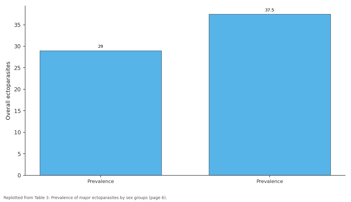
Overall ectoparasites: Prevalence vs Prevalence
Replotted from Table 3 (page 6) of the original paper. Every bar is the paper's own table value.
View source evidence ↗Figures & tables
From the original paper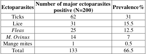 Table 1
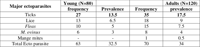 Table 1
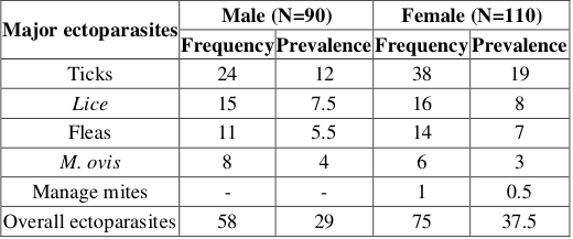 Table 3
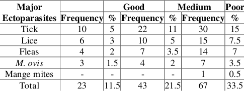 Table 4
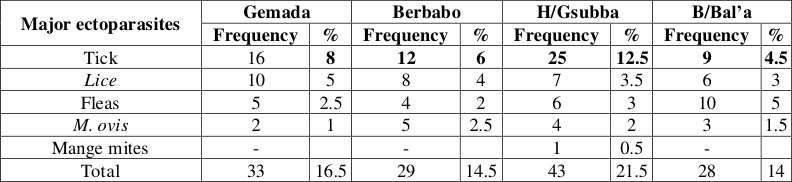 Table 5
Introduction
Ethiopia is believed to have the largest livestock population in Africa. This livestock sector has been contributing a considerable portion to the economy of a country, and still promising to rally around the economic development of the country (CSA, 2019). With an estimated population of 7.8 million equines, 1 million camels, 47.5 million cattle, 39.6 million chickens, 26.1 million sheep and 21.7 million goats. Small ruminants play a significant role in maintaining household stability by providing meat, milk, skin, and wool, generating cash income, and play traditional social and religious roles (Hiwot et al., 2020; Alemayehu and Fletcher, 1995) [17,2] . Among the small ruminants in Ethiopia, sheep are the dominant livestock, providing up to 63% of cash income and 23% of the food subsistence value obtained from livestock production (Zelalem and Fletcher, 1993) [39] . Regardless of the large size of the sheep population in the country and the huge potential therein the productivity per animal and the contribution of this sub-sector to the national economy is relatively low due to multitude of constraining factors including malnutrition, diseases, improper health care, and other management problems. In the diverse agro-climatic zones of Ethiopia, small ruminants are important source of income for rural communities and are one of the nation's major sources of foreign currency from exports. The country has, however, benefited little from this enormous resource owing to a multitude of problems, disease being the most important Infectious and parasitic diseases are common traits that affect productivity (Haileleul, 2017; Tesfaheywet, 2012) [42,43] .
Parasitic infections pose a serious health threat and limit the productivity of livestock due to the associated morbidity and mortality. Ethiopia is an ideal case for studying livestock diversity in the context of developing regions. Ethiopia can be considered as a center of livestock diversity, it is a route of sheep migration from Asia in to Africa, has large sheep population (CSA, 2006) [10], and diverse traditional sheep breeds spread across diverse ecology, communities, and production systems (Gizaw, 2008) [14] . At the national level, sheep/goat account for about 90% of the live animal/meat and 92% of skin and hide export trade value (Gizaw, 2008) [14] . At the farm level, sheep contribute as much as 22-63% to the net cash income derived from livestock production in the crop-livestock production system and are a main start of the pastoral livelihoods in the lowland pastoral system (Zelalem and Fletcher, 1993) [39] . Globally, sheep are the species with the highest number of recorded breeds, contributing 25% to the total mammalian breeds. Seventy percent of the mammalian breeds for which no risk status data are available are found in the developing world. Lack of such information, including for breeds in Ethiopia, is serious constraint to effective prioritization and planning of breed conservation measures including sustainable breeding strategies (Lyle, 1999) [22] . Production of sheep for milk, wool, hair skin, and manure is an attractive agricultural enterprise for Ethiopian farmers because of relatively low cost of breeding stock, the high reproductive rate of sheep, input, and maintenance cost to live in various conditions form desert to humid rain forest. In Ethiopia, sheep are the dominant livestock providing up to 63% of cash income and 23% of food substance value obtained from livestock production. Despite the large size of the sheep population, the productivity per animal and the contribution of this subsector to the nation's economy is relatively low. Internal and external parasitic infection and management problems are known to be the main factors that affect productivity (Gizaw, 2008) [34] . Ectoparasites feed on the outside of their host. They affect sheep and can cause severe irritation and hide damage. They are not associated with heavy mortalities in small ruminants but they are important causes of thriftiness and loss of production in affected animals. The common ectoparasites of veterinary importance in sub-Saharan Africa are lice, ticks, mange mites, sheep keds, and fleas (Kusiluka, and Kambarage, 1996) [21] . External parasites limit production in sheep in many ways the major ones are: attachment to the host causes irritation of the skin with subsequent ulceration and secondary infections, feeding in body tissue such as blood, skin, and hair, causes discomfort, weight loss, and disease transmission. In addition to this they cause huge economic losses through skin damage rendering it unsuitable for the leather industry (Lyle, 1999) [22] . Ethiopia used to get the second-largest foreign currency earnings from the export of skins and hides. This has been deteriorating due to the decrease in skin quality. Thirty years ago tanneries in Ethiopia used to produce 70% of processed skins with grades 1-3. About 10-20% of the skins were graded as poor quality. Currently, only 10-15% is in the good category while the rest are downgraded or rejected due to the increase in external parasite infestations (Hamto, 2010) [15] . This study aims to fill such gap and hence been carried out in sheep in and around West Shoa Zone Gindeberet Woreda. Therefore the objectives of this study were: to identify major eco parasites of sheep and to determine their prevalence in the study area and to assess the magnitudes of these parasites in relation to sex, age, and body condition.
Literature review 2.1 Ectoparasites Host Relationship
Ectoparasites are organisms, which inhabit the skin or outgrowth of the skin of the host for various periods (Hopla et al., 1994) [18] . The presence of external parasites on the host is termed as infestation. The association between arthropod ectoparasite and vertebrate hosts may take on variety of forms. In some cases the parasite may be totally dependent on the host, alternatively, the parasite may feed, or live only occasionally on the host, alternatively, the parasite may feed, or live only occasionally on the host, without being dependent on it (Wall and Shearer, 1997) [34] . The host provides a number of important resources for the parasite, most vitally the host supplies a source of food, which may be blood, lymph, tear or sweat, or the debris of the skin, hair, or feather. The host's body also provides the environment on which ectoparasites live, generating warmth, and moisture and within the skin or hair' a degree of protection from the external environment. The host may also provide transportation from place to place for the parasite, a site at which to mate and, in many cases, the means of transmission from host to host (Wall and Shearer, 1997) [34] . Despite the benefit of a close association with the host, there is a considerable variation in the amount of time spent on the host by various types of ectoparasites. Some ectoparasites such as many species of lice live in continuous association with their host throughout their life cycle and are therefore, highly dependent on the host. The majority of ectoparasites, however, has only intermittent contact with their hosts, and are free-living for the major portion of their life cycles. In some cases, ectoparasites, such as many species of mite, are highly host-specific; only one host species is exploited and, in some instances, the parasite can exist only on one defined area of the host. Other species are able to exploit a wider range of hosts (Wall and Shearer, 1997) [34] .
Major Ectoparasites of Sheep
Wide range of skin parasites can affect sheep, with resultant loss or damage to fleece and economic loss associated with impaired production. Some parasites are important in their own right and some are more important as vectors of other infections. Ectoparasites that can potentially infest the skin of sheep include lice, mites, sheep ked, ticks, and blowfly larvae (FAO, 2001) [12] . The general clinical signs, which are irritation, fleece loss, and dermatitis, are common to many infestations, and laboratory examinations may be required to establish an accurate diagnosis. Some non-parasitic diseases, such as Dermatophytosis or Scrapie have behavioral signs and skin lesions which resemble parasitic infestation. Fleece loss, especially during winter months, has a severe effect on affected sheep and their resistance to cold stress (Appleyard and Bailie, 1984) [4] .
Lice
Lice are obligates of the class insect, the species are hostspecific. Two suborders of lice are recognized Mallophaga and Anoplura. The Mallophaga are biting lice which have mouth parts adapted specially for chewing the epithelial debris of the skin. The Anoplura are sucking lice that have pointed heads and mouthparts. These lice are adapted for sucking tissue fluids and blood from the host (Radostits et al., 1994., Jubb et al., 1993) [26,19] . The Anoplura are also vectors of Rickettsia, (Typhus) and Borrelia (Relapsing fever) in human beings (Cox, 1986) [8] . There are approximately 540 valid species of sucking lice recognized, all of which are obligate hematophagous ectoparasites of mammals. By virtue of blood feeding, sucking lice are generally more at transmitting pathogens to animals than are chewing lice. Different species of chewing lice parasitize both mammals and birds but sucking lice parasitize only mammals (Hopla et al., 1994) [18] . The general clinical signs, which are irritation, fleece loss, and dermatitis, are common to many infestations, and laboratory examinations may be required to establish an accurate diagnosis. Some non-parasitic diseases, such as Dermatophytosis or Scrapie have behavioral signs and skin lesions which resemble parasitic infestation. Fleece loss, especially during winter months, has a severe effect on affected sheep and their resistance to cold stress (Appleyard and Bailie, 1984) [4] . Lice usually are unable to survive for more than 1-2 days off their host and tend to remain with a single host animal throughout their lives. Most species of louse are highly host specific but there are reports of naturally occurring transmission of the goat louse Damalinia caprea to sheep and experimental transmission of Damalina ovis to goats (Jubb et al., 1993) [19] . Many species specialize in infesting only one part of their host body (Wall and Shearer, 1997) [34] and transfer to new hosts is by body contact, particularly under condition of close confinement (Sewell and Brockes by, 1990; Peter, 1995) [49,25] . To allow them to survive as permanent ectoparasites, lice show a number of adaptations that enable them to maintain a life of intimate contact with their hosts.
Life cycle of lice
Lice of the two span of about a month the female lays 200-300 operculate eggs ('nits'). These are whitish and are glued to the hair where they may be seen with the naked eye. There is no true metamorphosis and from the egg hatches a nymph, similar to, though much smaller than, the adult. After three mounts, the fully grown adult is present. The whole cycle from egg to adult is present. The whole cycle from egg to adult takes two to three weeks (Radostits et al., 1994) [26] .
Pediculosis in sheep
The suckling lice in sheep are essentially parasites of the haired regions of the body, invading the wooly areas only when the population is expanding rapidly. They are not very active and have a gregarious habit, feeding in swarms. The biting louse of sheep is active and usually found in wooly areas (Urquhart et al., 1996) [32] . Though Linognatus spp. can cause anemia, it is Damalina usually considered to by the more pathogenic. Reduction in the value of the wool clip is economically the most important consequence of ovine pediculosis (Georgi, 1985) [13] . There are a number of morphologically similar host-specific species, the species that commonly affect Damalina ovis on sheep; Damalina caprea, Damalinia limbata and Damalina crassiceps on goats. The sheep-chewing lice, Damalina ovis, is one of the most common lice found on sheep, is D. ovis has typical life cycle. The female deposits about two eggs, attached to the wool or hair next to the skin by a viscid substance, every three days. The egg hatch in 9-10 days, and the nymph matures in about 21 days (Bay and Harris, 1988) [5] . Infestations with Damalina ovis occur over all areas of the body but the upper sides of the animal are favored. This species moves rapidly over the wool fiber but is usually found near the skin (Bay and Harris, 1988) [5] . Being highly active, Damalina ovis is usually considered to be most pathogenic in sheep and it can cause great irritation so that the sheep are restless and have their grazing interrupted. Exuded serum from the bite wounds causes wool matting. Rubbing leads to wool loss. Wounds may attract blowflies (Wall and Shearer, 1997) [34] . Like sheep kids, D. ovis is also associated with the development of cockle (Health et al., 1996) [16] . Out of more than 50 species of Linognathus described, the species that parasitize sheep includes; the face louse Linognathus ovilus, Linognathus africanus, and the foot louse Linognathus pedalis (Wall and Shearer, 2001) [35] . The adult female lays a single egg per day. Eggs hatch in 10-15 days; giving rise to nymph which requires about 2 weeks to pass through three nymphal stages. The egg to adult-life cycle requires about 20-40 days. The face louse, L. ovillus, usually occurs in colonies on the ear and face of sheep. The preferred sites for L. pedalis are the feet, legs, and scrotum. At high densities, however, both species may spread over the entire body. L. pedalis can survive for several days off the host. So the infestation may be picked up of contaminated pasture (Wall and Shearer, 1997) [34] . The damage caused is due to irritation which interferes with feeding causing decreased weight gain, and scratching results in wool loss, cuts, and bruises. Excretion of the lice soils the wool (Bay and Harris, 1988) [5] .
Epidemiology of Pediculosis
Lice cannot live away from their hosts for more than a few days. Consequently spread of infestation occurs mainly by direct contact among hosts and generally close bodily contact is necessary for the transfer of louse infestation (Urquhart et al. 1996; Jubb, et al., 1993) [32,19] . Pediculosis shows a seasonal periodicity with very low numbers in the summer when conditions are hot and becoming worse in winter. This is probably because the lice, which are sensitive to heat and low humidity cannot survive in the body fleece where temperature could be very high (Radostits et al., 1994; Jubb et al., 1993) [26,19] .
Pathogenesis of Pediculosis
Though Linognatus spp. Can cause anemia, it is Damalina which is considered to be the more pathogenic. Being very active it can cause great irritation (Urquhart, et al., 1996) [32] . All species cause irritation of the skin and stimulate scratching, rubbing, and licking leading to restlessness, damage to fleece and skins, and loss of milk production (Souls by, 1982; Radostits et al., 1994) [30,26] .
Melophagus ovinus
Commonly called the sheep 'ked', M. ovinus is a hairy, wingless insect approximately 5-7 mm long with a short head and broad, flattened, brownish thorax and abdomen. It has strong eggs provided with claws and is a permanent ecto-parasite (Urquhart, et al., 1996) [32] . Kids spread by contact and long-woo-led breeds appear to be particularly susceptible. The irritation caused by these parasites also results in animals biting and rubbing with resultant damage to the fleece (Charles, 1991) [7] .
Life Cycle of Melophagus
Adults live for several months and the larvae produced by the females adhere to the wool. These are immobile and pupate immediately, the 3-4mm long brown pupae being easily visible on the fleece. Adult kids emerge in approximately three weeks in summer, but this period may be extended considerably during winter. Ked population builds up slowly since only one larva is produced by each female every 10-12 days, up to a total of 15. Adult and pupae can only live for short periods of their hosts. M. ovinus is the vector of the non-pathogenic Trypanosoma melophagium. Heavy infestations of keds are most commonly seen in autumn and winter, which might lead to loss of condition and anemia. Keds spread by contact and long-woo-led breeds appear to be particularly susceptible. The irritation caused by these parasites also results in animals biting and rubbing with resultant damage to the fleece (Urquhart, et al., 1996) [32] .
Mites
Mites are tiny arthropods, usually less than 1 mm in size, and can be difficult to see and identify without the aid of a microscope or at least a hand lens. There are more than 200 families of mites and many thousands of species. Most mites are free living and feed on plant juices or prey up on other arthropods. Some mites have evolved to become important ectoparasite pets of animals. species of mites have even become ectoparasites, pets of animals. Some species of mites have even become ectoparasites, invading the ears, bronchi, and lungs, nose, and other tissues of animals. More than 50 species of mites live on or in the bodies of domestic animals. In general, mites can affect the health of animals in four ways: they damage tissues and cause dermatitis, cause blood or body fluid loss, cause allergic reactions and create conditions for secondary bacterial infection (Lyle, 1999) [22] . Scabies, a chronic contagious dermatitis of sheep and other domestic animals, is characterized by encrustation, denudation, and itching of the skin and is caused by five species of mites. Although most species of domestic animals are susceptible to scabies, a high degree of host specificity prevents transfer of parasites transfer between host species. This fact validates the separate control and eradication programs for scabies of sheep and scabies of cattle (Lyle, 1999) [22] . The parasitic mites are small, most being less than 0.5mm long, though a few blood-sucking species may attain several millimeters when fully engorged. With few exceptions they are in prolonged contact with the skin of the host, causing various forms of the condition generally known as manage. Although, like the ticks, mites are obligate parasites, they differ from them in important respect that most species spend the entire life cycles, from egg to adult, on the host so that transmission is mainly by contact. Unlike the ticks, once infection is established, pathogenic populations can build up on an animal without further acquisitions (Lyle, 1999) [22] .
Life Cycle of Mites
The generalized life cycle of mites can be described as follows. Mites' female lays eggs. The eggs hatch and sixlegged larvae emerge. These larvae feed and molt to the eight-legged nymph stage. Later, after feeding the nymphs molt and become adult male or female mites. This entire life cycle can take as little as eight days to as long as four weeks, depending on the species of mite, and temperature and humidity (Yakhchali and Robert, 1999) [38] .
Epidemiology of Mites
Clinically affected and carrier animals are source of infection. Transmission occurs by direct contact and contaminated fomites can be source of infection. Overcrowding of animals in houses, markets, dips, and communal grazing land facilitates rapid spread of the parasites. Kids and lambs are more severely affected than adult animals. Moist conditions and undercurrent infections increase the susceptibility of animals to manage mites (Kusiluka and Kambarage, 1996) [21] . The major species that cause mange in sheep belongs to the four genera of mite, namely Sarcoptes, Psoroptes, Chorioptes, and Demodex from which Sarcoptes and Demodex are burrowing while Psoroptes and Chorioptes are non-burrowing in their nature (Charles, 1991) [7] .
Ticks
Ticks are obligate, blood-feeding ectoparasites of vertebrates, particularly mammals and birds, and the most important group of ectoparasites, primarily because they feed on blood and tissue fluids in order to develop and because of the wide range of pathogenic agents that they transmit. In addition, they cause local irritation at the site of feeding, blood loss from severe infestations, wounds as sites for secondary infection, and tick paralysis (Wall and Shearer, 2001) [35] . Ticks are divided into two families: Argasidae (soft-bodied ticks), a relatively small group comprising 170 species, and Ixodidae (hard ticks); a larger group comprising over 650 species. Hard ticks are more common ectoparasites of mammals, in part because of their widespread distribution and prolonged association with the host while bloodfeeding. Ticks are primarily parasites of wild animals and only about 10% of species feed on domestic animals, primarily sheep and cattle (Wall and Shearer, 2001) [35] . Ixodid ticks are one of the most economically important ectoparasite of livestock in tropical and sub-tropical parts of the world. Because of the direct and indirect effect on their host, ticks are considered to be not only a significant threat to successful livestock production but also serious interference with economy of the country (Zenebe, 2005) [41] . More than 60 species of ticks infesting both domestic and wild domestic animals have been recorded in Ethiopia (Feseha, 1983) [44] . Among these about 37 species and subspecies are very wide spread and important parasites of livestock (Dalgliesh et al., 1990) [45] . In Ethiopia, tick and tick-borne diseases cause considerable losses to the livestock economy, ranking third among the prevalent parasitic diseases, after trypanosomes and endo-parasitism (Pegram et al., 1981; Zeleke and Bekele, 2004) [24,40] . Ticks undergo four life stages: egg, larva (3 pairs of legs), nymph (4 pairs of legs and no genital pore), and adult (4 pairs of legs and a genital pore). The life cycle of ticks vary widely. Some species pass their entire life on the host, others pass different stages of the life cycle on successive hosts, and others are parasitic only at the certain stages (William et al., 2001) [46] . Hard ticks require three blood meals for development and to complete the life cycle. Each stage blood feeds once, detaches from the host, and molts to the subsequent life stage on the ground. Often the larva, nymph, and adult feed on different hosts (i.e. three host ticks). Some species of hard ticks are one-host ticks (all stages feed on the same individual host). Most of the life cycle of one-host ticks occurs on the host with only gravid females, egg masses, and host-seeking larvae present on the ground. Females and immature hard ticks become greatly distended when bloodfed; females, for instance, often ingest more than 100 times their body weight. Blood meals are used for molting to the next stage or production of eggs. Eggs are laid in a mass of 100-10,000 in 3-30 days (depending on species and temperature) they are deposited on the soil, in a crevice, or beneath leaves. Males generally obtain small blood meals and expand little in size. Hard ticks feed relatively slowly and remain on the host 3-14 days before detaching. After feeding as immature, molting occurs after an interval that varies between species and with temperature (Wall and Shearer, 2001) [35] .
Life cycle of ticks
In the hard ticks mating takes place on the host, except with Ixodes where may also occur when the ticks are still on the vegetation. Male ticks remain on the host and will attempt to mate with many females whilst they are feeding. They transfer a sack of sperm (= sperm theca) to the females. The females mate only once before they are ready to engorge fully with blood. When they finally engorge they detach from the host and have enough sperm stored to fertilize all their eggs. Females' hard ticks lay many eggs (2000 to 20000) in a single batch. Female aramid ticks lay repeated small batches of eggs. Eggs of all ticks are laid in the physical environment, never on the host (Walker et al., 2003) [33] .
Three-host tick life cycle
This is the most common type of life cycle. Larvae develop in the eggs until ready to hatch, usually in several weeks. Larvae feed once on a host, then detach from the host and hide in sites such as soil or vegetation. They molt to nymphs. Nymphs feed once and molt in the same way as larvae. From the nymphal molt either a female or male hatches. The female feeds once and lays one huge batch of eggs. The depleted female then dies. The male may take several small feeds, mate, and then die. Ticks that have recently hatched from eggs or from molting have soft bodies and are inactive for one to two weeks until the external body wall hardens. The life cycle of three-host ticks is slow, from six months to several years (Walker et al., 2003) [33] .
One and two-host tick life cycles
This is a less common type of life cycle but it occurs in the entire Boophilus sub-genus of the Rhipicephalus genus and in other genera. Eggs are laid on soil. Larvae hatch after several weeks of development and crawl on to vegetation to quest for a host. When they have completed feeding they remain attached to the host and mounting occurs there. The nymphs then feed on the same host and also remain attached. After another moult, the adults hatch and then feed on the same host. The adults will change position on the same host for mating. Thus all three feedings of any individual tick occur on the same individual host. The life cycle of one-host life cycle is similar but only the larvae and nymphs feed on the same individual host, and the adults will feed on another host. Hyalomma detritus detritum and Rhipicephalus evertsi evertsi have two host life cycles (Walker et al., 2003) [33] .
Materials and Methods
Materials and Methods
Study area
The study was conducted from June 20, 2017, to January 2018 in and around West Shoa Gindeberet Woreda which is located in Oromia Regional Administration at 192 km West of Addis Ababa. The town has altitude of about 8°28'-10°65'W and longitude of about 26°42'-33°37'W and an elevation ranging from 1300-4500 M.A.S.L. The study area receives a mean annual rainfall of about 1800 mm which comes from long and short rainy seasons. The average minimum and maximum annual temperature ranges between 12.5 and 25.7 °C, respectively. The climatic condition of the area is weyenadega. Agriculture is the livelihood for more than 90% of the population in a rural community of the zone. The main agricultural system is mixed crop livestock production and animals are mainly produced in an extensive system. Gindeberet district has a livestock population of 35545 bovines, 6300 goats, and 12400 sheep, and 12540 horses, 6785 donkeys, and 4378 mules. The livestock production is characterized by extensive production system in which indigenous cattle are kept under traditional management (CSA, 2015).
Study Population
The study animals were sheep kept under the traditional extensive management system of indigenous breeds with different ages, sexes, and body conditions found in and around the Gindeberet district, Western Shoa zone.
A cross-sectional study was employed to assess the ectoparasites of sheep in the area, their prevalence, and the magnitude of these parasites in relation to age, sex, and body condition. A total of 200 sheep were selected randomly. The animal sex groups (male, female), age groups (Adult and young), and body condition (good, medium, and poor) were recorded.
Sampling and Sample Size Determination
The total sample size required for the study was calculated according to a formula given by Thursfield (2005) [31] . Therefore, a total sample size of 200 sheep was surveyed. Sampling techniques were used to determine based on the expected prevalence (50%) of sheep and the 5% desired absolute precision or level of precision and 95% confidence level value. Accordingly, 384 animals were supposed to be done; however, we were able to manage to examine 200 sheep.
Method of data collection
A questionnaire survey was carried out to estimate the prevalence of the major ectoparasites of sheep in and around West Shoa Zone Gindeberet Woreda. Cross-sectional and retrospective studies were conducted from June 20, 2017, up to October 2017 to determine the prevalence of the major ectoparasites of sheep in Gindeberet Woreda western, Ethiopia i.e. for the year 2010 Ethiopian Calendar). For the purpose of the prevalence major ectoparasites to identifying by visual observation sheep in Gindeberet Woreda. The total sample size of sheep in observation N=200 and N=133 were included in the study. The sheep-level characteristics incorporated in the questionnaire were ticks, lice, M. ovinus, fleas, and mange mites.
data analysis method
All data was entered in to Microsoft Excel 2003 and analyzed with SPSS version 20 for Windows program. For the analysis of different attributable factors and problems, recorded data were entered into spread sheet of Microsoft Excel. The appropriate statistical analysis was used to analyze were the tests used to analyze the data pertaining to the sex, age, and body condition, and overall prevalence of ectoparasites. Significance difference was considered when p-value is less than 0.05.
Results
Results
Overall prevalence of ectoparasites survey finding and major ectoparasite
Out of 200 sheep examined for ectoparasites 133(66.5%) sheep were infested with major ectoparasites (Table 1). The major ectoparasites identified were ticks 31%, followed by lice 15.5%, fleas 12.5%, Melophagus ovinus 7%, and Mange mites 0.5% (Table 1).
| Major ectoparasites | Young (N=80) | — | — | Adults (N=120) |
|---|---|---|---|---|
| frequency | Prevalence | Frequency | prevalence | |
| Ticks | 27 | 13.5 | 35 | 17.5 |
| Lice | 13 | 6.5 | 18 | 9 |
| Fleas | 10 | 5 | 15 | 7.5 |
| M. ovinus | 6 | 3 | 8 | 4 |
| Mange mites | - | - | 1 | 0.5 |
| Total Ecto parasite | 63 | 32.5 | 70 | 34 |
Table text extracted from the original PDF; superscript letters are shown inline. The original table image is below.
View original table image
| Ectoparasites | Number of major ectoparasites positive (N=200) | Prevalence% |
|---|---|---|
| Ticks | 62 | 31 |
| Lice | 31 | 15.5 |
| Fleas | 25 | 12.5 |
| M. Ovinus | 14 | 7 |
| Mange mites | 1 | 0.5 |
| Total | 133 | 66.5 |
Table text extracted from the original PDF; superscript letters are shown inline. The original table image is below.
View original table image
The commonest sites of ticks' attachment sites were the feet, especially the area just below the fetlock joint, head/ ear, belly, and tail, while for lice attachment was the skin of neck, shoulder, flank, and rump. For M. ovinus infested sheep the sites commonly parasitized were the neck shoulder, rump, and belly.
Prevalence of ectoparasites in different age groups
The prevalence of major ectoparasites regarding to the two age groups of study sheep was higher in adult sheep 34%) and in young sheep (32.5%), while lower prevalence both age groups was observed (Table 2) of these the most prevalent ectoparasites were ticks with the prevalence of 13.5% in young (lamb) and (17.5%) in adult followed by lice with prevalence of (6.5%) in young and 9% in adults.
The least prevalent ectoparasite was mange mites with a prevalence of (0.5%) in adults but no prevalence in young (Table 2).
Prevalence of ectoparasites by sex groups
The overall prevalence of ectoparasites of sheep based on sex was 29% in males and 37.5% in females respectively of these the most prevalent ectoparasite was tick with a prevalence of 19% in females and 12% in males. The second most prevalent ectoparasite was lice7.5% in females while in males 8%. The least prevalent ectoparasite was mange mites with a prevalence of 0.5% in males and without observed prevalence in females (Table 3).
| Major ectoparasites | Male (N=90) | — | Female (N=110) | — |
|---|---|---|---|---|
| Frequency | Prevalence | Frequency | Prevalence | |
| Ticks | 24 | 12 | 38 | 19 |
| Lice | 15 | 7.5 | 16 | 8 |
| Fleas | 11 | 5.5 | 14 | 7 |
| M. ovis | 8 | 4 | 6 | 3 |
| Manage mites | - | - | 1 | 0.5 |
| Overall ectoparasites | 58 | 29 | 75 | 37.5 |
Table text extracted from the original PDF; superscript letters are shown inline. The original table image is below.
View original table image
Discussion
The high prevalence of ectoparasites was recorded in this study (66.5%), which is suggestive of the importance of these health problems in the sheep population of the study area, poor management, and low level of awareness of sheep owners on the effect of ectoparasites are believed to have contributed to the widespread occurrence of the parasites. Tick infestations were the most prevalent ectoparasites recorded with a prevalence of 31% from the 62 sheep positive for ticks. Ticks were observed significantly affecting poor body condition (15%) sheep more than medium (11%) and good (5%) body condition sheep. Of these was observed among body condition of the study animals for the occurrence of ectoparasites in sheep (Table 4). This might have resulted from the nature of the ticks, which can infect different types of animals of all ages and sex groups. When compared to observations by Sertse, (2004) [27] (3.8%) in Amhara region and Ali (2009) (6.6%) in eastern Ethiopia the prevalence of ticks in the study area was higher but when compared with observations by Yakob et al., (2008) [37] in southern Ethiopia (31.78%) and Abunna et al., (2000) [1] in Mieso (89.7%) it shows decreased figures. Differences in the environmental conditions (study season and design) could have contributed for this variation. Indeed, low tick prevalence may be related to unfavorable climatic conditions (intermittent and weak rainfall in the Jimma area during the study period coupled to relatively moderate temperatures).
| Major Ectoparasites | — | Good | — | Medium | — | Poor |
|---|---|---|---|---|---|---|
| Frequency | % | Frequency | % | Frequency | % | |
| Tick | 10 | 5 | 22 | 11 | 30 | 15 |
| Lice | 6 | 3 | 10 | 5 | 15 | 7.5 |
| Fleas | 4 | 2 | 7 | 3.5 | 14 | 7 |
| M. ovis | 3 | 1.5 | 4 | 2 | 7 | 3.5 |
| Mange mites | - | - | - | - | 1 | 0.5 |
| Total | 23 | 11.5 | 43 | 21.5 | 67 | 33.5 |
Table text extracted from the original PDF; superscript letters are shown inline. The original table image is below.
View original table image
The overall lice prevalence obtained in this study is lower than the findings of Sertse, (2004) [27] 39.8% in Amhara region prevalent ectoparasites recorded with a prevalence of 15.5% from the 31 sheep-positive Yacob et al., (2008) [37] 25.7% in southern Ethiopia, Sodo, and Wondimu, (2010) [36] 75.5% in Arsi zone. Such differences in prevalence with the above observations may arise from differences in agro climate, management and health care of animals, know-how farmers about ectoparasites in the study sites, and the sensitivity of the diagnostic method used to reveal ectoparasites. Louse infestations may indicate some other underlying problems such as malnutrition and chronic diseases (Wall and Shearer, 1997) [34] . The possible reasons for such low prevalence of lice in the study area include; medium sanitation, medium feeding and management, and improper utilization of veterinary services. Flea infestation important ectoparasite problem encountered, in the study area with an overall prevalence of 12.5% from the 25 sheep positive for fleas. The prevalence of fleas in this study was found to be lower than the observations made by Bekele et al., (2011) [6] in Woomera, (32.31%), Yakob et al., (2008) [37] and the greater than in Wolayita Sodo (11.21%), Sertse, 2004 [27] in Amhara Region (0.2%) and Ali, 2009 in Eastern Ethiopia (0.4%) compared the result of observation.
M. ovinus was one the ectoparasites observed on sheep accounting for (7%) from the 14 sheep positive for M. ovinus overall prevalence. Although infestation with M. ovinus does produce any marked changes in health of sheep, their presence leads to irritation and staining of the wool by the feces of the ked. The irritation results in animal biting and rubbing with resultant damage to the fleece and development of a vertical ridging of the skin called 'cockle'
(Urquhart et al., 1996; Wall and Shearer, 1997) [32,34] .
The prevalence of M. ovis in this study was lower than the observations made by Amin Ali, (2009) [3] (11.25%) in eastern Ethiopia, in the Amhara region by Sertse, (2004) [27] (12.1%), in Tigray by Kassaye [26] in the hot, humid tropics the parasite is restricted to cooler highlands, and infestations may be lost when sheep are moved to hot dry areas. Kettle, (1984) [47] suggested as an account for this fact; temperature may play an important role in the dynamics of the Keds. In addition to environmental temperature differences, the variation in prevalence of sheep ked might have resulted from differences in the breeds of sheep which might contribute to different to differences in woolliness of coats, since heavily wool-led sheep are more suitable for ked colonization.
In the current study, the overall prevalence of mange mite was 0.5% from the 1 sheep positive for mange mite overall prevalence. The prevalence was less than observations made in southern Ethiopia (1.99%) by Sheferaw et al., (2010) [29] , in northern Ethiopia (11.8%) by Demissie et al., (1999) [11], and in Tigray (0.95%) by Kassaye and Kebede, (2010) [20] . According to Pangui (1994) [23] high temperature, humidity, and sunlight favor mange mite infestation. The higher temperature, humidity, and sunlight that prevail in lowland and midland may have accounted for the differences in prevalence.
Conclusion and Recommendations
Ectoparasites are among the major causes of sheep production constraints and quality deterioration of exported skin in Ethiopia. The prevalence of ectoparasites in sheep in the present study is high and can potentially result in high economic losses through decreased production and skin damage. Ticks were the most prevalent ectoparasites followed by lice whereas mange was the least prevalent parasite species. The rate of ectoparasitic infestation is high in adults and poor body-conditioned animals as compared to the young age and sheep in good body conditions respectively. Therefore, based on the above conclusion the following recommendations are forwarded: The ectoparasitic infestation in the sheep population of the study area should be reduced by using appropriate ectoparasite control methods and improving the management system.
Strategic prevention and control of ectoparasites should be established through regular and scheduled uses of acaridae sprays and dipping techniques. The farming community should be aware about the effects of these parasites and the possible control and prevention methods. Further study should be done to have full picture of the distribution and seasonal occurrences of these ectoparasites and the associated economic losses.
Acknowledgment
Above all, we would like to praise the Almighty God who helped me in every aspect of our lives and the conditions for our success. Secondly, our great pleasure goes to express our sincere gratitude and appreciation to the Veterinary Teaching Hospital of Addis Ababa University and Modjo Veterinary Clinic staff members, SPANA, Biomedical, Microbiology, Parasitology, Clinical pathology, and VTH multipurpose laboratory workers who are involved directly or indirectly in this research work.
Figures and tables from the paper
| Major ectoparasites | Gemada | — | Berbabo | — | H/Gsubba | — | B/Bal’a | — |
|---|---|---|---|---|---|---|---|---|
| Frequency | % | Frequency | % | Frequency | % | Frequency | % | |
| Tick | 16 | 8 | 12 | 6 | 25 | 12.5 | 9 | 4.5 |
| Lice | 10 | 5 | 8 | 4 | 7 | 3.5 | 6 | 3 |
| Fleas | 5 | 2.5 | 4 | 2 | 6 | 3 | 10 | 5 |
| M. ovis | 2 | 1 | 5 | 2.5 | 4 | 2 | 3 | 1.5 |
| Mange mites | - | - | 1 | 0.5 | - | |||
| Total | 33 | 16.5 | 29 | 14.5 | 43 | 21.5 | 28 | 14 |
Table text extracted from the original PDF; superscript letters are shown inline. The original table image is below.
View original table image
References
23 in the original paper- Abunna F, Kasasa D, Shelima F, Megersa F. Survey of tick infestation in small ruminants of Miesso district, West Harergie, Oromia Region, Ethiopia. Tropical Animal Health and Production. 2000 Aug;41:969-972.
- Alemayehu Z, Fletcher I. Small ruminant productivity in the central Ethiopian mixed farming system. Institution of Agriculture proceeding. 1995;4:19411947.
- Bekele J, Tariku M, Abebe R. External parasite in small ruminants in Wolmera district of Oromia Region, Central Ethiopia. Journal of Animal Veterinary Advances. 2011;10(4):518-523.
- Demissie A, Siraw B, Teferi K, Tsertse T, Mammo G, Mekonnen D, et al. Mange: A Disease of Growing Threat for the production of small ruminants in the Amhara National Regional State. Journal of Animal and Veterinary Advances. 1999;18(5):6-49.
- Hamto D. Estimation of weight and age of sheep and goats. ESGPIP Technical Bulletin No. 2010;23:8-10.
- Health AC, Bishop DM, Cole DJWP, Feffer AT. The development of cockle, a sheep pelts defect, in relation to Bovicola ovis, the sheep biting louse. Veterinary Parasitology. 1996;67:259-267.
- Wodajo HD, Gemeda BA, Kinati W, Mulem AA, Eerdewijk AV, Wieland B. Contribution of small ruminants to food security for Ethiopian smallholder farmers, Small Ruminant Research. 2020 Mar 1;184:106064, ISSN: 0921-4488, https://doi.org/10.1016/j.smallrumres. 2020.106064.
- Hopla CE, Dureden LA, Keirans JK. Ectoparasites and Classification. Revue Scientifique et Technique -Office International Des Épizooties. 1994;13(4):985-1012.
- Kassaye E, Kebede E. Epidemiological study on manage mite, lice and sheep keds of small ruminants in Tigray region, northern Ethiopia. Ethiopian Veterinary Journal. 2010;14(2):51-65.
- Lyle G. Sheep Scabies. Navajo Sheep Project. 1999;5(3):43-45.
- Panqui L. Mange in domestic animals and methods of control. OIF -Review. 1994;13(4):1227-1247.
- Pegram R, Hoogstral H, Wassef Y. 'Ticks (Ixodidae) of Ethiopia: Distribution, ecology and host relationship of tick species affecting livestock', Bulletin of Entomological Research. 1981;71(3):339-359.
- Chandran D. Bovine babesiosis: A general review. International Journal of Veterinary Sciences and Animal Husbandry. 2021;6(3):40-44.
- Sheferaw D, Degefu H, Banteyirgu D. Epidemiological study of small ruminant mange mites in three Agroecological zones of Wolaita, Southern Ethiopia. Ethiopia Journal of Veterinary. 2010;14(1):31-38.
- Urquhart G, Armour J, Duncan J, Dunn A, Jennings FW. Veterinary Parasitology, 2 nd Ed., Black well Science Ltd, UK, P. 141-205. Veterinary Journal. 1996;9(1):9-16.
- Yacob T, Yalew A, Dinka A. Ectoparasite prevalence in sheep and in goats in and around Wolaita Soddo, Southern Ethiopia. Review of Medical and Veterinary. 2008;159(8-9):450-454.
- Yakhchali M, Robert M. Factors affecting the occurrence of mites and ticks. Veterinarski Archv. 1999;76(5):431-442.
- Zeleke M, Bekele T. Species of ticks on camels and their seasonal dynamics in Eastern Ethiopia. Tropical Animal Health and Production. 2004;36:225-231.
- Negussie H, Gizaw D, Tessema TS, Nauwynck HJ. Equine herpesvirus-1 Myeloencephalopathy, an emerging threat of working equids in Ethiopia. Transboundary and Emerging Diseases. 2017 Apr;64(2):389-397.
- Tesfaheywet Z. Helminthosis of sheep and goats in and around Haramaya, South Eastern Ethiopia. Journal of Veterinary Medicine and Animal Health. 2012;4(3):4855.
- Dalgliesh RJ, Jorgensen WK, VOS DAJ. Australian frozen vaccines for the control of babesiosis and anaplasmosis in cattle: A review. Tropical Animal Health and Production. 1990;22(1):44-52.
- Srinivas S, Watanabe T, Lin CS, William CM, Tanabe Y, Jessell TM, et al. Cre-reporter strains produced by targeted insertion of EYFP and ECFP into the ROSA26 locus. BMC developmental biology. 2001;1(1):1-8.
- Sennuga SO, Solarin LWI, Adeoye WA, Alabuja FO. Extension's role in improving livestock production: Information needs, institutions and opportunities. International Journal of Agriculture and Nutrition. 2022;4(2):43-51.